


Abstract
We performed a systematic review and metaanalysis of the performance of 68Ga-DOTA–conjugated somatostatin receptor–targeting peptide (68Ga-DOTA-SST) PET in the detection of pheochromocytomas and paragangliomas (PPGLs). Methods: PubMed and Embase were searched until May 8, 2018. We included studies that reported the detection rate of 68Ga-DOTA-SST PET in patients with PPGLs. Detection rates were pooled using a random-effects model. Subgroup analyses and metaregression were performed to explore the cause of heterogeneity. Results: Thirteen studies were included for qualitative synthesis. Per-lesion detection rates of 68Ga-DOTA-SST PET were consistently higher (ranging from 92% to 100%) than other imaging modalities, including 18F-fluorohydroxyphenylalanine (18F-FDOPA) PET, 18F-FDG PET, and 123/131I-metaiodobenzylguanidine (123/131I-MIBG) scintigraphy. However, in patients with polycythemia/paraganglioma syndrome, the detection rate of 68Ga-DOTA-DOTATATE PET was 35%. Nine studies (215 patients) with no specific inclusion criteria for subtype were quantitatively synthesized. The pooled detection rate was 93% (95% confidence interval [CI], 91%–95%), which was significantly higher than that of 18F-FDOPA PET (80% [95% CI, 69%–88%]), 18F-FDG PET (74% [95% CI, 46%–91%]), and 123/131I-MIBG scan (38% [95% CI, 20%–59%], P < 0.001 for all). A greater prevalence of head and neck paragangliomas was associated with higher detection rates of 68Ga-DOTA-SST PET (P = 0.0002). Conclusion: 68Ga-DOTA-SST PET exhibited superior performance for lesion detection, over other functional imaging modalities, in patients with PPGLs, with the exception of polycythemia/paraganglioma syndrome. This might suggest 68Ga-DOTA-SST PET as a first-line imaging modality for the primary staging of PPGL or the restaging of PPGL with unknown genetic status.
Pheochromocytomas and paragangliomas (PPGLs) are tumors arising from sympathetic lineage-derived cells in adrenal medulla and extraadrenal thoracic and abdominal paraganglia or from the parasympathetic nervous system in the head and neck (1). Functional imaging plays an important role in the confirmation of diagnosis, staging or restaging, selection of targeted radionuclide therapy, and response evaluation in patients with PPGLs (2). 18F-fluorohydroxyphenylalanine (18F-FDOPA) PET is one of the standard diagnostic work-up for nonmetastatic PPGLs in the current guidelines (2–4). In a metaanalysis, the pooled lesion-based sensitivity and specificity of 18F-FDOPA PET were 79% and 95%, respectively (5). However, the diagnostic performance of 18F-FDOPA PET is largely influenced by tumor location and genetic status (6). 18F-FDG PET is recommended in metastatic PPGLs with succinate dehydrogenase A–D (collectively, SDHx) mutation and unknown or negative genetic mutations (2,3). A previous metaanalysis showed that the pooled sensitivity and specificity of 18F-FDG PET for metastatic PPGLs at a per-lesion level is 83% and 74%, respectively (7). 123I-metaiodobenzylguanidine (123I-MIBG) scintigraphy has excellent sensitivity and specificity on a per-patient basis (8,9); however, its lesion-based diagnostic accuracy is limited (2).
Because PPGLs express high levels of somatostatin receptor (SSTR) (10–12), 68Ga-DOTA–conjugated somatostatin receptor–targeting peptides (68Ga-DOTA-SST) PET have shown an excellent lesion-based accuracy in detection of PPGLs (13–21). Recent publications suggest that 68Ga-DOTA-SST PET provides a high detection rate across a wide range of mutations (22–25). However, because of the small number of subjects in individual studies, it is difficult to conclude a higher level of evidence.
Therefore, we performed a systematic review and metaanalysis to evaluate the performance of 68Ga-DOTA-SST PET for lesion detection in patients with PPGLs.
MATERIALS AND METHODS
This systematic review and metaanalysis adhered to the Preferred Reporting Items for Systematic Reviews and Meta-Analyses guidelines (26). The protocol was registered to the International Prospective Register of Systematic Reviews (registration no. CRD42018085906). The research question for this metaanalysis was as follows: “What is the performance of 68Ga-DOTA-SST PET for lesion detection in patients with PPGL, compared with histopathologic results or best value comparator (BVC; a combination of imaging, clinical, or biologic studies)?”
Search Strategy
A computerized search on PubMed and Embase databases was performed until May 8, 2018. The search query included key words of “pheochromocytoma/paraganglioma,” “68Ga-DOTA-SST PET,” and their related terms, as follows: (paraganglioma OR paragangliomas OR paragangliom* OR pheochromocytoma OR pheochromocytomas OR pheochromocytoma* OR feochromocytoma*) AND (Gallium OR Ga) AND (DOTA* OR somatostatin) AND (“PET” OR PET). Reference lists of the retrieved articles were also checked to identify additional relevant articles. The search was not limited to any particular language.
Study Selection
Studies were included based on “Patient/Intervention/Comparator/Outcome/Study design” (PICOS) criteria (26): (1) “patients” with PPGL, (2) 68Ga-DOTA-SST PET as “intervention,” (3) histopathology or BVC as “comparator,” (4) detection rate as “outcome,” and (5) “study design” as original articles. The following exclusion criteria were applied: (1) population ≤ 5; (2) nonoriginal articles; (3) papers irrelevant to the research question; and (4) overlapping study populations. When study populations overlapped, we selected the publication with the largest population for the metaanalysis. Two independent reviewers performed the literature search and selection process. Disagreement was resolved via discussion.
Data Extraction and Quality Assessment
Study and clinicopathologic characteristics were extracted using a standardized form. The methodologic quality of included studies was assessed using the Quality Assessment of Diagnostic Accuracy Studies-2 (QUADAS-2) tool (27). Data extraction and quality assessment were independently performed by 2 reviewers; any disagreements were resolved by discussion.
Data Synthesis and Analysis
The primary outcome was per-lesion detection rate of 68Ga-DOTA-SST PET in patients with PPGLs. The secondary outcome was a comparison of the pooled estimates with those of other functional imaging modalities (18F-FDOPA PET, 18F-FDG PET, or MIBG scintigraphy) and to assess heterogeneity among the included articles.
The detection rate for each study was based on proportions reported in the study or calculated on the basis of the number of total lesions and number of lesions detected on PET. Of note, we recalculated the detection rate in one study after excluding one patient with medullary thyroid cancer (18). One study assessed metastasis on a per-site basis; it was analyzed on a per-lesion basis because the sites were subdivided into abdomen, bones, liver, lungs, and mediastinum (15).
The proportions were metaanalytically pooled using random-effects models with logit transformation. Statistical analyses were performed using “meta” and “metafor” packages in R software (version 3.4.3; R Foundation for Statistical Computing). Publication bias was evaluated with the funnel plot and Egger’s test (28). Heterogeneity was evaluated by the Higgins I2 test (29). Subgroup analyses and metaregression were performed to investigate the possible causes of heterogeneity using several clinically relevant covariates.
RESULTS
Literature Search
The detailed study selection process is shown in Figure 1. A total of 382 articles were retrieved by the initial systematic search. After the removal of 93 duplicate articles and exclusion of 261 papers during screening of the titles and abstracts, there were 28 potentially eligible articles. Full-text reviews were performed, and 15 were excluded for the following reasons: neuroendocrine tumor other than PPGL (n = 7) (30–35), population ≤ 5 (n = 3) (36–38), overlapping study population (n = 2) (39,40), insufficient information for detection rate (n = 1) (41), and nonoriginal articles (n = 3) (42–44). Thus, 13 studies were included in the qualitative synthesis. We further excluded 4 studies that had exclusive patient populations: SDHB mutation (22), SDHx mutation in pediatric patients (24), sporadic type (23), and polycythemia/paraganglioma syndrome (45); inclusion of those studies might hinder generalization of the results. Therefore, 9 studies (215 patients) with no specific inclusion criteria for subtype were included in the metaanalysis (13–21), with the assumption that this pooled population might reflect patients with unknown genetic status in clinical practice.

Flow diagram showing study selection process.
Characteristics of Included Studies
Study and clinicopathologic characteristics are described in Tables 1 and 2, respectively. Seven studies used histopathology and BVC as the reference standard (13,17–21,45), whereas 6 used only BVC (14–16,22–24). The imaging modalities used for BVC included CT, MRI, 18F-FDG PET, 18F-FDOPA PET, and MIBG scintigraphy. 68Ga-DOTA-SST PET was performed for primary staging in 4 (15,18–20), restaging in 2 (22,45), and staging or restaging in 7 studies (13,14,16,17,21,23,24). Radioligands were DOTATATE in 9 (13–15,18,21–24,45), DOTATOC in 2 (16,17), and DOTANOC in 2 studies (19,20).
Study Characteristics
Clinicopathologic Characteristics
Quality Assessment
The quality of the studies was considered moderate to good, with 12 of 13 studies satisfying at least 4 of the 7 QUADAS-2 domains (Fig. 2). Regarding the patient selection domain, 3 studies had an unclear risk of bias because they were retrospective, and it was not reported whether patients were consecutively enrolled (16–18). There was a high concern of applicability in 4 studies, as they only included patients with a specific genetic status or phenotypic subtype (15,22,24,45). Regarding the index test domain, there was an unclear risk of bias in 3 studies, as it was unclear whether the index test was interpreted without knowledge of the reference standard (14,18,21). For all studies, the concern for applicability was low. Regarding the reference standard domain, 4 studies showed an unclear risk of bias, as it was unclear whether reference standard interpretation was masked to the index test results (14,19–21). There was an unclear concern for applicability in 10 studies because the BVCs were solely based on imaging modalities, without clinical or biochemical follow-up (13–18,21–24). Regarding the flow and timing domain, 3 studies had an unclear risk of bias, as the PET–reference standard interval was not provided (14,19,20).
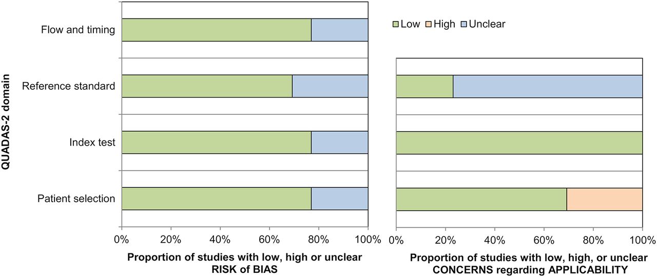
Quality assessment of 13 included studies.
Qualitative Synthesis
The detection rates of 68Ga-DOTA-SST PET and other imaging modalities (18F-FDOPA PET, 18F-FDG PET, and 123/131I-MIBG scanning) are illustrated in Figure 3. 68Ga-DOTA-SST PET consistently showed a higher detection rate than 18F-FDOPA PET, 18F-FDG PET, and 123/131I-MIBG scintigraphy, with the exception of one study regarding polycythemia/paraganglioma syndrome (45). In that study, 68Ga-DOTA-SST PET showed the lowest detection rate of 35% (95% confidence interval [CI], 24%–48%), whereas the detection rate for 18F-FDOPA PET was 99% (95% CI, 93%–100%). In the studies included, patients with SDHx mutation (22,24) and sporadic type (15), 68Ga-DOTA-SST PET showed the highest detection rates among the functional imaging modalities.
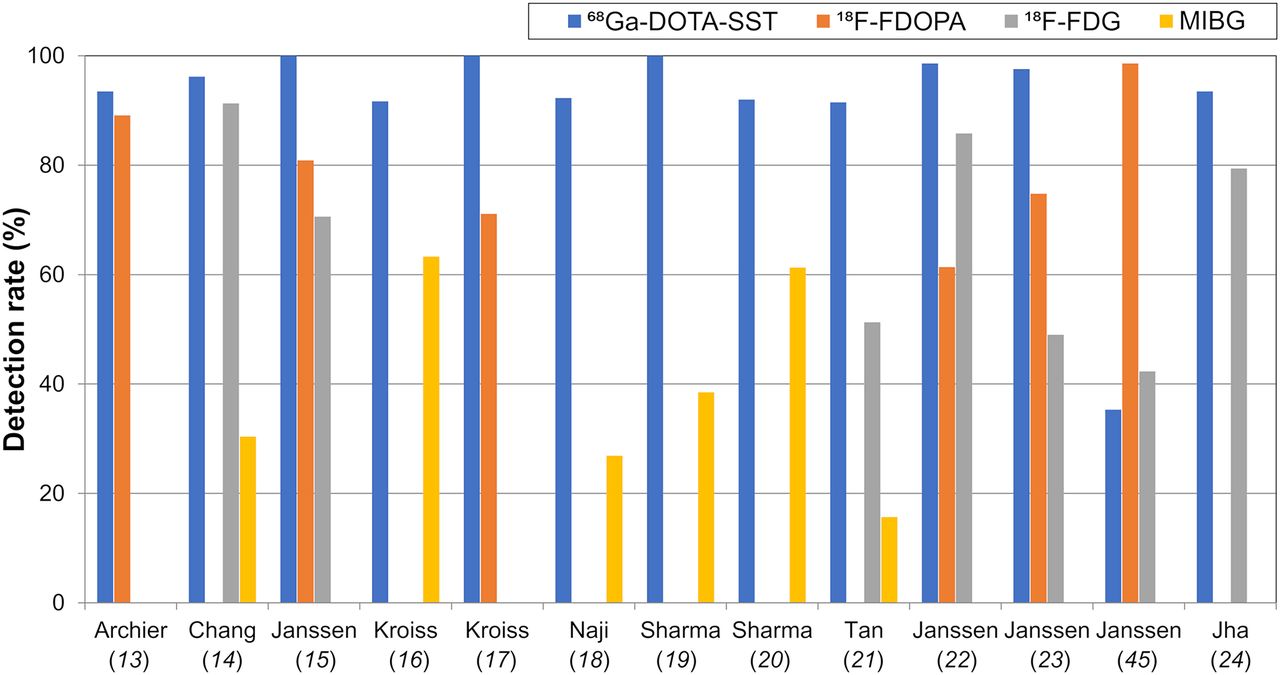
Comparison of detection rates among functional imaging modalities in 13 included studies.
Quantitative Synthesis
The per-lesion detection rate in 9 studies included in the quantitative synthesis ranged from 92% to 100%, with a pooled estimate of 93% (95% CI, 91%–95%) (Fig. 4). On the basis of the Higgins I2 statistics (I2 = 26%), no significant heterogeneity was present. There was significant publication bias, according to the funnel plot and Egger’s test (P = 0.0809) (Supplemental Fig. 1; supplemental materials are available at http://jnm.snmjournals.org). The pooled detection rate of 68Ga-DOTA-SST PET was significantly higher than that of 18F-FDOPA PET (80% [95% CI, 69%–88%], P = 0.0003), 18F-FDG PET (74% [95% CI, 46%–91%], P < 0.0001), or 123/131I-MIBG scintigraphy (38% [95% CI, 20%–59%], P < 0.0001). There was no difference in the detection rates of 68Ga-DOTA-SST PET among the multiple subgroups stratified by reference standard, clinical setting, or radioligand (Table 3). A greater proportion of head and neck paragangliomas was significantly associated with higher detection rates of 68Ga-DOTA-SST PET (P = 0.0002), whereas other variables, including the proportions of multifocal or metastatic disease, SDHx mutation, sporadic type, catecholamine-secretory PPGLs, age, and tumor size, were not significant in metaregression analyses (Fig. 5; Table 4).
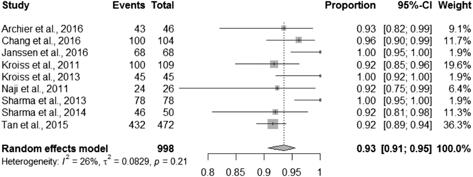
Forest plot showing pooled proportion of detection rate of 68Ga-DOTA-SST PET.
Subgroup Analyses for Detection Rates

Bubble plot for detection rate of 68Ga-DOTA-SST PET and the proportion of head and neck paragangliomas shows that it is a significant factor affecting heterogeneity (P = 0.0002).
Results of Metaregression Analyses
DISCUSSION
In the present systematic review and metaanalysis, we evaluated the performance of 68Ga-DOTA-SST PET for lesion detection in patients with PPGLs. The pooled detection rate was 93%, which was significantly higher than the detection rates of other functional imaging modalities. Accurate lesion detection is important for PPGLs, as these are typically surgically amenable; complete resection of lesions is needed, especially for catecholamine-secreting tumors.
18F-FDOPA PET is one of the standard imaging modalities in nonmetastatic PPGLs (2–4). However, the difficulty in synthesis and the requirement of a nearby cyclotron precludes the wider use of 18F-FDOPA. Furthermore, the diagnostic performance of 18F-FDOPA PET is lower in extraadrenal paraganglioma and SDHx-related metastatic disease (6). The role of 18F-FDG PET in PPGLs is limited for metastatic disease. MIBG scintigraphy requires complicated patient preparation (including thyroid blockade and discontinuation of certain drugs) and a long delay between injection and imaging. 123I might not be available in every facility, whereas 131I suffers from low image quality and unfavorable dosimetry. In contrast, 68Ga-DOTA-SST PET imaging exhibits both practical advantages (no patient preparation, easy synthesis, and wide availability due to 68Ge/68Ga generator) and superior detection rates, relative to any other functional imaging modalities. The high cost of 68Ge/68Ga generators can be a potential drawback of 68Ga-DOTA-SST PET imaging. However, increasing demand for 68Ga-labeled radiotracers and recent approval of the SST analog kit by the U.S. Food and Drug Administration will make 68Ge/68Ga generators more readily available. Further, more effective planning, such as imaging centralization and a referral system, would help reduce the cost of 68Ga imaging.
For metaanalysis, we excluded 4 studies that exclusively included patients with specific subtypes. If we assume that the study samples included in our quantitative synthesis are representative of a PPGL population with unknown genetic status, it may be suggested that 68Ga-DOTA-SST PET can serve as a first-line imaging modality for the primary staging of PPGLs, or the restaging of PPGLs with unknown genetic status. However, in 4 of the included studies (13–15,18), a substantial portion of patients was found to have the SDHx mutation; these proportions ranged from 27% to 80%, which are higher than the proportions in general PPGL populations (46). A higher prevalence of multifocal or metastatic disease, which is related to SDHx mutation, was also observed. Therefore, caution is necessary regarding the general application of our pooled estimate. On the basis of on our metaregression analyses, the performance of 68Ga-DOTA-SST PET may not be affected by the prevalence of metastasis, SDHx mutation, or sporadic type. Our study also suggested that 68Ga-DOTA-SST PET might exhibit a superior detection rate relative to 18F-FDOPA or 18F-FDG PET and serve as a functional imaging modality of choice in PPGLs with metastasis, SDHx mutation, or sporadic type.
68Ga-DOTA-SST ligands have the highest affinity for SSTR2, with different affinities for other SSTR subtypes (12). 68Ga-DOTATATE predominantly binds to SSTR2, 68Ga-DOTATOC binds to SSTR2 and SSTR5, and 68Ga-DOTANOC has a high affinity throughout SSTR2–5. No difference in detection performance was observed between the radioligands in our subgroup analysis; however, the low number of studies limited its significance. Of note, higher detection rates of 68Ga-DOTA-SST PET were reported in studies that showed greater prevalence of head and neck paragangliomas. These tumors are parasympathetic in origin and usually do not secrete catecholamine; thus, they differ from pheochromocytomas or paragangliomas in the thorax and abdomen (1). Our findings are consistent with the recent guideline that recommends 68Ga-DOTA-SST PET as the first-line imaging tool for head and neck paraganglioma (4). We suspect that the difference in overexpressed SSTR subtypes between the 2 kinds of PPGLs might affect the diagnostic performance of 68Ga-DOTA-SST PET. Paragangliomas overexpress SSTR2 predominantly (11,12), whereas a single in vitro study showed that pheochromocytomas overexpress SSTR3 predominantly and SSTR2 to a lesser extent (10).
It should be noted that 68Ga-DOTATATE showed poor diagnostic performance in patients presenting with polycythemia/paraganglioma syndrome, whereas 18F-FDOPA PET exhibited the highest detection rate (45). The reason for this disparate diagnostic performance remains unclear; however, we speculate that a lack of SSTR expression, inactivation of SSTR, or overexpression of other SSTR subtypes (non-SSTR2) could explain such behavior. Similarly, in a recent study by Taieb et al. (38), 68Ga-DOTATATE PET showed an inferior lesion detection rate, compared with 18F-FDOPA PET, in MYC-associated factor X–related pheochromocytoma; however, only 3 subjects were evaluated. Further research is needed to clarify these discrepancies.
There are some limitations in our review. First, the number of included studies is small. Even after a systematic search without any language restriction, we could identify only 8 suitable studies for quantitative synthesis. Nevertheless, metaanalysis is an appropriate method to generate a higher level of evidence in rare diseases, such as PPGLs, for which large cohort studies are not feasible. Second, approximately half of the included studies were retrospective in nature. Pooling results based on predominantly retrospective studies might lead to overestimation of the outcomes. Third, there were heterogeneities in scanners, image acquisition, and reconstruction protocols among the studies. Lastly, our pooled estimates were not based on studies that assessed patients with specific genetic mutations. No genetic test was performed in half of the included studies in our quantitative synthesis. Therefore, our results might not be applicable to specific genetic subtypes of PPGLs.
CONCLUSION
68Ga-DOTA-SST PET demonstrated an excellent lesion detection rate in patients with PPGLs. The pooled detection rate of the 8 included articles was 93%, which was significantly higher than the detection rate of other functional imaging modalities. Greater prevalence of head and neck paragangliomas was associated with higher detection rates of 68Ga-DOTA-SST PET. However, in patients with polycythemia/paraganglioma syndrome, 68Ga-DOTA-SST PET exhibited a poor detection rate.
DISCLOSURE
No potential conflict of interest relevant to this article was reported.
Footnotes
↵* Contributed equally to this work.
Published online Jul. 20, 2018.
- © 2019 by the Society of Nuclear Medicine and Molecular Imaging.
REFERENCES
- Received for publication March 20, 2018.
- Accepted for publication July 9, 2018.


Jump to section
Related Articles
Cited By...
- Choice Is Good at Times: The Emergence of [64Cu]Cu-DOTATATE-Based Somatostatin Receptor Imaging in the Era of [68Ga]Ga-DOTATATE
- Diagnostic Performance of 124I-Metaiodobenzylguanidine PET/CT in Patients with Pheochromocytoma
- Subclinical phaeochromocytoma: a diagnostic and management challenge
- Imaging of Pheochromocytoma and Paraganglioma
- A Clinical Challenge: Endocrine and Imaging Investigations of Adrenal Masses
- Possible pitfalls in the workup of ectopic ACTH secretion illustrated by four rare cases