


Abstract
Imaging of expression of therapeutic targets may enable stratification of patients for targeted treatments. The use of small radiolabeled probes based on the heavy-chain variable region of heavy-chain–only immunoglobulins or nonimmunoglobulin scaffolds permits rapid localization of radiotracers in tumors and rapid clearance from normal tissues. This makes high-contrast imaging possible on the day of injection. This mini review focuses on small proteins for radionuclide-based imaging that would allow same-day imaging, with the emphasis on clinical applications and promising preclinical developments within the field of oncology.
The discovery of key pathways that drive disease progression has led to the identification of new targetable molecules. When such molecules are located on the cell membrane or in the extracellular space, monoclonal antibodies (mAbs) can often block or activate these pathways. These mAbs typically show a slow systemic clearance, allowing a triweekly treatment regimen. Several mAbs have been used for PET imaging—the so-called immuno-PET approach (1). This approach allows study of the pharmacokinetics and receptor occupancy of the therapeutics, as well as the presence and accessibility of the molecular target in the individual patient. Because these mAbs are produced in large quantities by the pharmaceutical industry as therapeutics, they are typically available as a targeting moiety for the development of an imaging agent at very low cost. A major disadvantage is the long blood circulation time, with half-lives of up to 28 d, requiring delayed scanning time points typically between 4 and 6 d (Fig. 1A). Even at that time, appreciable quantities of the tracer remain in the blood, resulting in low sensitivity due to high background uptake and low specificity due to an enhanced permeability and retention effect, especially for targets with a low expression level.

Maximum-intensity-projection PET images from patients with metastatic HER2-positive breast cancer, imaged with mAb trastuzumab (A), antibody-derived sdAb (B), or scaffold protein Affibody molecule (C) at various times after injection. Arrows indicate tumor lesions. (Adapted from (7,21,42) and with permission of (42).)
To overcome the slow clearance and extravasation, mAbs have been engineered to smaller fragments such as antigen-binding, variable, and single-chain variable fragments; diabodies; and minibodies (2), with a molecular weight of 25–110 kDa. Although this size reduction increases the clearance rate of nonbound tracers, the clearance and extravasation rates remain overall too low to allow same-day imaging with sufficient contrast. Attractive alternative targeting proteins, with a further reduction in size, are Camelid single-domain antibody fragments (sdAbs) and several nonimmunoglobulin protein scaffolds such as Affibody molecules (Affibody AB), anticalins, designed ankyrin repeat proteins (DARPins; Molecular Partners AG), and fibronectin type III (FN3, Adnectin; Adnexus) (Fig. 2; Table 1) (3). Their low molecular weight enables fast penetration in tumor tissues, and their lack of a fragment crystallizable region further improves clearance. They allow a same-day imaging approach, much like the current practice for 18F-FDG PET, and their application results in a 4- to 6-fold lower radiation exposure than for immuno-PET, making them attractive for routine use. This approach is promising for assessing target expression levels in individual patients to identify those who will likely benefit from targeted treatments.
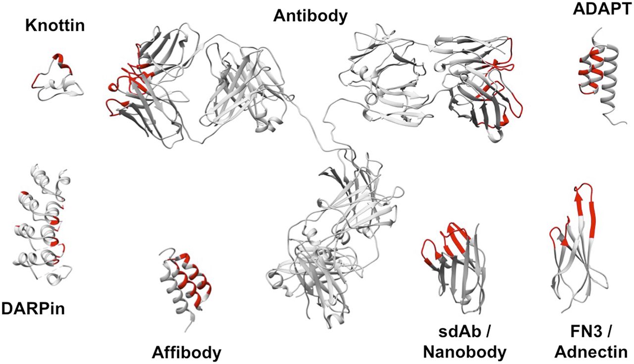
Schematic illustration of structures of various targeting vehicles discussed in this review. Most frequent variable stretches and residues are colored red. Structures are sized relative to each other. Representative protein-data-bank files were retrieved from Research Collaboratory for Structural Bioinformatics Protein Data Bank and manipulated using Chimera software: 1IGT (Antibody), 5MY6 (sdAb/Nanobody), 2KZJ (Affibody), 4HRM (DARPin), 2N8B (Knottin), 3QWQ (FN3/Adnectin), and 1GJT (ADAPT).
Overview of Radioactively Labeled Targeting Proteins That Have Been Used In Vivo for Same-Day Imaging in Last 5 Years
GENERATION AND TYPICAL CHARACTERISTICS OF SMALL PROTEINS
sdAbs, also referred to as Nanobodies (Ablynx), represent the smallest functional antibody fragments (12–15 kDa), consisting of the heavy-chain variable region of an immunoglobulin type that is naturally present in Camelidae. sdAbs are typically highly stable and bind antigens quickly and with high specificity and affinity. Because of their small size, sdAbs show improved tissue penetration compared with mAbs (4). sdAbs are generated by immunizing camelids with the antigen of choice. The sdAb gene fragments are amplified from isolated lymphocytes, providing a library of potential binders. Affinity-matured target-specific sdAbs are selected via protein display and biopanning and are finally produced in microbial hosts.
Besides antibody fragments, which are derived from different types of antibody classes, different scaffold proteins have also been engineered as small imaging probes (5). Scaffold proteins fall into 2 structural classes. The first is domain-sized compounds (6–20 kDa) such as Affibody (Affibody, Inc.) molecules, albumin-binding domain–derived affinity proteins (ADAPTs), Affilin scaffold proteins (Scil Proteins GmbH), Anticalins (Pieris, Inc.), Atrimer trivalent proteins (Anaphore, Inc.), DARPins (Dyax, Inc., Shire Inc.), FN3 scaffolds (Molecular Partners, Inc.), Fynomer platforms (Janssen), Kunitz domains, or Pronectin (Protelica), FN3-based sequences (Protelica, Inc.). The second structural class of scaffold protein comprises constrained peptides (2–4 kDa) such as vimers (Avimers [Avidia, Inc., now Amgen]), bicyclic peptides (Bicycle Therapeutics, Inc.), and cystine knot peptides (3). For both classes of scaffold protein, a library of potential binders is typically generated by random or targeted mutagenesis of the parent scaffold protein at residues that are not essential for protein folding. From this, target-specific binders are selected via phage display, yeast surface display, or ribosome/messenger RNA display. Most sdAbs and engineered scaffold proteins are easily produced in microbial systems, are stable and soluble, and show good binding affinity and specificity (3). So far, several sdAbs and scaffold proteins, selected against different targets, have been used as probes for SPECT and PET imaging (4,5).
SDABS IN RADIONUCLIDE IMAGING
sdAbs have been successfully applied as probes for radionuclide imaging, of which the human epidermal growth factor receptor (HER2)–targeting sdAbs are most dominantly explored. HER2 is an interesting therapeutic target, as it is overexpressed in several types of cancer, including breast, ovarian, and gastric. The 68Ga-labeled HER2-targeting sdAb 2Rs15d enabled high-contrast PET imaging of HER2-positive breast cancer at 1 h after injection in SKOV-3 xenografts (tumor-to-blood ratio, 29 ± 8) (6). A good manufacturing practice–grade version was evaluated in a phase I PET study in HER2-positive breast cancer patients (Fig. 1B). The compound was safe, with fast urinary clearance, resulting in a biologic half-life of 1 h and only 10% of injected activity remaining in the blood at 1 h after injection. Biodistribution data showed background uptake in the liver, kidneys, and to a lesser extent bowel and salivary glands. Uptake was high in both primary HER2-positive tumors and metastasis, with standardized uptake value (SUVmean) of up to 11.8 and 6.0, respectively, at 1 h after injection (Fig. 1B). The effective dose was 0.043 mSv/MBq, resulting in an average of 4.6 mSv per patient, with the critical organ being the urinary bladder wall (0.406 mGy/MBq) (7). The same HER2-targeting sdAb was also 18F-labeled and tested preclinically, with a high tumor-to-blood ratio (13 ± 2 as early as 1 h after injection). Uptake in the kidneys was, however, only half that measured for 68Ga-labeled 2Rs15d (8) and thus might further decrease the radiation exposure. Finally, 2Rs15d was successfully radiolabeled with 131I and showed superior tumor uptake and remarkably low kidney retention in HER2-positive tumor xenografted mice (9). Consequently, this version is now being evaluated as a theranostic drug in a first clinical breast cancer trial (NCT02683083). Another HER2-targeting sdAb, 5F7, was radiolabeled with both 18F and 125I and also demonstrated high-contrast imaging in preclinical studies (10–12).
A promising target is the prostate-specific membrane antigen (PSMA), as it is expressed on virtually all metastatic prostate cancers. A PSMA-targeted sdAb (PSMA30) radiolabeled with 99mTc demonstrated fast and specific uptake in PSMA-positive xenografts (tumor-to-blood ratio of 8.7 at 90 min after injection) (Fig. 3A (13)). Another PSMA-targeting sdAb (JVZ-007) was labeled with 111In for SPECT/CT imaging. Excellent tumor targeting as early as 3 h after injection was observed (tumor-to-blood ratio of 48 ± 5), in the absence of nonspecific uptake (14). Given the current success of clinical imaging using the available PSMA-binding peptides, the question remains of whether these sdAbs have enough added value to bring them to the clinic.

CD20 is expressed in over 90% of B-cell non-Hodgkin lymphomas and is clinically used for targeted radionuclide therapy with mAbs (90Y-ibritumomab [Zevalin; Spectrum Pharmaceuticals, Inc.]). Recently, Krasniqi et al. selected a lead CD20-targeting sdAb 9079 and confirmed its potential for same-day PET imaging in a preclinical model (tumor-to-blood ratio of approximately 4) (Fig. 3B (15)).
Traditionally, targets selected for molecular imaging and therapy were those overexpressed on the tumor cells themselves. More recently, interest has been raised about targets expressed on the tumor stroma since they can play important roles in tumor angiogenesis, invasion, and immune escape. Such targets are typically less abundantly present in the tumor, which demands a sensitive and very specific technique. It is expected that tracers with slow clearance will not be able to accurately discern such expression from remaining background signals, making the use of sdAb and scaffold proteins the ideal choice. A promising sdAb is one that targets the macrophage mannose receptor (MMR). MMR is highly expressed on protumorigenic tumor-associated macrophages, which play an important role in tumor growth, tumor angiogenesis, metastasis, and immune suppression. Being able to visualize and quantify the presence of MMR (and thus tumor-associated macrophages) in tumor tissue might be a helpful prognostic tool for the treatment of cancer. To this end, Movahedi et al. selected and preclinically validated a lead anti-MMR sdAb (Fig. 3A (16)). A 18F-labeled variant was successfully developed as a PET tracer for MMR expression in mice (17), confirming its potential for clinical translation.
Other promising targets within tumor stroma are the immune checkpoints. Success with immune checkpoint blockade with cytotoxic T-lymphocyte–associated protein-4 and programmed death 1 or programmed death ligand 1 (PD-L1) antibody therapy has, however, been seen in only a subset of all cancer patients. Biomarker imaging might help to select patients for such therapies. Anti-PD-L1 sdAbs were developed that could allow the assessment of PD-L1 expression in the tumor microenvironment (Fig. 3A) (18,19).
AFFIBODY MOLECULES IN RADIONUCLIDE IMAGING
HER2-targeting Affibody molecules were the first scaffold proteins evaluated for radionuclide molecular imaging. The second-generation Affibody molecule ABY-025 labeled with 111In for SPECT imaging and 68Ga for PET imaging were evaluated in phase I/II clinical studies for assessment of HER2 expression in breast cancer metastases (Fig. 1C) (20,21). ABY-025 was found to be safe in humans, and no anti-Affibody antibodies were found after repeated administration. The effective radiation doses were 0.15 ± 0.02 mSv/MBq (21 mSv/patient) for 111In and 0.028 ± 0.003 mSv/MBq (5.6 mSv/patient) for 68Ga (20,22). Injection of 500 μg of 68Ga-ABY-025 provided better sensitivity and specificity than 100 μg, mainly because of reduced retention in the liver (21). Comparison with immunohistochemical staining of biopsy material demonstrated that the measurement of the maximal uptake value at 2–4 h after injection permits clear discrimination between metastases with 3+ and 2+ levels of HER2 expression (21). Furthermore, the spleen can be used as an within-image reference tissue to calculate tumor-to-spleen ratios, permitting a simple and robust discrimination between metastases with high and low HER2 expression using both PET and SPECT (23) and thus making the diagnosis independent of external hardware calibrations.
High renal reabsorption of Affibody molecules complicates their use for radionuclide therapy. However, internalization of anti-HER2 Affibody molecules is slow after binding to malignant cells but rapid in proximal tubuli. The peptide-based chelator GGGC provides nonresidualizing 188Re labeling, thereby resulting in good tumor retention but rapid washout from kidneys (24). Extrapolation to humans suggests that the absorbed dose to tumors would exceed that to the kidney by approximately 3.4-fold. An alternative might be an Affibody-based pretargeting. Two approaches were evaluated: one mediated by a bioorthogonal reaction between transcyclooctene and tetrazine (25) and another mediated by an interaction between 2 complementary peptide nucleic acids (26). Both methodologies provided appreciably higher uptake of radiometals in tumors than in kidneys, with the peptide nucleic acid–mediated approach resulting in better tumor retention.
Resistance to trastuzumab therapy may be associated with overexpression of HER3 and insulin-like growth factor type 1 receptor (IGF-1R) (27). Imaging of an emerging expression of these receptors might suggest an onset of resistance and a need to modify treatment. Expression of these receptors is also essential in other malignancies (e.g., prostate and ovarian carcinomas), and multiple therapeutics targeting HER3 or IGF-1R are under development. A challenge in imaging of both receptors is a modest expression level in malignant cells (typically below 40,000 receptors per cell) and expression in normal tissues. Affibody molecules were selected for both molecular targets. To mimic the clinical situation, binders with approximately equal affinity to human and murine receptors were generated. An anti-IGF-1R Affibody molecule, ZIGF1R:4551, was labeled using 99mTc(CO)3 and demonstrated receptor-specific uptake in both IGF-1R–expressing tumors and IGF-1R–expressing tissues (Fig. 4A) (28). An anti-HER3 Affibody molecule, Z08698, with affinity of 50 pM, was previously labeled with 111In and 99mTc for SPECT imaging. To enable straightforward quantification, HEHEHE-Z08698-NOTA was labeled with positron-emitting 68Ga, showing accumulation in tumor xenografts that was proportional to HER3 expression level (Fig. 4B) (29).

Epidermal growth factor receptor (EGFR) is the molecular target for several mAbs and tyrosine kinase inhibitors. Detection of overexpression may help to predict outcomes for some treatment regimens for non–small cell lung cancer and head-and-neck squamous cell carcinoma. One of the challenges in imaging EGFR expression is that it is expressed in liver and some other tissues. ZEGFR:2377, having equal affinity to murine and human EGFR (dissociation constant, 0.8–0.9 nM), was selected, and an injection of 30–50 μg partially saturated receptors in healthy tissues but not in tumors. Labeling of DOTA-ZEGFR:2377 with 68Ga and 57/55Co was assessed. Evaluation of conjugates in A431 xenografts demonstrated that labeling of ZEGFR:2377 with 57/55Co provided significantly higher tumor-to-organ ratios than did labeling with 68Ga (43). Importantly, a tumor-to-liver ratio of 3.1 ± 0.5 (3 h after injection) was obtained (Fig. 4A).
Carbonic anhydrase IX (CAIX) is overexpressed by hypoxic cells, and imaging of CAIX expression may be used to identify radioresistant hypoxic tumors. In addition, CAIX is expressed by normoxic renal cell carcinoma and may be used to distinguish between malignant and benign renal tumors. A panel of anti-CAIX Affibody molecules labeled with 99mTc and 125I was evaluated in SK-RC-52 renal cell carcinoma (30). The best hypoxia tracer was 99mTc-(HE)3-ZCAIX:2. 125I-ZCAIX:4 was best suited for imaging of renal cell carcinoma, with a tumor-to-kidney ratio of 2.1 ± 0.5 (Fig. 4A).
Inhibition of platelet-derived growth factor receptor β (PDGFRβ) in tumor stroma (pericytes of neovasculature and cancer-associated fibroblasts) normalizes tumor interstitial pressure and improves drug uptake and efficacy. An anti-PDGFRβ-Affibody molecule, DOTA-Z09591, was labeled with 68Ga and evaluated as a potential companion diagnostic (31). In mice, 68Ga-DOTA-Z09591 provided clear visualization of U-87 MG xenografts (36,000 PDGFRβ receptors per cell) at 2 h after injection (Fig. 4B).
To evaluate PD-L1 expression before programmed death 1– or PD-L1–targeting therapy and for response monitoring, a PD-L1–specific Affibody molecule with a dissociation constant of 1 nM was selected (32). This NOTA-conjugated ZPD‐L1_1 was labeled with 18F using 18F-AlF. 18F-AlF-NOTA-ZPD‐L1_1 enabled specific imaging of PD-L1 in murine models, with a tumor-to-blood ratio exceeding values obtained using radiolabeled antibodies several days after injection (Fig. 4B).
PRECLINICAL STUDIES WITH OTHER PROTEIN SCAFFOLDS
Knottins
Inhibitor cystine knots, also known as knottin peptides, are small polypeptides consisting of 30–50 amino acids with a molecular weight of about 4 kDa. They are characterized by their structural motif with 3 cystine bridges, forming a knot, and can be found in a wide range of animals, plants, and fungi (3).
Kimura et al. reported a knottin peptide that binds to αvβ6, an integrin overexpressed in many cancers. It demonstrated fast and specific tumor targeting in mice bearing αvβ6-expressing tumors. However, low tumor-to-liver, tumor-to-kidney, and tumor-to-gut ratios were also observed (33).
Jiang et al. reported the potential use of a 7C knottin peptide as a SPECT probe for imaging of integrin αvβ3–positive tumors. Despite the fact that no SPECT imaging data were shown, a biodistribution study on mice revealed fast and specific accumulation of 111In-labeled 7C in αvβ3-positive human subcutaneously growing U87MG glioblastoma at 0.5 h after injection (34).
More recently, the 18F-labeled divalent integrin αvβ3-targeting knottin peptide 3-4A, containing 2 separate integrin-binding paratopes, was evaluated in subcutaneously growing U87MG xenografts (35). Images revealed clear uptake in tumors after 30 min, with low background signal, except in kidneys (Fig. 5B).

DARPins
DARPins are scaffold proteins with a size of 14–18 kDa, which are derived from natural ankyrin repeat proteins. Synthetic DARPin libraries have been designed to select DARPins against different targets, mainly for nonradioactive therapeutic applications (3).
Goldstein et al. described SPECT imaging of HER2 expression using 125I-/111In-labeled G3 DARPin (Fig. 5A). High tumor uptake was measured for both radionuclides in mice bearing human BT474 HER2–positive xenografts at 4 h after injection. However, the lower uptake of 111In-compound in normal tissues led to higher tumor-to-background ratios than for its 125I counterpart (36).
FN3 Scaffolds
A small anti-CD20 receptor protein based on the 10-kDa human FN3 has been generated for PET imaging of B-cell lymphomas (37). 64Cu-DOTA-FN3CD20 was evaluated in human CD20-transgenic mice and showed a high specific accumulation in the spleen and other CD20-expressing organs as soon as 1 h after injection. In nontransgenic mice bearing human CD20 tumors, 64Cu-FN3CD20 showed specific tumor targeting with low healthy-tissue uptake as soon as 4 h after injection (37).
Recently, synthesis of a human and cynomolgus cross-reactive anti-PD-L1 FN3 scaffold was described. In vivo PET imaging with an 18F-labeled scaffold (18F-BMS-986192) clearly visualized PD-L1 tumors in mice at 2 h after injection (Fig. 5B) (38). Cynomolgus PET imaging showed specific binding of 18F-BMS-986192 in the spleen, with fast blood clearance through the kidneys and bladder. Currently, this compound is being tested in a single-center substudy of the CheckMate 511 clinical trial (NCT02714218/EudraCT 2015-004920-67) to evaluate its potential as a biomarker in metastatic melanoma patients (38).
ADAPTs
ADAPTs use a 46-amino-acid scaffold of an albumin-binding domain (5.2 kDa) of streptococcal protein G. The anti-HER2 ADAPT6 was selected for its high-affinity binding (1.1 nM) and its loss of binding to albumin to facilitate fast blood clearance. ADAPT6 was site-specifically DOTA-conjugated at the N terminus (39). 111In/68Ga-DOTA-ADAPT6 showed high uptake in SKOV-3 xenografts, and as soon as 1 h after injection, PET enabled clear discrimination between xenografts with high and low expression. Reabsorption of ADAPT6 in the kidneys was high and could not be reduced using injection of cationic amino acids or Gelofusine (Hausmann Laboratories Ltd.). Further studies demonstrated that targeting properties of ADAPTs might be appreciably improved by modification of labeling chemistry and N terminus composition. An optimized 111In-labeled variant, DOTA-Cys59-ADAPT6, had a tumor-to-blood ratio of 277 ± 35 at 4 h after injection. Use of the nonresidualizing 125I-HPEM label provided a tumor-to-kidney ratio of 13 ± 3, opening a way for radionuclide therapy (Fig. 5A) (40).
The radiolabeling chemistry might influence the properties of targeting proteins by modifying critical amino acids or the local charge and lipophilicity of the protein surface. This might affect on-target and off-target interactions, excretion, and intracellular retention of the radionuclide after internalization (41). In this way, changes in radiolabeling chemistry might have important effects on the imaging contrast obtained and optimal imaging time point. Therefore, a meticulous optimization of labeling chemistry is crucial for high-sensitivity imaging using small proteins.
CONCLUSION
sdAbs and Affibody proteins have clinically confirmed the safety, low radiation exposure, and high potential of molecular imaging in cancer patients. Although studies with larger patient cohorts are required to establish their exact clinical value, the early-phase data for HER2-specific probes are encouraging. Also, several emerging sdAb- and scaffold protein–based probes that target other clinically relevant targets have been developed preclinically, and their clinical translation may impact future patient care.
DISCLOSURE
Marleen Keyaerts has received travel and accommodation expenses from Bayer NV. Nick Devoogdt and Matthias D’Huyvetter are cofounders of CamelIDs. Nick Devoogdt has received funding from Boehringer-Ingelheim, Complix. Ahmet Krasniqi, Marleen Keyaerts, Nick Devoogdt, and Matthias D’Huyvetter have patents on sdAb imaging and therapy. Fredrik Frejd is an employee of Affibody AB. Fredrik Frejd, Anna Orlova, and Vladimir Tolmachev have patents on Affibody development and applications. Ahmet Krasniqi has a doctoral grant from Agentschap Innoveren & Ondernemen (IWT.141388); Marleen Keyaerts is a senior clinical investigator; and Matthias D’Huyvetter is a postdoctoral fellow of the Research Foundation–Flanders (FWO). Research was funded by CancerPlan Action (Federal Public Service Health, Food Chain Safety, and Environment, Belgium), Kom-op-tegen-Kanker, FWO, Swedish Cancer Society, and Swedish Research Council. No other potential conflict of interest relevant to this article was reported.
Footnotes
Published online Mar. 15, 2018.
- © 2018 by the Society of Nuclear Medicine and Molecular Imaging.
REFERENCES
- Received for publication December 22, 2017.
- Accepted for publication March 6, 2018.


Jump to section
Related Articles
Cited By...
- Immuno-PET/CT Imaging of Trop2 with [18F]AlF-RESCA-T4 Differentiates Lung Cancer from Inflammation
- Theranostics Is Not Radiotheranostics
- Protein Fitness Prediction is Impacted by the Interplay of Language Models, Ensemble Learning, and Sampling Methods
- Phase I Trial of 99mTc-(HE)3-G3, a DARPin-Based Probe for Imaging of HER2 Expression in Breast Cancer
- Phase I Trial of 131I-GMIB-Anti-HER2-VHH1, a New Promising Candidate for HER2-Targeted Radionuclide Therapy in Breast Cancer Patients
- Phase I Study of 99mTc-ADAPT6, a Scaffold Protein-Based Probe for Visualization of HER2 Expression in Breast Cancer
- Efficacy of Affibody-Based Ultrasound Molecular Imaging of Vascular B7-H3 for Breast Cancer Detection
- The Future of Nuclear Medicine as an Independent Specialty