


Abstract
We investigated cytologic radiation damage in thyroid cancer after 131I therapy using micronucleus assay (MNA) of B lymphocytes exclusively, as opposed to our previous study in which MNA of all lymphocyte subsets was used. Methods: We studied 22 thyroid cancer patients treated with 3.7 GBq of 131I. Peripheral lymphocytes were harvested, and B lymphocytes were isolated by an immunomagnetic method and assayed for the frequency of micronuclei. Results: The frequency of micronuclei among B cells after 131I therapy was significantly increased relative to that in untreated control subjects, and the 131I-induced increase in micronuclei frequency among B cells was significantly greater than that among all lymphocytes. Conclusion: Compared with the MNA of all lymphocytes, the MNA among specifically B cells may more sensitively detect cytologic radiation damage associated with 131I therapy of thyroid cancer.
Dosimetric studies have estimated the radiation dose to blood and bone marrow after treatment with a large amount of radioiodine (1). Chromosomal damage to peripheral lymphocytes in vivo after radioiodine therapy has been reported (2,3). In a previous study using micronucleus assay (MNA), we too demonstrated cytologic radiation damage to lymphocytes in thyroid cancer patients after 131I therapy (4). To our knowledge, however, the cytologic effects of large therapeutic doses of radioiodine on specific subpopulations of lymphocytes have not been examined.
Because B cells are more sensitive to radiation in vitro than are other lymphocyte subpopulations (5,6), Vral et al. (7) suggested that MNA of B cells might be particularly useful for assessment of cytologic radiation damage in vivo. Therefore, this study used MNA to compare radiation-induced cytologic damage to B lymphocytes with that to peripheral lymphocytes generally in thyroid cancer patients after therapeutic administration of 3.7 GBq of 131I. Chromosomal damage to lymphocytes induced in vivo by 131I should augment the cells with micronuclei (8). A comparison of the increased frequency of micronuclei among B lymphocytes and peripheral lymphocytes irradiated in vivo by 131I may sensitively estimate cytologic radiation damage in thyroid cancer patients.
MATERIALS AND METHODS
Patients
From May 1996 to Feb 2002, 22 patients were admitted for radioiodine treatment of differentiated thyroid carcinoma at the Department of Radiology of Toyama Medical and Pharmaceutical University in Japan. There were 9 men and 13 women, ranging in age from 40 to 71 y (mean, 56.9 y). All patients were diagnosed by biopsy and underwent total thyroidectomy. After giving informed consent, the 22 patients, who received 3.7 GBq of 131I, were studied. The thyroid-stimulating hormone concentration was higher than 30 μU/mL in these patients on the day before therapy.
Lymphocyte Isolation and Culture
Peripheral blood lymphocytes were collected from each patient before and 1 wk after therapy. The lymphocytes were cultured according to the method of Fenech and Morley (8). Pokeweed mitogen (Gibco BRL) was used to stimulate the lymphocytes to proliferate in culture. A solution of cytochalasin B (Aldrich Chemical Co.) was added 44 h after the commencement of the culture. The cultures were terminated 72 h after initiation.
External Irradiation
In an in vitro study, lymphocytes were collected from 4 young, healthy volunteers and were irradiated with electron beams (3 Gy/min, 5 MeV, Metatron, MD 2/40; Toshiba) at doses ranging from 0.5 to 2 Gy and were assessed for cytologic damage by MNA.
MNA
Anti-CD 19 (anti-Pan B) antibody-conjugated Dynabeads M-450 magnetic beads (Dynal AS) were incubated with lymphocytes at 4°C for 15 min. The rosetted CD 19–positive B cells were isolated using a magnet. Dynabeads were detached from B cells by incubation with polyclonal antibody DETACHaBEAD (Dynal AS) at 36°C for 60 min. After incubation, B cells were isolated by a magnet (9). The acridine orange fluorescent staining procedure was applied to the MNA in this study according to the method of Hayashi et al. (10). Micronuclei were stained yellowish green with green fluorescence (Fig. 1). The number of micronuclei per 500 binucleated cells was scored visually.
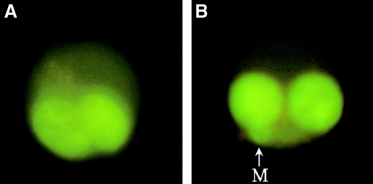
Photomicrographs of binucleated cells, including one without (A) and one with (B) a micronucleus (M).
The Previous Study
We reevaluated the increased number of peripheral lymphocyte micronuclei found after therapy in the 25 thyroid cancer patients of our previous study (4).
Statistical Analysis
The number of B-cell micronuclei before and after therapy was measured. The 22 patients represented a new patient group, whose data were compared with those of another group of 25 similar thyroid cancer patients analyzed in our previous study. Data are expressed as mean ± SD. The Student paired or unpaired t test was used to analyze differences between means. Probability values of <0.01 were considered significant.
RESULTS
Radiation Damage to Lymphocytes
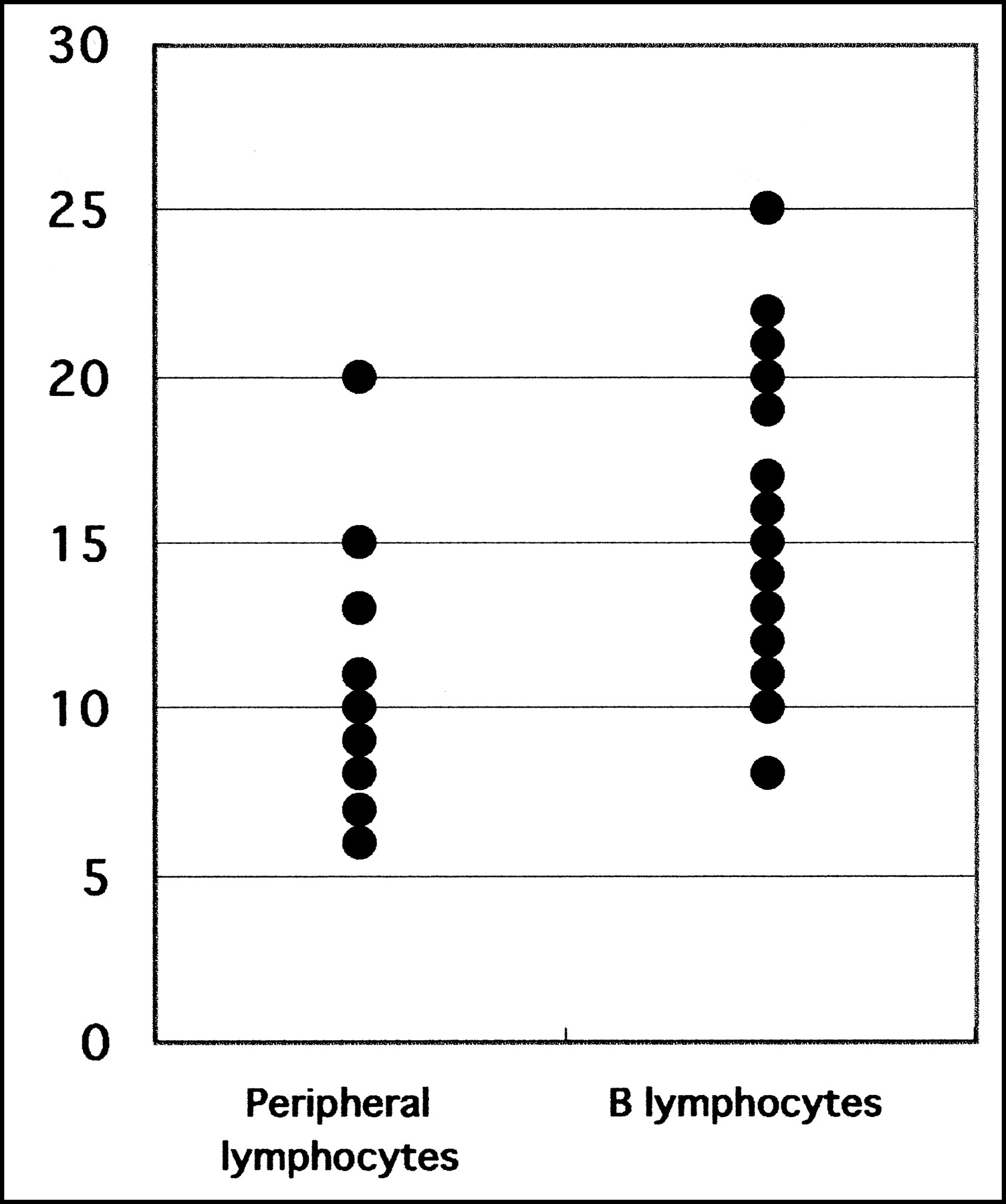
The number of micronuclei per 500 binucleated B lymphocytes was 6.8 ± 1.9 before and 22.5 ± 5.4 after 131I therapy, a statistically significant increase (P < 0.001). The number of micronuclei per 500 binucleated peripheral lymphocytes was 5.4 ± 1.4 before and 15.7 ± 2.7 after 131I therapy in our previous study (Fig. 2). The increased number of B lymphocyte and peripheral lymphocyte micronuclei after therapy was 15.6 ± 4.8 and 10.0 ± 3.1, and these differences were statistically significant (P < 0.001) (Fig. 3). The percentage increases in the frequencies of micronuclei were 247% ± 105% for B cells and 212% ± 132% for all lymphocytes. Although these differences were not statistically significant, a trend toward higher values was noted in the former.

The frequency of micronuclei among peripheral lymphocytes and B lymphocytes before and after 131I therapy.

The increased number of micronuclei among peripheral lymphocytes and B lymphocytes.
Dose Estimation
The number of B-cell micronuclei was measured after electron irradiation in vitro at absorbed doses of 0.5–2 Gy, and the resulting data fit a straight line, y = 35.7x + 6.3 (r = 0.95). Based on this dose–response relationship and the observed frequency of B-cell micronuclei in vivo, the absorbed dose among the 22 thyroid cancer patients treated with 131I was 0.45 ± 0.14 Gy.
DISCUSSION
MNA was used to assess radiation-induced cytologic damage to B lymphocytes in thyroid cancer patients after therapeutic administration of 3.7 GBq of 131I. In this study, B lymphocytes (CD19) were collected by an immunomagnetic technique (Dynabeads) after being cultured from peripheral blood lymphocytes. This Dynabeads method for separation of cultured lymphocytes is simple and fast (9). Our findings showed that B lymphocytes exposed to 131I have statistically higher frequencies of micronuclei than do controls. We also revealed that the degree of micronucleus induction among B lymphocytes after 131I therapy is higher than that among peripheral lymphocytes. Thus, radiation-induced damage to B lymphocytes after 131I therapy may be greater than that to peripheral lymphocytes in vivo.
In an in vitro study, Wuttke et al. confirmed that B cells are more radiosensitive than T cells (11). Vral et al. also demonstrated the highly radiosensitive behavior of B cells with respect to micronucleus production in vitro (7). On the basis of immune function assessment (12) and apoptosis (13), B cells appear to be more radiosensitive than other lymphocyte subpopulations. However, the radiosensitivity of B lymphocytes to radiotherapy in vivo has not been clarified until now. In a previous study (4), we used phytohemagglutinin to stimulate peripheral lymphocytes, which are a mixture of mainly 3 groups of cells: B lymphocytes, T lymphocytes, and natural killer cells. Generally, phytohemagglutinin stimulates T cells, and phytohemagglutinin-stimulated lymphocytes can be considered predominantly T cells. Our finding of a higher degree of micronucleus induction in vivo for B lymphocytes than for peripheral lymphocytes appears to be consistent with the in vitro results noted above. These results suggest that B lymphocytes may express high radiosensitivity in vivo after 131I therapy.
We evaluated an external-irradiation study to compare the micronucleus incidence in B lymphocytes irradiated in vivo with that in B lymphocytes irradiated in vitro and to estimate the biologically equivalent absorbed dose to B lymphocytes in vivo. We determined that cytologic damage to B lymphocytes in vivo after 131I therapy may be equivalent to the damage observed after a mean external-irradiation dose of 0.45 Gy in vitro. In our previous study, using similar methods, we estimated that peripheral lymphocytes receive a dose of 0.33 Gy (4). Our estimated radiation dose was somewhat higher than the previous estimation, possibly because of the limited number of control subjects available for in vitro studies; B lymphocytes were collected from only 4 young, healthy volunteers, and peripheral lymphocytes from 3 patients. One may argue that the total dose from low-dose-rate radiation exposure in vivo with 131I is considerably underestimated when compared with high-dose-rate external-radiation exposure in vitro (14). Therefore, both doses may be underestimated.
The estimated radiation dose with 131I therapy in thyroid cancer may vary. Monsieurs et al., using lymphocyte MNA, showed that the mean equivalent dose was 0.32 Gy (15). M’Kacher et al., using a similar method, estimated the biologic dose to be 0.54 Gy (3). For the patients in our study, using B-lymphocyte MNA, the mean estimated internal radiation exposure appeared to be in a range similar to these results.
Internal radiation damage after 131I therapy in thyroid cancer was slightly greater to B lymphocytes than to peripheral lymphocytes, based on the comparative cytologic studies using MNA. These results suggest that B-lymphocyte MNA could be more sensitive for assessing cytologic radiation-induced chromosomal damage. It is true that the small number of subjects limits our results and that we believe the dose to have been underestimated. Nevertheless, this approach remains the most sensitive and practical method presently available to evaluate cytologic radiation damage in thyroid cancer after 131I therapy.
CONCLUSION
Compared with MNA of all lymphocytes, MNA of specifically B cells may more sensitively detect cytologic radiation damage associated with 131I therapy of thyroid cancer and may facilitate estimation of the radiation doses absorbed with this therapy.
Acknowledgments
The authors thank Shuichi Tonami and Shousuke Kato for their technical assistance and John S. Gelblum for his kind assistance in preparing the manuscript.
Footnotes
Received May 30, 2003; revision accepted Jan. 5, 2004.
For correspondence or reprints contact: Naoto Watanabe, MD, Department of Radiology, Toyama Medical and Pharmaceutical University, Sugitani 2630, Toyama City, Japan 930-0194.
E-mail: nw31456{at}ms.toyama-mpu.ac.jp
In this issue


Jump to section
Related Articles
Cited By...
- Investigation of the Importance of Applying Various Methods of Calculation in Determining the Blood-Absorbed Dose for Patients with Differentiated Thyroid Carcinoma
- Iodine Biokinetics and Dosimetry in Radioiodine Therapy of Thyroid Cancer: Procedures and Results of a Prospective International Controlled Study of Ablation After rhTSH or Hormone Withdrawal
- Blood Dosimetry and Dose-Rate Effects After Radioiodine Therapy of Differentiated Thyroid Cancer