
Main menu

  • Home
  • Content
    • Current
    • Ahead of print
    • Past Issues
    • JNM Supplement
    • SNMMI Annual Meeting Abstracts
    • Continuing Education
    • JNM Podcasts
  • Subscriptions
    • Subscribers
    • Institutional and Non-member
    • Rates
    • Journal Claims
    • Corporate & Special Sales
  • Authors
    • Submit to JNM
    • Information for Authors
    • Assignment of Copyright
    • AQARA requirements
  • Info
    • Reviewers
    • Permissions
    • Advertisers
  • About
    • About Us
    • Editorial Board
    • Contact Information
  • More
    • Alerts
    • Feedback
    • Help
    • SNMMI Journals
  • SNMMI
    • JNM
    • JNMT
    • SNMMI Journals
    • SNMMI

User menu

  • Subscribe
  • My alerts
  • Log in
  • Log out
  • My Cart

Search

  • Advanced search
Journal of Nuclear Medicine
  • SNMMI
    • JNM
    • JNMT
    • SNMMI Journals
    • SNMMI
  • Subscribe
  • My alerts
  • Log in
  • Log out
  • My Cart
Journal of Nuclear Medicine

Advanced Search

  • Home
  • Content
    • Current
    • Ahead of print
    • Past Issues
    • JNM Supplement
    • SNMMI Annual Meeting Abstracts
    • Continuing Education
    • JNM Podcasts
  • Subscriptions
    • Subscribers
    • Institutional and Non-member
    • Rates
    • Journal Claims
    • Corporate & Special Sales
  • Authors
    • Submit to JNM
    • Information for Authors
    • Assignment of Copyright
    • AQARA requirements
  • Info
    • Reviewers
    • Permissions
    • Advertisers
  • About
    • About Us
    • Editorial Board
    • Contact Information
  • More
    • Alerts
    • Feedback
    • Help
    • SNMMI Journals
  • View or Listen to JNM Podcast
  • Visit JNM on Facebook
  • Join JNM on LinkedIn
  • Follow JNM on Twitter
  • Subscribe to our RSS feeds
Research ArticleClinical Investigation

Delayed Imaging Improves Lesion Detectability in [99mTc]Tc-PSMA-I&S SPECT/CT in Recurrent Prostate Cancer

Christoph Berliner, Lisa Steinhelfer, Maythinee Chantadisai, Markus Kroenke, Daniel Koehler, Randi Pose, Peter Bannas, Sophie Knipper, Matthias Eiber and Tobias Maurer
Journal of Nuclear Medicine May 2023, 265252; DOI: https://doi.org/10.2967/jnumed.122.265252
Christoph Berliner
1Department of Diagnostic and Interventional Radiology and Nuclear Medicine, Universitätsklinikum Hamburg–Eppendorf, Hamburg, Germany;
2Department of Nuclear Medicine, Universitätsklinikum Essen, Essen, Germany;
  • Find this author on Google Scholar
  • Find this author on PubMed
  • Search for this author on this site
Lisa Steinhelfer
3Department of Nuclear Medicine, Klinikum Rechts der Isar, Technische Universität München, München, Germany;
  • Find this author on Google Scholar
  • Find this author on PubMed
  • Search for this author on this site
Maythinee Chantadisai
3Department of Nuclear Medicine, Klinikum Rechts der Isar, Technische Universität München, München, Germany;
  • Find this author on Google Scholar
  • Find this author on PubMed
  • Search for this author on this site
Markus Kroenke
3Department of Nuclear Medicine, Klinikum Rechts der Isar, Technische Universität München, München, Germany;
  • Find this author on Google Scholar
  • Find this author on PubMed
  • Search for this author on this site
Daniel Koehler
1Department of Diagnostic and Interventional Radiology and Nuclear Medicine, Universitätsklinikum Hamburg–Eppendorf, Hamburg, Germany;
  • Find this author on Google Scholar
  • Find this author on PubMed
  • Search for this author on this site
Randi Pose
4Martini-Klinik Prostate Cancer Center, University Hospital Hamburg–Eppendorf, Hamburg, Germany; and
  • Find this author on Google Scholar
  • Find this author on PubMed
  • Search for this author on this site
Peter Bannas
1Department of Diagnostic and Interventional Radiology and Nuclear Medicine, Universitätsklinikum Hamburg–Eppendorf, Hamburg, Germany;
  • Find this author on Google Scholar
  • Find this author on PubMed
  • Search for this author on this site
Sophie Knipper
4Martini-Klinik Prostate Cancer Center, University Hospital Hamburg–Eppendorf, Hamburg, Germany; and
  • Find this author on Google Scholar
  • Find this author on PubMed
  • Search for this author on this site
Matthias Eiber
4Martini-Klinik Prostate Cancer Center, University Hospital Hamburg–Eppendorf, Hamburg, Germany; and
  • Find this author on Google Scholar
  • Find this author on PubMed
  • Search for this author on this site
Tobias Maurer
4Martini-Klinik Prostate Cancer Center, University Hospital Hamburg–Eppendorf, Hamburg, Germany; and
5Department of Urology, University Hospital Hamburg–Eppendorf, Hamburg, Germany
  • Find this author on Google Scholar
  • Find this author on PubMed
  • Search for this author on this site
  • Article
  • Figures & Data
  • Supplemental
  • Info & Metrics
  • PDF
Loading

Article Figures & Data

Figures

  • Tables
  • Additional Files
  • Figure
    • Download figure
    • Open in new tab
    • Download powerpoint
  • FIGURE 1.
    • Download figure
    • Open in new tab
    • Download powerpoint
    FIGURE 1.

    Flowchart of included patients. Total of 222 of 300 patients in our institutional databases met inclusion criteria for analysis. We excluded 78 patients (incomplete availability of imaging data [n = 67], staging images provided as PSMA PET/MRI [n = 10], or staging images provided as choline PET/CT [n = 1]).

  • FIGURE 2.
    • Download figure
    • Open in new tab
    • Download powerpoint
    FIGURE 2.

    Examples of early (A–D) and late (E–H) [99mTc]Tc-PSMA-I&S SPECT/CT imaging. (A–D) In 74-y-old patient who had RP in 2004 and BCR in 2016 (PSA, 0.66 ng/mL), PSMA PET (A and B) shows local recurrence in right prostate fossa with intense PSMA expression (SUVmax, 12.7 [asterisk]); 4 h after [99mTc]Tc-PSMA I&S injection, SPECT/CT (C and D) morphologically identifies known local recurrent cancer (asterisk) but uptake is not above background, with overall score of 2. (E–H) In 66-y-old patient who had RP in 2012 and BCR in 2018 (PSA, 0.59 ng/mL), PSMA PET (E and F) shows right internal iliac lymph node metastasis with intense PSMA expression (SUVmax, 5.0 [arrow]); 18 h after [99mTc]Tc-PSMA-I&S injection, SPECT/CT (G and H) confirms metastasis with significant uptake above background (score 4, arrow).
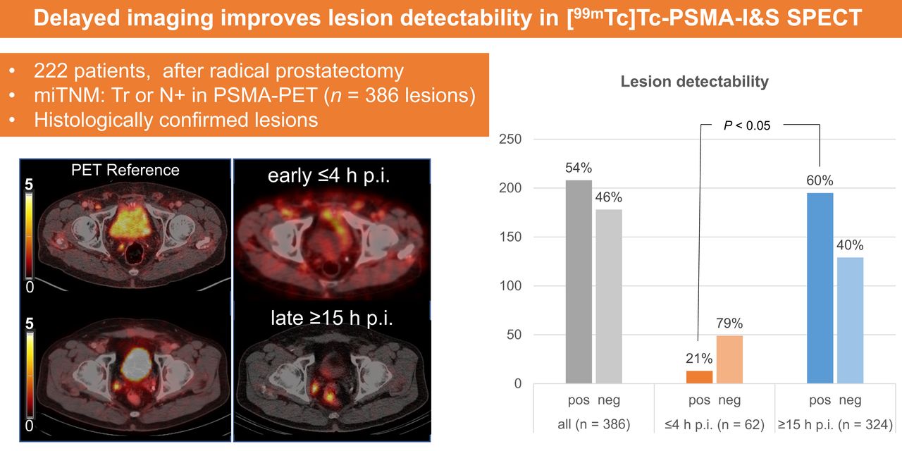
  • FIGURE 3.
    • Download figure
    • Open in new tab
    • Download powerpoint
    FIGURE 3.

    Overall and size-dependent lesion-based detectability by early (≤4 h) vs. late (≥15 h) [99mTc]Tc-PSMA-I&S SPECT/CT imaging.

Tables

  • Figures
  • Additional Files
    • View popup
    TABLE 1.

    Characteristics of 222 Patients Treated with RGS Between 2014 and 2020 at 2 Centers*

    ParameterEarly SPECT (≤4 h after injection; n = 44)Late SPECT (≥15 h after injection; n = 178)P
    Year of initial RP2010 (2005–2013)2014 (2010–2016)<0.001
    PSA at RP (ng/mL)10 (6–16)9 (6–15)0.8
    Lymph node yield at RP13 (8–20)13 (8–20)0.6
    Positive lymph nodes at RP0.6
     032 (73)115 (65)
     13 (6.8)19 (11)
     23 (6.8)9 (5.1)
     ≥32 (4.5)5 (2.8)
     Unknown4 (9.1)30 (17)
    Surgical margin status0.05
     R029 (66)130 (73)
     R19 (20)41 (23)
     RX/NA6 (14)7 (3.9)
    RT after RP0.1
     No RT15 (34)59 (33)
     RT after RP28 (64)119 (67)
     NA1 (2.3)0 (0)
    Time from RP to SPECT (mo)70 (22–128)46 (22–88)0.2
    Age at PSMA RGS (y)72 (65–75)66 (61–70)<0.001
    PSA before SPECT (ng/mL)1.4 (0.7–3.0)1.0 (0.5–2.0)0.09
    • * Patients received [99mTc]Tc-PSMA-I&S SPECT/CT before surgery and presented with BCR after RP with histopathology-confirmed positive lesions at PSMA PET/CT.

    • NA = not assigned; RT = radiotherapy.

    • Qualitative data are number and percentage; continuous data are median and IQR.

    • View popup
    TABLE 2.

    PSMA PET/CT and SPECT/CT Imaging Characteristics of 222 Patients with BCR After RP Treated with RGS Between 2014 and 2020 at 2 Centers Within Early and Late [99mTc]Tc-PSMA-I&S SPECT/CT Groups

    ParameterEarly SPECT (≤4 h after injection; n = 44)Late SPECT (≥15 h after injection; n = 178)P
    Lesions on PSMA PET0.7
     130 (68%)94 (53%)
     211 (25%)53 (30%)
     32 (4.5%)16 (9.0%)
     41 (2.3%)7 (3.9%)
     ≥50 (0%)8 (4.5%)
    Maximum lesion size on PET/CT (mm)8 (7–12)9 (6–12)0.7
    Maximum lesion SUV on PET9 (6–17)8 (5–16)0.1
    Ratio of maximum lesion SUV to background on PET12 (7–22)8 (5–16)0.02
    miTNM-Tr0 (0%)28 (16%)0.01
    miTNM-N134 (77%)112 (63%)0.1
    miTNM-N210 (23%)53 (30%)0.5
    miTNM-M1a0 (0%)15 (8.4%)0.1
    [99mTc]Tc-PSMA-I&S activity (MBq)550 (416–702)752 (695–786)<0.001
    Corrected [99mTc]Tc-PSMA-I&S activity at time of SPECT (MBq)488 (370–632)99 (91–108)<0.001
    Interval between [99mTc]-Tc-PSMA-I&S tracer injection and SPECT imaging (h)1.0 (1.0–2.0)18.0 (17.0–18.0)<0.001
    Lesions on SPECT<0.001
     033 (75%)45 (25%)
     19 (20%)90 (51%)
     21 (2%)27 (15%)
     31 (2%)13 (7%)
     40 (0%)2 (1%)
     ≥50 (0%)1 (1%)
    Maximum lesion size on SPECT/CT (mm)11.5 (7.8–17.1)9.0 (7.0–13.0)0.07
    • Qualitative data are number and percentage; continuous data are median and IQR.

    • View popup
    TABLE 3.

    Uni- and Multivariable Logistic Regression Models Regarding Visibility of Lesions in [99mTc]Tc-PSMA-I&S SPECT/CT Imaging

    UnivariableMultivariable
    VariableORCI, 2.5%CI, 97.5%PORCI, 2.5%CI, 97.5%P
    Gleason grade group at RP
     I–IIRef.
     III–V0.340.742.420.3
    pT stage at RP
     pT2Ref.
     pT3a/b0.650.341.210.2
    pN stage at RP
     pN0/XRef.
     pN10.530.271.050.07
    Margin status at RP
     R0Ref.
     R11.00.512.060.9
    RT after RP
     NoRef.
     Yes0.820.431.490.5
    Time from RP to SPECT (continuous)0.990.991.00.8
    Age at SPECT (continuous)0.990.951.030.6
    PSA at SPECT (continuous)1.211.031.470.031.250.991.660.09
    Lesions on PET (continuous)2.031.383.210.0011.571.022.670.07
    Maximum lesion size on PET (continuous)1.131.051.220.0021.060.971.170.2
    Maximum lesion SUV on PET (continuous)1.101.051.16<0.0011.151.071.27<0.001
    Activity of injected [99mTc]Tc-PSMA-I&S (continuous)1.001.001.00<0.0010.990.991.00.6
    Interval between injection and SPECT (continuous)1.161.111.22<0.0011.271.171.40<0.001
    • OR = odds ratio; RT = radiotherapy.

Additional Files

  • Figures
  • Tables
  • Supplemental Data

    Files in this Data Supplement:

    • Supplemental Data
PreviousNext
Back to top

In this issue

Journal of Nuclear Medicine: 66 (5)
Journal of Nuclear Medicine
Vol. 66, Issue 5
May 1, 2025
  • Table of Contents
  • Table of Contents (PDF)
  • About the Cover
  • Index by author
  • Complete Issue (PDF)
Print
Download PDF
Article Alerts
Sign In to Email Alerts with your Email Address
Email Article

Thank you for your interest in spreading the word on Journal of Nuclear Medicine.

NOTE: We only request your email address so that the person you are recommending the page to knows that you wanted them to see it, and that it is not junk mail. We do not capture any email address.

Enter multiple addresses on separate lines or separate them with commas.
Delayed Imaging Improves Lesion Detectability in [99mTc]Tc-PSMA-I&S SPECT/CT in Recurrent Prostate Cancer
(Your Name) has sent you a message from Journal of Nuclear Medicine
(Your Name) thought you would like to see the Journal of Nuclear Medicine web site.
Citation Tools
Delayed Imaging Improves Lesion Detectability in [99mTc]Tc-PSMA-I&S SPECT/CT in Recurrent Prostate Cancer
Christoph Berliner, Lisa Steinhelfer, Maythinee Chantadisai, Markus Kroenke, Daniel Koehler, Randi Pose, Peter Bannas, Sophie Knipper, Matthias Eiber, Tobias Maurer
Journal of Nuclear Medicine May 2023, 265252; DOI: 10.2967/jnumed.122.265252

Citation Manager Formats

  • BibTeX
  • Bookends
  • EasyBib
  • EndNote (tagged)
  • EndNote 8 (xml)
  • Medlars
  • Mendeley
  • Papers
  • RefWorks Tagged
  • Ref Manager
  • RIS
  • Zotero
Share
Delayed Imaging Improves Lesion Detectability in [99mTc]Tc-PSMA-I&S SPECT/CT in Recurrent Prostate Cancer
Christoph Berliner, Lisa Steinhelfer, Maythinee Chantadisai, Markus Kroenke, Daniel Koehler, Randi Pose, Peter Bannas, Sophie Knipper, Matthias Eiber, Tobias Maurer
Journal of Nuclear Medicine May 2023, 265252; DOI: 10.2967/jnumed.122.265252
Twitter logo Facebook logo LinkedIn logo Mendeley logo
  • Tweet Widget
  • Facebook Like
  • Google Plus One
Bookmark this article

Jump to section

  • Article
    • Visual Abstract
    • Abstract
    • MATERIALS AND METHODS
    • RESULTS
    • DISCUSSION
    • CONCLUSION
    • DISCLOSURE
    • Footnotes
    • REFERENCES
  • Figures & Data
  • Supplemental
  • Info & Metrics
  • PDF

Related Articles

  • PubMed
  • Google Scholar

Cited By...

  • Diagnostic Efficacy of Various Imaging Modalities Across Different Stages of Prostate Cancer: A Network Meta-Analysis of Diagnostic Studies
  • 61Cu-PSMA-Targeted PET for Prostate Cancer: From Radiotracer Development to First-in-Human Imaging
  • Strong Correlation Between SUVmax on PSMA PET/CT and Numeric Drop-In {gamma}-Probe Signal for Intraoperative Identification of Prostate Cancer Lesions
  • Google Scholar

More in this TOC Section

  • First-in-Human Study of 18F-Labeled PET Tracer for Glutamate AMPA Receptor [18F]K-40: A Derivative of [11C]K-2
  • Detection of HER2-Low Lesions Using HER2-Targeted PET Imaging in Patients with Metastatic Breast Cancer: A Paired HER2 PET and Tumor Biopsy Analysis
  • [11C]Carfentanil PET Whole-Body Imaging of μ-Opioid Receptors: A First in-Human Study
Show more Clinical Investigation

Similar Articles

Keywords

  • prostate carcinoma
  • scintigraphy
  • radioguided
  • salvage surgery
  • biochemical recurrence
  • PET
SNMMI

© 2025 SNMMI

Powered by HighWire