
Main menu

  • Home
  • Content
    • Current
    • Ahead of print
    • Past Issues
    • JNM Supplement
    • SNMMI Annual Meeting Abstracts
    • Continuing Education
    • JNM Podcasts
  • Subscriptions
    • Subscribers
    • Institutional and Non-member
    • Rates
    • Journal Claims
    • Corporate & Special Sales
  • Authors
    • Submit to JNM
    • Information for Authors
    • Assignment of Copyright
    • AQARA requirements
  • Info
    • Reviewers
    • Permissions
    • Advertisers
  • About
    • About Us
    • Editorial Board
    • Contact Information
  • More
    • Alerts
    • Feedback
    • Help
    • SNMMI Journals
  • SNMMI
    • JNM
    • JNMT
    • SNMMI Journals
    • SNMMI

User menu

  • Subscribe
  • My alerts
  • Log in
  • Log out
  • My Cart

Search

  • Advanced search
Journal of Nuclear Medicine
  • SNMMI
    • JNM
    • JNMT
    • SNMMI Journals
    • SNMMI
  • Subscribe
  • My alerts
  • Log in
  • Log out
  • My Cart
Journal of Nuclear Medicine

Advanced Search

  • Home
  • Content
    • Current
    • Ahead of print
    • Past Issues
    • JNM Supplement
    • SNMMI Annual Meeting Abstracts
    • Continuing Education
    • JNM Podcasts
  • Subscriptions
    • Subscribers
    • Institutional and Non-member
    • Rates
    • Journal Claims
    • Corporate & Special Sales
  • Authors
    • Submit to JNM
    • Information for Authors
    • Assignment of Copyright
    • AQARA requirements
  • Info
    • Reviewers
    • Permissions
    • Advertisers
  • About
    • About Us
    • Editorial Board
    • Contact Information
  • More
    • Alerts
    • Feedback
    • Help
    • SNMMI Journals
  • View or Listen to JNM Podcast
  • Visit JNM on Facebook
  • Join JNM on LinkedIn
  • Follow JNM on Twitter
  • Subscribe to our RSS feeds
Research ArticleFEATURED BASIC SCIENCE ARTICLE

Development of a Lensless Radiomicroscope for Cellular-Resolution Radionuclide Imaging

Justin S. Klein, Tae Jin Kim and Guillem Pratx
Journal of Nuclear Medicine March 2023, 64 (3) 479-484; DOI: https://doi.org/10.2967/jnumed.122.264021
Justin S. Klein
Department of Radiation Oncology, Stanford University, Stanford, California
  • Find this author on Google Scholar
  • Find this author on PubMed
  • Search for this author on this site
Tae Jin Kim
Department of Radiation Oncology, Stanford University, Stanford, California
  • Find this author on Google Scholar
  • Find this author on PubMed
  • Search for this author on this site
Guillem Pratx
Department of Radiation Oncology, Stanford University, Stanford, California
  • Find this author on Google Scholar
  • Find this author on PubMed
  • Search for this author on this site
  • Article
  • Figures & Data
  • Supplemental
  • Info & Metrics
  • PDF
Loading

Article Figures & Data

Figures

  • Additional Files
  • Figure
    • Download figure
    • Open in new tab
    • Download powerpoint
  • FIGURE 1.
    • Download figure
    • Open in new tab
    • Download powerpoint
    FIGURE 1.

    Conceptual, physical, and schematic diagram of the LRM. (A) Cells are grown on a glass slide and treated with α- or β-emitting radiopharmaceuticals. The slide is inverted and placed in direct contact with a CMOS imaging sensor. Emitted particles deposit energy in adjacent CMOS pixels, producing a detectable electronic signal. (B) The LRM consists of an imaging sensor (CMOS) that is read out through a compact Raspberry Pi computer (RPI). For live-cell imaging, sensor temperature is regulated by a proportional-integral-derivative (PID) controller connected to a thermocouple (TC) and thermoelectric cooler (TEC). The computer is also connected to an LED, which can be switched on for brightfield imaging. (C) The β-microscope imaging chamber, filled with medium. (D) Light-tight β-microscope enclosure. The LED used for brightfield imaging is housed in the upper cylinder. Attached to the bottom of the enclosure are the thermoelectric cooler, heat sync and fan.

  • FIGURE 2.
    • Download figure
    • Open in new tab
    • Download powerpoint
    FIGURE 2.

    LRM physical characterization and calibration. (A) Brightfield image of 1-μm polystyrene sphere with inset line profile showing full width at half maximum. (B) Fourier ring correlation analysis plot showing spatial resolution of RLM.
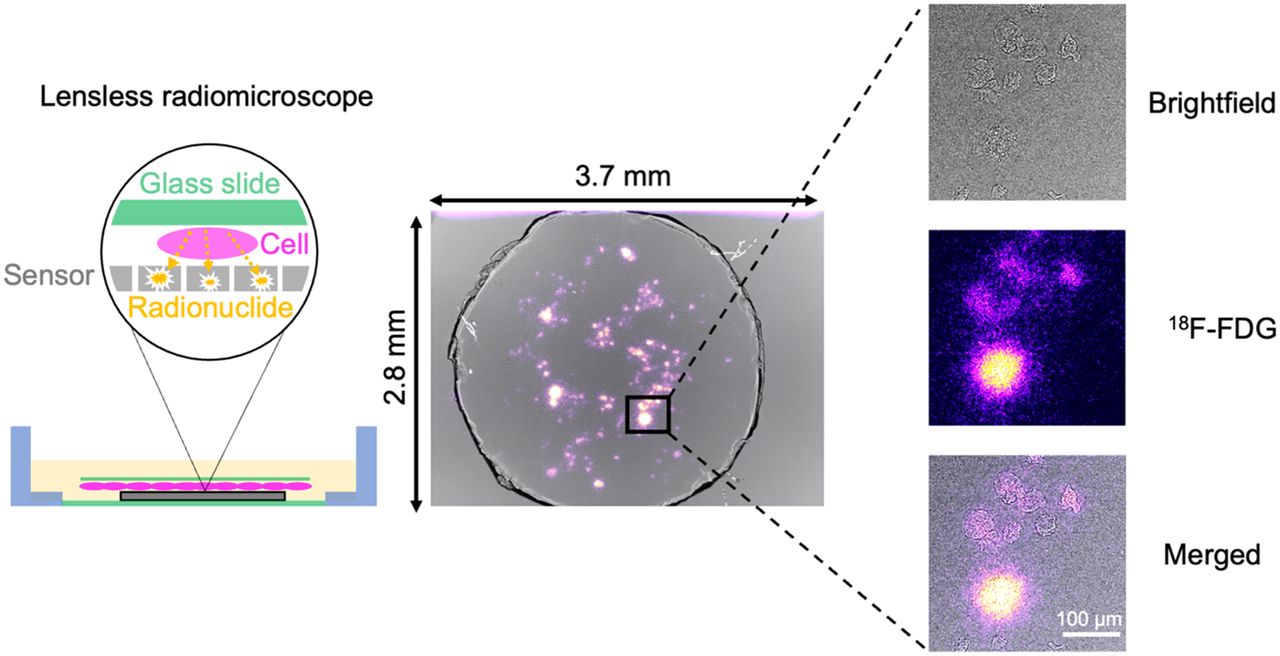
  • FIGURE 3.
    • Download figure
    • Open in new tab
    • Download powerpoint
    FIGURE 3.

    High-resolution β-imaging of 18F-FDG in breast cancer cells. MDA-MB-231 cells were imaged using brightfield and β-modes. Images are cropped to 600 × 600 μm from the full 3.7 × 2.8 mm field of view. Total imaging time for β-imaging was 65 min. Scale bar is 50 μm.

  • FIGURE 4.
    • Download figure
    • Open in new tab
    • Download powerpoint
    FIGURE 4.

    18F-FDG β-imaging of breast cancer cells treated with radiation. β-images of cells receiving 0 Gy (A) and 3 Gy (B). Scale bar is 200 μm. (C) Scatter plot of activity vs. area for treated and untreated cells. (D) Bar graphs of activity per cell, cell size, and cell activity per area.
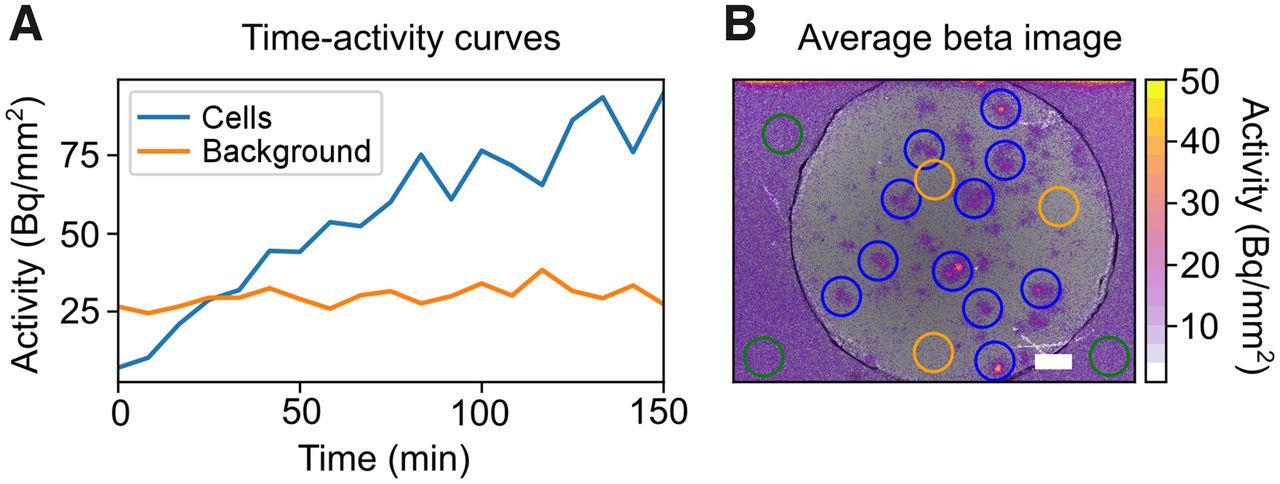
  • FIGURE 5.
    • Download figure
    • Open in new tab
    • Download powerpoint
    FIGURE 5.

    Dynamic 18F-FDG imaging of cells. (A) Time–activity curves measured from cell and background ROIs. (B) Cumulative uptake of 18F-FDG by MDA-MB-231 cells, with ROIs shown for cells (blue) and background (orange). Scale bar is 200 μm.

  • FIGURE 6.
    • Download figure
    • Open in new tab
    • Download powerpoint
    FIGURE 6.

    α-imaging of Po-210 needle source. The needle is visible as a dark shadow in the brightfield image. α-emission is readily localized to the needle source in the merged image. Scale bar is 500 μm.

Additional Files

  • Figures
  • Supplemental Data

    Files in this Data Supplement:

    • Supplemental Data
    • Supplemental Video 1
    • Supplemental Video 2
PreviousNext
Back to top

In this issue

Journal of Nuclear Medicine: 64 (3)
Journal of Nuclear Medicine
Vol. 64, Issue 3
March 1, 2023
  • Table of Contents
  • Table of Contents (PDF)
  • About the Cover
  • Index by author
  • Complete Issue (PDF)
Print
Download PDF
Article Alerts
Sign In to Email Alerts with your Email Address
Email Article

Thank you for your interest in spreading the word on Journal of Nuclear Medicine.

NOTE: We only request your email address so that the person you are recommending the page to knows that you wanted them to see it, and that it is not junk mail. We do not capture any email address.

Enter multiple addresses on separate lines or separate them with commas.
Development of a Lensless Radiomicroscope for Cellular-Resolution Radionuclide Imaging
(Your Name) has sent you a message from Journal of Nuclear Medicine
(Your Name) thought you would like to see the Journal of Nuclear Medicine web site.
Citation Tools
Development of a Lensless Radiomicroscope for Cellular-Resolution Radionuclide Imaging
Justin S. Klein, Tae Jin Kim, Guillem Pratx
Journal of Nuclear Medicine Mar 2023, 64 (3) 479-484; DOI: 10.2967/jnumed.122.264021

Citation Manager Formats

  • BibTeX
  • Bookends
  • EasyBib
  • EndNote (tagged)
  • EndNote 8 (xml)
  • Medlars
  • Mendeley
  • Papers
  • RefWorks Tagged
  • Ref Manager
  • RIS
  • Zotero
Share
Development of a Lensless Radiomicroscope for Cellular-Resolution Radionuclide Imaging
Justin S. Klein, Tae Jin Kim, Guillem Pratx
Journal of Nuclear Medicine Mar 2023, 64 (3) 479-484; DOI: 10.2967/jnumed.122.264021
Twitter logo Facebook logo LinkedIn logo Mendeley logo
  • Tweet Widget
  • Facebook Like
  • Google Plus One
Bookmark this article

Jump to section

  • Article
    • Visual Abstract
    • Abstract
    • MATERIALS AND METHODS
    • RESULTS
    • DISCUSSION
    • CONCLUSION
    • DISCLOSURE
    • Footnotes
    • REFERENCES
  • Figures & Data
  • Supplemental
  • Info & Metrics
  • PDF

Related Articles

  • PubMed
  • Google Scholar

Cited By...

  • No citing articles found.
  • Google Scholar

More in this TOC Section

  • RadioFlow Cytometry Reveals That [18F]FDG Uptake in K-RAS Lung Cancer Is Driven by Immune Cells: An Analysis on a Single-Cell Level
  • Comparison Between Brain and Cerebellar Autoradiography Using [18F]Flortaucipir, [18F]MK6240, and [18F]PI2620 in Postmortem Human Brain Tissue
  • Preclinical Evaluation of 226Ac as a Theranostic Agent: Imaging, Dosimetry, and Therapy
Show more FEATURED BASIC SCIENCE ARTICLE

Similar Articles

Keywords

  • radionuclide imaging
  • in vitro assays
  • 18F-FDG
  • CMOS detector
SNMMI

© 2025 SNMMI

Powered by HighWire