


Article Figures & Data
Figures
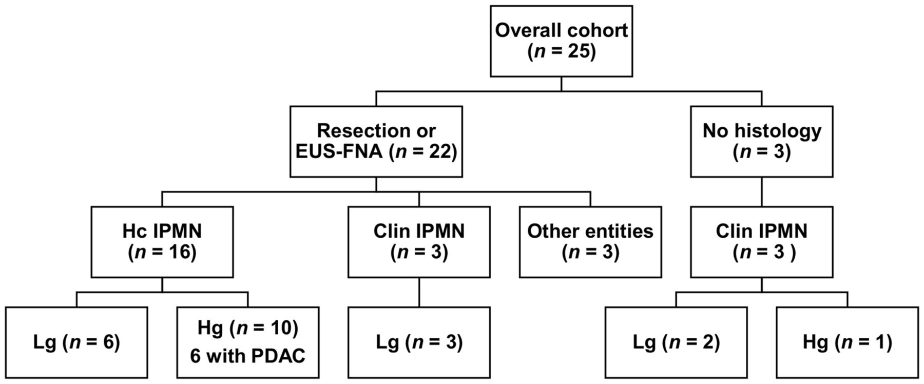
- FIGURE 1.
Histologic diagnoses and clinical classification of 25 patients with suspected IPMN who underwent 68Ga-FAPI PET/CT. clin IPMN = clinical IPMN; EUS-FNA = endoscopic ultrasound–guided fine-needle aspiration; hc IPMN = histologically confirmed IPMN; hg = high grade; lg = low grade; PDAC = pancreatic ductal adenocarcinoma.
- FIGURE 2.
Biodistribution analysis (SUVmax and SUVmean ± SD) of 25 patients with suspected IPMN based on static PET imaging at 1 h after injection of 68Ga-labeled FAPI-74.
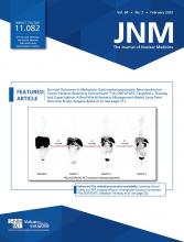
- FIGURE 3.
(A and B) Boxplots of SUVmax (A) and SUVmean (B) of different types of cystic pancreatic lesions. (C and D) Boxplots of SUVmax (C) and SUVmean (D) sorted by given or not given indication for surgery. Boxes represent the interquartile range (IQR) and whiskers the range of 1.5 IQR; horizontal line within box indicates the median and cross the mean. Data outliers are shown separately within graph. clin hg-IPMN = clinical high-grade IPMN; clin lg-IPMN = clinical low-grade IPMN; hc hg-IPMN = histologically confirmed high-grade IPMN; hc lg-IPMN = histologically confirmed low-grade IPMN; hc others = histologically confirmed other entities.
- FIGURE 4.
(A–C) Time–activity curves displaying averaged 68Ga-FAPI-74 uptake (relative to peak) kinetics of histologically confirmed menacing IPMN (hc men-IPMN) (A), histologically confirmed low-grade IPMN (hc lg-IPMN) (B), and clinical low-grade IPMN (clin lg-IPMN) (C). (D) Box plot displaying time to peak values of histologically confirmed menacing IPMN, histologically confirmed low-grade IPMN, and clinical low-grade IPMN as measured by dynamic 68Ga-FAPI-74 PET imaging. (E and F) Box plots displaying K1 (E) and k2 (F) values of histologically confirmed menacing IPMN, histologically confirmed low grade IPMN, and clinical low-grade IPMN as calculated by kinetic modeling of dynamic 68Ga-FAPI-74 PET imaging data. Boxes represent the interquartile range (IQR) and whiskers the range of 1.5 IQR; horizontal line within box indicates the median and cross the mean. Data outliers are shown separately within graph.
- FIGURE 5.
(A and B) Receiver-operating-characteristic (ROC) curves depicting sensitivity and specificity of quantitative static (SUVmax and SUVmean) and dynamic (TTP) 68Ga-FAPI-74 PET parameters for differentiation of histologically confirmed menacing IPMN and low-grade IPMN (A) and of lesions with and without indication for surgery (B). AUC = area under the curve.
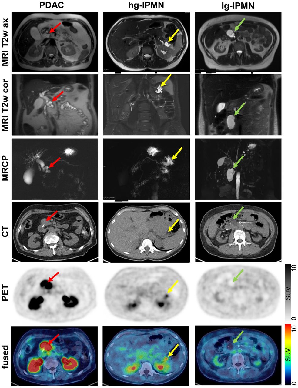
- FIGURE 6.
Representative axial and coronal T2-weighted MRI (MRI T2w ax and MRI T2w cor, respectively), MR cholangiopancreatography (MRCP), axial CT (CT ax), axial PET (PET ax), and fused images of a patient with hc hg-IPMN with progression into PDAC, a patient with hg-IPMN without PDAC, and patient with lg-IPMN. Red, yellow, and green arrows indicate pathologies.


Tables
- TABLE 1.
Clinical Characteristics and Histologic Diagnoses of 25 Patients with Suspected IPMN and 68Ga-FAPI-74 PET/CT
Patient Sex Age (y) Cyst size (mm) IPMN type Additional information Surgery/histology Histologic diagnosis 1 M 52 29 BD Whipple lg-IPMN 2 F 52 57 BD Excision lg-IPMN 3 M 76 40 BD Cytology lg-IPMN 4 F 42 30 BD Whipple lg-IPMN 5 M 71 60 BD Whipple lg-IPMN 6 F 67 44 BD Distal pancreatectomy lg-IPMN 7 M 56 18* MD Mural nodule Enucleation hg-IPMN 8 M 79 10* MD Whipple hg-IPMN 9 M 53 20* MD Distal pancreatectomy PDAC 10 F 64 11* MD Whipple PDAC 11 M 68 32 BD MD with dliatation to 4.8 mm, jaundice Whipple PDAC 12 F 44 10* Mixed type Distal pancreatectomy hg-IPMN 13 F 57 50* MD Pancreatectomy PDAC 14 M 78 90 Mixed type Solid components Therapy refused None 15 F 74 25 BD Size progressing Distal pancreatectomy hg-IPMN 16 F 80 42 BD Distal pancreatectomy PDAC 17 F 83 45 BD Distal pancreatectomy PDAC 18 M 63 30 BD Cytology (nonconclusive) None 19 M 77 60 BD Cytology None 20 F 64 30 BD Cytology (nonconclusive) None 21 F 57 23 BD Size progressing None None 22 M 74 10 BD None None 23 F 54 38 Cytology SCN 24 F 38 38 Distal pancreatectomy SCN 25 M 62 21 Distal pancreatectomy PanIN ↵* Main duct diameter.
BD = branch duct; MD = main duct; SCN = serous cystic neoplasia; PanIN = pancreatic intraepithelial neoplasia.
- TABLE 2.
Threshold and Specificity at Fixed Sensitivities of 90% and 80% for Differentiation Between Histologically Confirmed Low-Grade IPMN and Histologically Confirmed Menacing IPMN and Given Versus Not Given Indication for Surgery
Endpoint Parameter Threshold Sensitivity (%) 95% CI Specificity (%) 95% CI TN TP FN FP lg/men SUVmax 3.62 90.0 55.5–99.7 66.7 22.3–95.7 4 9 1 2 4.85 80.0 44.4–97.5 83.3 35.9–99.6 5 8 2 1 lg/men SUVmean 2.07 90.0 55.5–99.7 83.3 35.9–99.6 5 9 1 1 2.19 80.0 44.4–97.5 83.3 35.9–99.6 5 8 2 1 lg/men TTP 135.00 88.9 51.8–99.7 100.0 39.8–100.0 4 8 1 0 225.00 77.8 40.0–97.2 100.0 39.8–100.0 4 7 2 0 Surgery SUVmax 3.62 90.9 58.7–99.8 71.4 41.9–91.6 10 10 1 4 4.85 81.8 48.2–97.7 92.9 66.1–99.8 13 9 2 1 Surgery SUVmean 2.07 90.9 58.7–99.8 64.3 35.1–87.2 9 10 1 5 2.19 81.8 48.2–97.7 71.4 41.9–91.6 10 9 2 4 Surgery TTP 135 88.9 51.8–99.7 83.3 51.6–97.9 10 8 1 2 225 77.8 40.0–97.2 91.7 61.5–99.8 11 7 2 1 FN = false negative; FP = false positive; lg/men = histologically confirmed low-grade IPMN vs. histologically confirmed menacing IPMN; TN = true negative; TP = true positive; TTP = time to peak. For some settings only approximate sensitivities could be selected due to sparsity of data.
Supplemental Data
Files in this Data Supplement:
In this issue


Jump to section
Related Articles
Cited By...
- Diagnostic Potential of Supplemental Static and Dynamic 68Ga-FAPI-46 PET for Primary 18F-FDG-Negative Pulmonary Lesions
- Immunohistochemical FAP Expression Reflects 68Ga-FAPI PET Imaging Properties of Low- and High-Grade Intraductal Papillary Mucinous Neoplasms and Pancreatic Ductal Adenocarcinoma
- Tumor Characterization by [68Ga]FAPI-46 PET/CT Can Improve Treatment Selection for Pancreatic Cancer Patients: An Interim Analysis of a Prospective Clinical Trial