
Main menu

  • Home
  • Content
    • Current
    • Ahead of print
    • Past Issues
    • JNM Supplement
    • SNMMI Annual Meeting Abstracts
    • Continuing Education
    • JNM Podcasts
  • Subscriptions
    • Subscribers
    • Institutional and Non-member
    • Rates
    • Journal Claims
    • Corporate & Special Sales
  • Authors
    • Submit to JNM
    • Information for Authors
    • Assignment of Copyright
    • AQARA requirements
  • Info
    • Reviewers
    • Permissions
    • Advertisers
  • About
    • About Us
    • Editorial Board
    • Contact Information
  • More
    • Alerts
    • Feedback
    • Help
    • SNMMI Journals
  • SNMMI
    • JNM
    • JNMT
    • SNMMI Journals
    • SNMMI

User menu

  • Subscribe
  • My alerts
  • Log in
  • My Cart

Search

  • Advanced search
Journal of Nuclear Medicine
  • SNMMI
    • JNM
    • JNMT
    • SNMMI Journals
    • SNMMI
  • Subscribe
  • My alerts
  • Log in
  • My Cart
Journal of Nuclear Medicine

Advanced Search

  • Home
  • Content
    • Current
    • Ahead of print
    • Past Issues
    • JNM Supplement
    • SNMMI Annual Meeting Abstracts
    • Continuing Education
    • JNM Podcasts
  • Subscriptions
    • Subscribers
    • Institutional and Non-member
    • Rates
    • Journal Claims
    • Corporate & Special Sales
  • Authors
    • Submit to JNM
    • Information for Authors
    • Assignment of Copyright
    • AQARA requirements
  • Info
    • Reviewers
    • Permissions
    • Advertisers
  • About
    • About Us
    • Editorial Board
    • Contact Information
  • More
    • Alerts
    • Feedback
    • Help
    • SNMMI Journals
  • View or Listen to JNM Podcast
  • Visit JNM on Facebook
  • Join JNM on LinkedIn
  • Follow JNM on Twitter
  • Subscribe to our RSS feeds
Research ArticleClinical (Oncology: Other)

CD8-Targeted PET Imaging of Tumor-Infiltrating T Cells in Patients with Cancer: A Phase I First-in-Humans Study of 89Zr-Df-IAB22M2C, a Radiolabeled Anti-CD8 Minibody

Michael D. Farwell, Raymond F. Gamache, Hasan Babazada, Matthew D. Hellmann, James J. Harding, Ron Korn, Alessandro Mascioni, William Le, Ian Wilson, Michael S. Gordon, Anna M. Wu, Gary A. Ulaner, Jedd D. Wolchok, Michael A. Postow and Neeta Pandit-Taskar
Journal of Nuclear Medicine May 2022, 63 (5) 720-726; DOI: https://doi.org/10.2967/jnumed.121.262485
Michael D. Farwell
1Department of Radiology, Perelman School of Medicine, University of Pennsylvania, Philadelphia, Pennsylvania;
2Abramson Cancer Center, Perelman School of Medicine, University of Pennsylvania, Philadelphia, Pennsylvania;
  • Find this author on Google Scholar
  • Find this author on PubMed
  • Search for this author on this site
Raymond F. Gamache
1Department of Radiology, Perelman School of Medicine, University of Pennsylvania, Philadelphia, Pennsylvania;
  • Find this author on Google Scholar
  • Find this author on PubMed
  • Search for this author on this site
Hasan Babazada
1Department of Radiology, Perelman School of Medicine, University of Pennsylvania, Philadelphia, Pennsylvania;
  • Find this author on Google Scholar
  • Find this author on PubMed
  • Search for this author on this site
Matthew D. Hellmann
3Parker Institute for Cancer Immunotherapy, Memorial Sloan Kettering Cancer Center, New York, New York;
4Department of Medicine, Memorial Sloan Kettering Cancer Center, New York, New York;
5Department of Medicine, Weill Cornell Medical College, New York, New York;
  • Find this author on Google Scholar
  • Find this author on PubMed
  • Search for this author on this site
James J. Harding
4Department of Medicine, Memorial Sloan Kettering Cancer Center, New York, New York;
5Department of Medicine, Weill Cornell Medical College, New York, New York;
  • Find this author on Google Scholar
  • Find this author on PubMed
  • Search for this author on this site
Ron Korn
6Imaging Endpoints, Scottsdale, Arizona;
  • Find this author on Google Scholar
  • Find this author on PubMed
  • Search for this author on this site
Alessandro Mascioni
7ImaginAb, Inc., Inglewood, California;
  • Find this author on Google Scholar
  • Find this author on PubMed
  • Search for this author on this site
William Le
7ImaginAb, Inc., Inglewood, California;
  • Find this author on Google Scholar
  • Find this author on PubMed
  • Search for this author on this site
Ian Wilson
7ImaginAb, Inc., Inglewood, California;
  • Find this author on Google Scholar
  • Find this author on PubMed
  • Search for this author on this site
Michael S. Gordon
8HonorHealth Research Institute, Scottsdale, Arizona;
  • Find this author on Google Scholar
  • Find this author on PubMed
  • Search for this author on this site
Anna M. Wu
7ImaginAb, Inc., Inglewood, California;
9Department of Molecular Imaging and Therapy, Beckman Research Institute of the City of Hope, Duarte, California;
  • Find this author on Google Scholar
  • Find this author on PubMed
  • Search for this author on this site
Gary A. Ulaner
10Molecular Imaging and Therapy, Hoag Family Cancer Institute, Newport Beach, California;
  • Find this author on Google Scholar
  • Find this author on PubMed
  • Search for this author on this site
Jedd D. Wolchok
3Parker Institute for Cancer Immunotherapy, Memorial Sloan Kettering Cancer Center, New York, New York;
4Department of Medicine, Memorial Sloan Kettering Cancer Center, New York, New York;
5Department of Medicine, Weill Cornell Medical College, New York, New York;
11Human Oncology and Pathogenesis Program, Memorial Sloan Kettering Cancer Center, New York, New York;
  • Find this author on Google Scholar
  • Find this author on PubMed
  • Search for this author on this site
Michael A. Postow
4Department of Medicine, Memorial Sloan Kettering Cancer Center, New York, New York;
5Department of Medicine, Weill Cornell Medical College, New York, New York;
  • Find this author on Google Scholar
  • Find this author on PubMed
  • Search for this author on this site
Neeta Pandit-Taskar
3Parker Institute for Cancer Immunotherapy, Memorial Sloan Kettering Cancer Center, New York, New York;
12Department of Radiology, Memorial Sloan Kettering Cancer Center, New York, New York; and
13Department of Radiology, Weill Cornell Medical College, New York, New York
  • Find this author on Google Scholar
  • Find this author on PubMed
  • Search for this author on this site
  • Article
  • Figures & Data
  • Supplemental
  • Info & Metrics
  • PDF
Loading

Article Figures & Data

Figures

  • Tables
  • Additional Files
  • Figure
    • Download figure
    • Open in new tab
    • Download powerpoint
  • FIGURE 1.
    • Download figure
    • Open in new tab
    • Download powerpoint
    FIGURE 1.

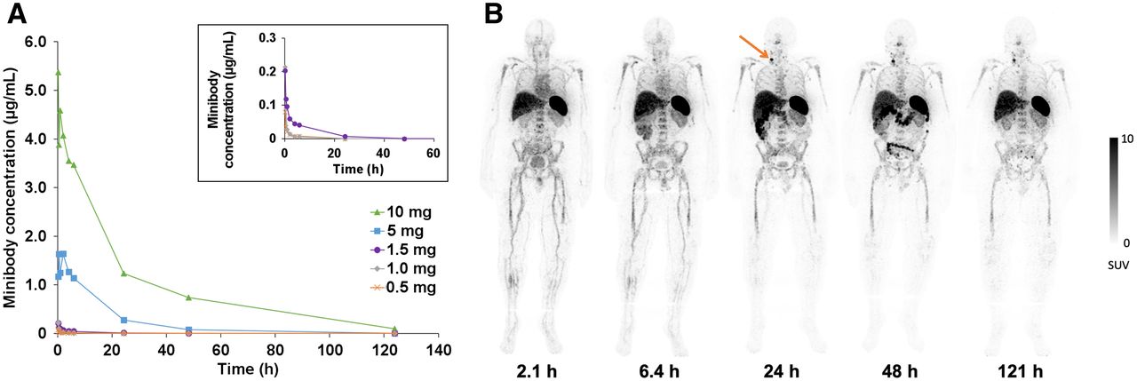
    Serum clearance and biodistribution of 89Zr-Df-IAB22M2C. (A) Serum clearance of 89Zr-Df-IAB22M2C based on enzyme-linked immunosorbent assay measurements (limit of detection = 5 ng/mL). No minibody was detected in serum at the 0.2-mg dose. (B) Whole-body PET images of a patient at various times after injection of 89Zr-Df-IAB22M2C (1.5-mg minibody dose) demonstrating the distribution of 89Zr-Df-IAB22M2C in normal tissues and uptake in a nodal metastasis in the right neck (arrow), with good visualization of uptake in the nodal metastasis at 24–48 h after injection.

  • FIGURE 2.
    • Download figure
    • Open in new tab
    • Download powerpoint
    FIGURE 2.

    89Zr-Df-IAB22M2C uptake in normal tissues and tumor lesions versus time. (A) 89Zr-Df-IAB22M2C uptake in CD8-rich reference tissues in patients administered 0.5 and 1.5 mg of minibody mass. (B) 89Zr-Df-IAB22M2C uptake in CD8-poor reference tissues in patients administered 0.5 and 1.5 mg of minibody mass. (C) Box and whisker plots of 89Zr-Df-IAB22M2C uptake in tumor lesions from all subjects (n = 15). Boxes outline first and third quartile values. Median SUVMAX values are indicated by horizontal line and mean SUVMAX values are indicated with +. Outlier values are indicated by dots. (D) 89Zr-Df-IAB22M2C mean tumor uptake in patients who received 0.5 and 1.5 mg of minibody mass. BM = bone marrow; LN = lymph nodes.

  • FIGURE 3.
    • Download figure
    • Open in new tab
    • Download powerpoint
    FIGURE 3.

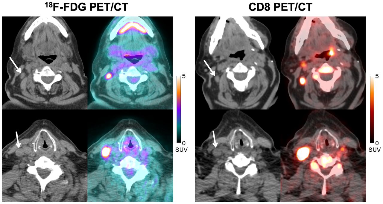
    A 77-y-old man with metastatic melanoma treated with pembrolizumab. CT and fused 18F-FDG PET/CT images (left) acquired at approximately 8 mo after initiation of immunotherapy demonstrate 2 18F-FDG–avid nodal metastases in right neck (SUVMAX = 8.0, top image; SUVMAX = 16.8, bottom image), which could represent viable metastases. Corresponding CT and fused CD8 PET/CT images (right) obtained at 1 mo after 18F-FDG PET/CT demonstrate significant tracer activity in both metastases (SUVMAX = 5.4, top image; SUVMAX = 14.6, bottom image), which suggests that some of the 18F-FDG activity could be due to tumor-infiltrating CD8+ T cells rather than tumor cells. Follow-up imaging over the next 6 mo demonstrated stable disease, supportive of this hypothesis.
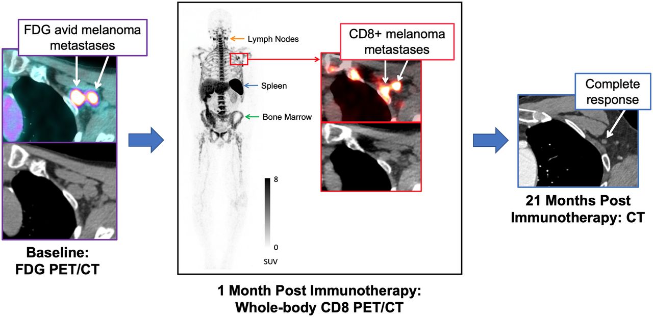
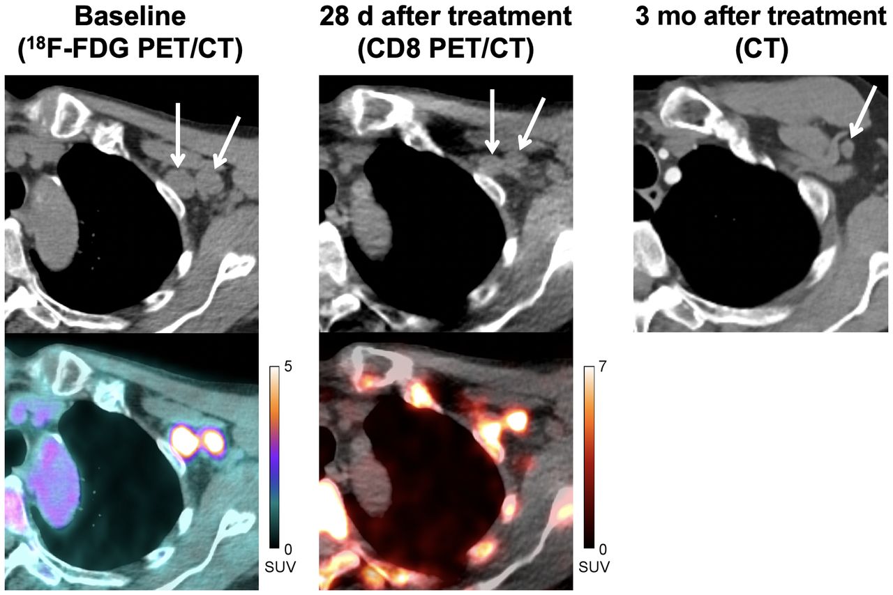
  • FIGURE 4.
    • Download figure
    • Open in new tab
    • Download powerpoint
    FIGURE 4.

    A 71-y-old man with locally advanced stage III melanoma treated with pembrolizumab. Baseline CT and fused 18F-FDG PET/CT images (left) demonstrate 2 18F-FDG–avid metastases in left axilla (SUVMAX = 10.0, medial node; SUVMAX = 7.6, lateral node). CT and fused CD8 PET/CT images (middle) obtained at 28 d after start of immunotherapy demonstrate increased tracer activity in both metastases (SUVMAX = 9.5, medial node; SUVMAX = 10.0, lateral node), suggestive of tumor infiltration by CD8+ T cells. Follow-up imaging with contrast-enhanced CT (right) demonstrated complete response to therapy.

Tables

  • Figures
  • Additional Files
    • View popup
    TABLE 1.

    Patient Characteristics

    CharacteristicAll patients (n = 15)
    Median age (y)64 (range, Embedded Image)
    Sex (n)
     Male9 (60)
     Female6 (40)
    Tumor type (n)
     Melanoma8 (53)
     Non–small cell lung carcinoma6 (40)
     Hepatocellular carcinoma1 (7)
    Treatment profile at the time of imaging (n)
     On immunotherapy (Embedded Image mo)3 (20)
     On immunotherapy (Embedded Image mo)5 (33)
     On targeted therapy (Embedded Image mo)2 (13)
     Discontinued prior treatment (Embedded Image mo)2 (13)
     Treatment naïve3 (20)
    • Data in parentheses are percentages unless otherwise indicated.

Additional Files

  • Figures
  • Tables
  • Supplemental Data

    Files in this Data Supplement:

    • Supplemental Data
PreviousNext
Back to top

In this issue

Journal of Nuclear Medicine: 63 (5)
Journal of Nuclear Medicine
Vol. 63, Issue 5
May 1, 2022
  • Table of Contents
  • Table of Contents (PDF)
  • About the Cover
  • Index by author
  • Complete Issue (PDF)
Print
Download PDF
Article Alerts
Sign In to Email Alerts with your Email Address
Email Article

Thank you for your interest in spreading the word on Journal of Nuclear Medicine.

NOTE: We only request your email address so that the person you are recommending the page to knows that you wanted them to see it, and that it is not junk mail. We do not capture any email address.

Enter multiple addresses on separate lines or separate them with commas.
CD8-Targeted PET Imaging of Tumor-Infiltrating T Cells in Patients with Cancer: A Phase I First-in-Humans Study of 89Zr-Df-IAB22M2C, a Radiolabeled Anti-CD8 Minibody
(Your Name) has sent you a message from Journal of Nuclear Medicine
(Your Name) thought you would like to see the Journal of Nuclear Medicine web site.
Citation Tools
CD8-Targeted PET Imaging of Tumor-Infiltrating T Cells in Patients with Cancer: A Phase I First-in-Humans Study of 89Zr-Df-IAB22M2C, a Radiolabeled Anti-CD8 Minibody
Michael D. Farwell, Raymond F. Gamache, Hasan Babazada, Matthew D. Hellmann, James J. Harding, Ron Korn, Alessandro Mascioni, William Le, Ian Wilson, Michael S. Gordon, Anna M. Wu, Gary A. Ulaner, Jedd D. Wolchok, Michael A. Postow, Neeta Pandit-Taskar
Journal of Nuclear Medicine May 2022, 63 (5) 720-726; DOI: 10.2967/jnumed.121.262485

Citation Manager Formats

  • BibTeX
  • Bookends
  • EasyBib
  • EndNote (tagged)
  • EndNote 8 (xml)
  • Medlars
  • Mendeley
  • Papers
  • RefWorks Tagged
  • Ref Manager
  • RIS
  • Zotero
Share
CD8-Targeted PET Imaging of Tumor-Infiltrating T Cells in Patients with Cancer: A Phase I First-in-Humans Study of 89Zr-Df-IAB22M2C, a Radiolabeled Anti-CD8 Minibody
Michael D. Farwell, Raymond F. Gamache, Hasan Babazada, Matthew D. Hellmann, James J. Harding, Ron Korn, Alessandro Mascioni, William Le, Ian Wilson, Michael S. Gordon, Anna M. Wu, Gary A. Ulaner, Jedd D. Wolchok, Michael A. Postow, Neeta Pandit-Taskar
Journal of Nuclear Medicine May 2022, 63 (5) 720-726; DOI: 10.2967/jnumed.121.262485
Twitter logo Facebook logo LinkedIn logo Mendeley logo
  • Tweet Widget
  • Facebook Like
  • Google Plus One
Bookmark this article

Jump to section

  • Article
    • Visual Abstract
    • Abstract
    • MATERIALS AND METHODS
    • RESULTS
    • DISCUSSION
    • CONCLUSION
    • DISCLOSURE
    • Footnotes
    • REFERENCES
  • Figures & Data
  • Supplemental
  • Info & Metrics
  • PDF

Related Articles

  • PubMed
  • Google Scholar

Cited By...

  • Approaches to Imaging Immune Activation Using PET
  • Exploring molecular imaging to investigate immune checkpoint inhibitor-related toxicity
  • FDG-PET associations with pathological response and survival with neoadjuvant immunotherapy for melanoma
  • Granzyme B PET/CT Imaging Evaluates Early Response to Immunotherapy in Gastric Cancer
  • Head-to-head comparison of nuclear imaging approaches to quantify tumor CD8+ T-cell infiltration
  • Single Chelator-Minibody Theranostic Agents for 89Zr PET Imaging and 177Lu Radiopharmaceutical Therapy of PSMA-Expressing Prostate Cancer
  • Making the effect visible - OX40 targeting nanobodies for in vivo imaging of activated T cells
  • Evolocumab as an immunomodulator in glioma: A window of opportunity trial evaluating PCSK9 inhibition to enhance surface MHC-I on tumor
  • Impact of human CD8+ T cell senescence on 89Zr radiolabelling and homing properties
  • Imaging With PET/CT of Diffuse CD8 T-Cell Infiltration of Skeletal Muscle in Patients With Inclusion Body Myositis
  • Two birds with one stone: human SIRP{alpha} nanobodies for functional modulation and in vivo imaging of myeloid cells
  • First-in-human immunoPET imaging of COVID-19 convalescent patients using dynamic total-body PET and a CD8-targeted minibody
  • Single-Domain Antibody Theranostics on the Horizon
  • MRI techniques for immunotherapy monitoring
  • Google Scholar

Similar Articles

Keywords

  • 89Zr-Df-IAB22M2C
  • PET imaging
  • CD8+ T cell
  • minibody
  • immunotherapy
SNMMI

© 2025 SNMMI

Powered by HighWire