


Article Figures & Data
Figures
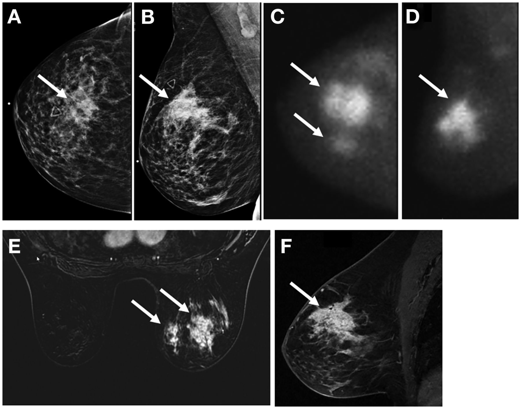
- FIGURE 1.
MBI with 300 MBq (8 mCi) of 99mTc-sestamibi for extent-of-disease evaluation in 59-y-old woman with palpable irregular mass (arrows) in right upper central breast. (A and B) Mass measured 3.3 cm on craniocaudal (A) and mediolateral oblique (B) mammograms. (C and D) MBI showed 10 cm of uptake on craniocaudal (C) and mediolateral oblique (D) views. (E and F) Postcontrast axial (E) and sagittal (F) MRI confirmed 10.2 cm of abnormal enhancement. After NAT and mastectomy, surgical pathology showed 8-cm treated tumor bed with 0.2 cm of residual invasive carcinoma.
- FIGURE 2.
A 38-y-old woman with right-breast triple-negative and node-negative invasive ductal carcinoma (arrows). (A and B) Pretreatment postcontrast sagittal fat-suppressed T1-weighted MRI shows irregular mass in right breast (A), and MBI with 300 MBq (8 mCi) of 99mTc-sestamibi shows intense uptake in mediolateral oblique view (B). (C and D) On posttreatment imaging, there is no residual enhancement on MRI (C) and no residual uptake on MBI (D). Surgical pathology showed pathologic complete response.
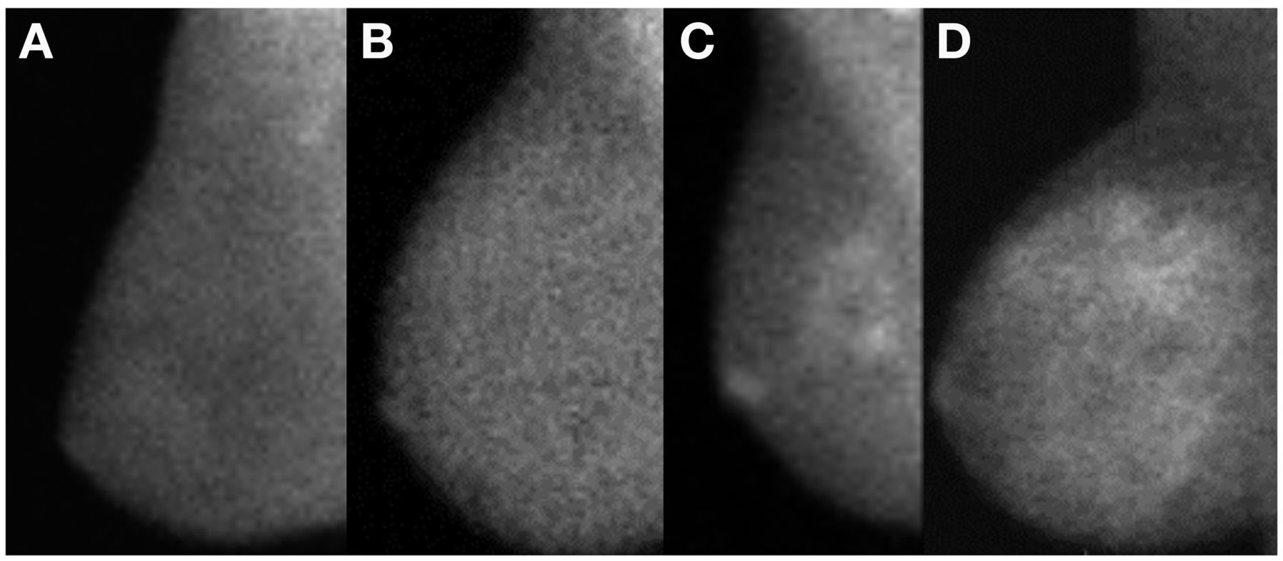
- FIGURE 3.
Different degrees of BPU on MBI (300 MBq [8 mCi] of 99mTc-sestamibi): photopenic (fibroglandular uptake less intense than fat uptake) (A), minimal to mild (fibroglandular uptake equal to, or just noticeably more intense than, fat uptake) (B), moderate (fibroglandular uptake more than mild but less than twice as intense as fat uptake) (C), and marked (fibroglandular uptake at least twice as intense as fat uptake) (D).

In this issue


Jump to section
Related Articles
Cited By...
- No citing articles found.