


Article Figures & Data
Figures
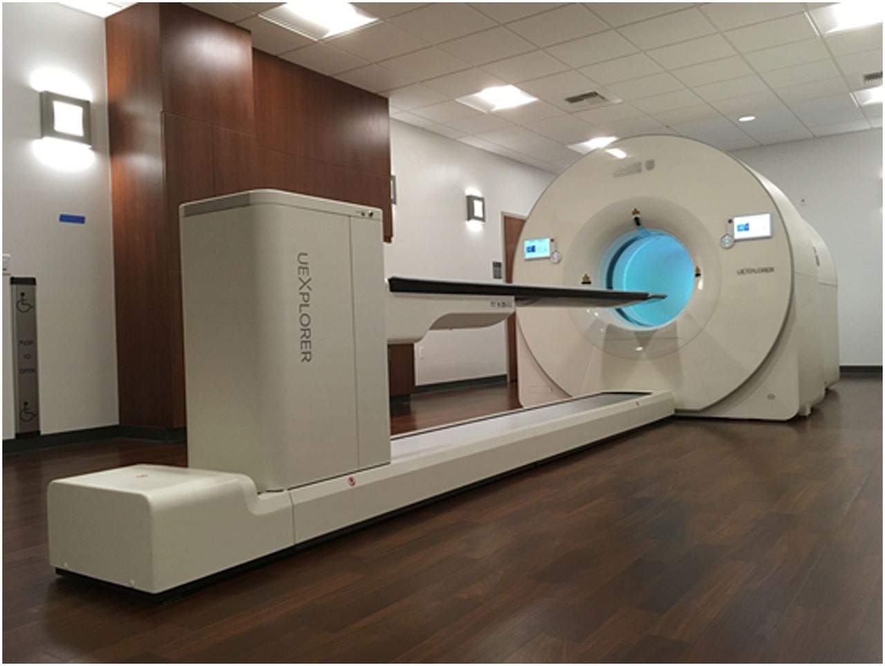
- FIGURE 1.
Photograph of uEXPLORER total-body PET/CT scanner installed at EXPLORER Molecular Imaging Center in Sacramento, CA.
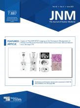
- FIGURE 2.
Axial sensitivity profiles for 70-cm (NEMA NU 2-2018) (A) and 170-cm (B) line source phantoms. Sinogram slice thickness is 1.444 mm.
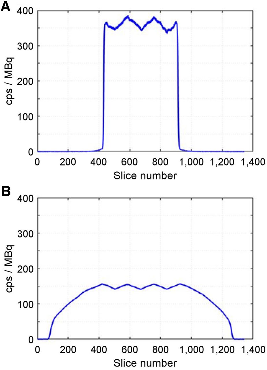
- FIGURE 3.
(A and B) 70-cm-long NEMA NU 2 scatter phantom (A) and 175-cm-long scatter phantom (B) assembled from multiple NEMA NU 2 phantoms on uEXPLORER PET/CT patient bed. (C and D) Measured count-rates with 70-cm-long (C) and 175-cm-long (D) scatter phantom. Count-rate measures are plotted vs. left vertical axes; scatter fractions are plotted vs. right vertical axes. Activity concentrations for A and B were computed by dividing total activity in phantom at each time-point by phantom volume (22 L for 70-cm-long phantom and 55 L for 175-cm-long phantom).
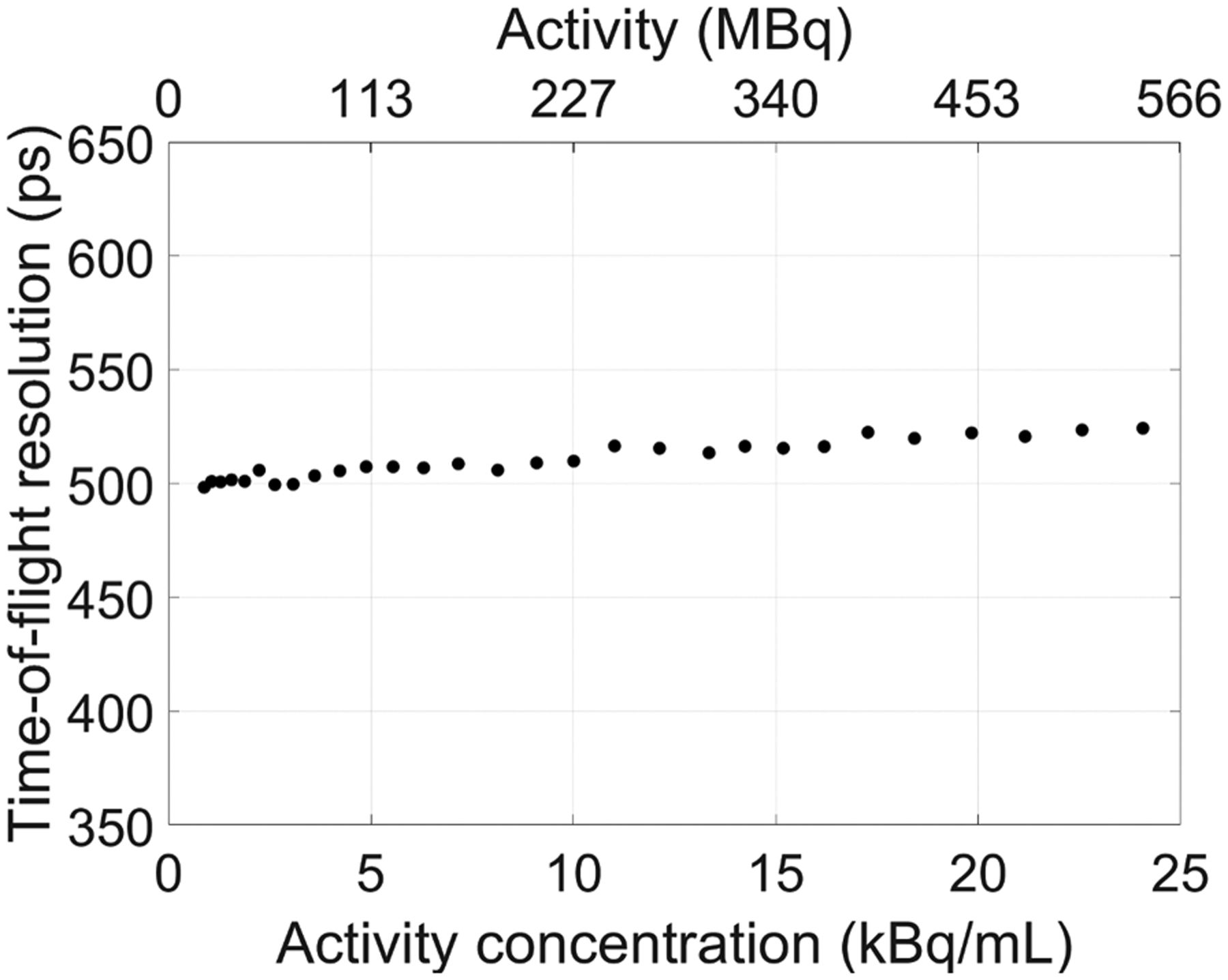
- FIGURE 4.
TOF resolution plotted vs. activity concentration using 70-cm-long NEMA NU 2 scatter phantom. TOF resolution of 505 ps at 5.3 kBq/mL was obtained.
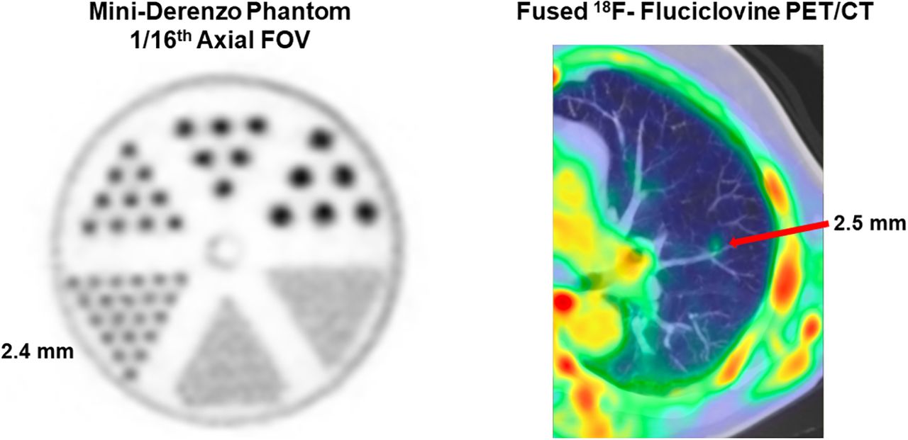
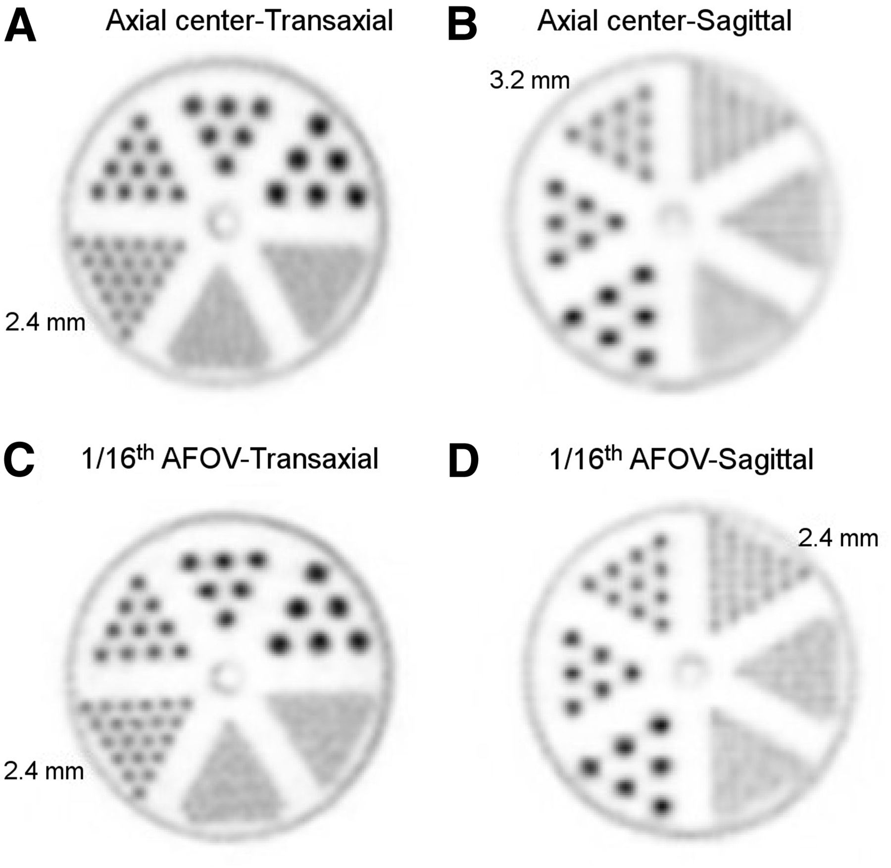
- FIGURE 5.
Reconstructed image slices of mini-Derenzo phantom imaged at axial center (A and B) and ⅟₁₆ AFOV (C and D) and with 2 orientations: transaxial (A and C) and sagittal (B and D). Image slice thickness is 1.172 mm.
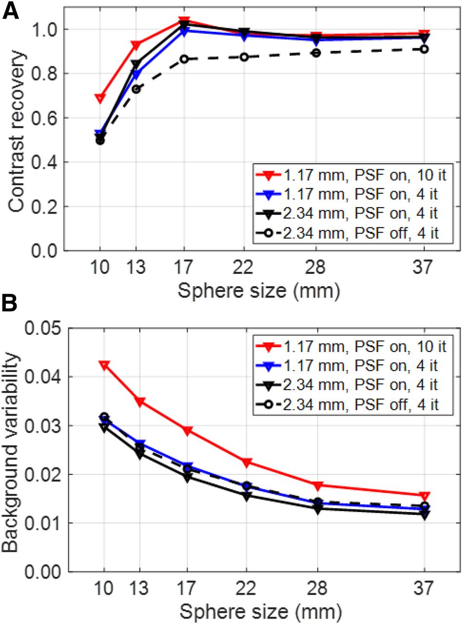
- FIGURE 6.
Contrast recovery (A) and background variability (B) measured with standard NEMA IQ phantom evaluation placed at center of AFOV and scanned for 30 min.
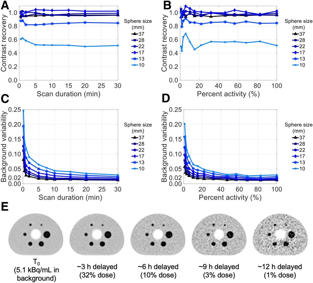
- FIGURE 7.
Contrast recovery (A and B) and background variability (C and D) as function of scan duration (A and C) and activity (B and D). Percentage activity is relative to initial activity in phantom at starting time of scans. (E) Transaxial image slices of 30-min scan at several imaging time-points reconstructed using clinical protocol. All images are decay-corrected and use same color scale: 0–20 kBq/mL.
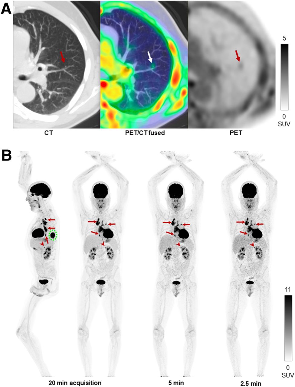
- FIGURE 8.
Human imaging examples of performance of uEXPLORER total-body PET scanner. (A) Axial slice from 18F-fluciclovine PET image (right), with corresponding fused image (middle) and CT image (left), of 68-y-old patient with castration-resistant metastatic prostate cancer, demonstrating clear visualization of 18F-flucicovine accumulation within 2.5-mm-diameter pulmonary nodule. (B) Maximum-intensity projection of representative clinical oncology 18F-FDG PET scan reconstructed with 20-, 5-, and 2.5-min durations, of 59-y-old patient with lung cancer. Images show primary tumor in left lower lobe of lung (dashed circle), with multiple variable-sized (0.8–6 cm) hilar, mediastinal, and lower esophageal nodal metastases (arrows) and ∼1-cm 18F-FDG–avid left adrenal nodule (arrowhead), which is visualized for all scan durations.


Tables
- TABLE 1
Spatial Resolution of 18F Point Sources Measured with Fourier-Rebinned Filtered Backprojection Reconstruction
Full width at half maximum (mm) Location Position Tangential Radial Axial Center AFOV 1 cm 3.0 3.0 2.8 10 cm 3.1 3.4 3.2 20 cm 4.0 4.7 3.2 ⅛ AFOV 1 cm 2.9 3.0 2.9 10 cm 3.2 3.6 3.1 20 cm 4.4 4.6 3.3 37-mm sphere 22-mm sphere Axial position Background activity (kBq/mL) Scan duration CRC Background variability CRC Background variability −63 cm (⅟₂₀ AFOV) 3.3 10.5 min 96.8% 1.4% 94.7% 2.8% −39 cm (³⁄₁₀ AFOV) 3.6 9.6 min 96.7% 1.7% 96.8% 2.4% 0 cm (center) 4.5 7.6 min 95.8% 1.6% 98.9% 1.9% +39 cm (⁷⁄₁₀ AFOV) 2.9 11.7 min 94.7% 1.6% 96.7% 2.4% +63 cm (⁸⁄₁₀ AFOV) 2.7 13.0 min 91.5% 1.8% 90.4% 2.5% Figure Image Injected dose Subject weight Time point Scan duration Singles/prompts/randoms Dead-time fraction 8A Transaxial slices 320 MBq 18F-fluciclovine 76 kg 4 min after injection 10-min 101/24.7/18.3 Mcps 14.4% 8B Maximum-intensity projection 188 MBq 18F-FDG 93 kg 90 min after injection 20, 5, and 2.5 min 19.6/4.4/2.2 Mcps 1.2% Count rates were extracted from list-mode tags; dead-time fractions were estimated according to dead-time fractions at equivalent activity concentration with 175-cm scatter phantom.
Supplemental Data
Files in this Data Supplement:


Jump to section
Related Articles
Cited By...
- In Vivo Positronium Lifetime Measurements with Intravenous Tracer Administration and a Long Axial Field-of-View PET/CT
- Quantitative Total-Body Imaging of Blood Flow with High-Temporal-Resolution Early Dynamic 18F-FDG PET Kinetic Modeling
- Is Long-Axial-Field-of-View PET/CT Cost-Effective? An International Health-Economic Analysis
- Feasibility of an Ultra-Low-Dose PET Scan Protocol with CT-Based and LSO-TX-Based Attenuation Correction Using a Long-Axial-Field-of-View PET/CT Scanner
- Feasibility of Ultra-Low-Activity 18F-FDG PET/CT Imaging Using a Long-Axial-Field-of-View PET/CT System
- In Vivo Intralesional Positronium Lifetime Imaging of Thyroid Cancer Using Clinically Routine I-124 PET/CT
- Early 10-Minute Postinjection [18F]F-FAPI-42 uEXPLORER Total-Body PET/CT Scanning Protocol for Staging Lung Cancer Using HYPER Iterative Reconstruction
- Total-Body Parametric Imaging Using Relative Patlak Plot
- Total-Body PET System Designs with Axial and Transverse Gaps: A Study of Lesion Quantification and Detectability
- Quantitative Accuracy Assessment of the NeuroEXPLORER for Diverse Imaging Applications: Moving Beyond Standard Evaluations
- Performance Characteristics of a New Generation 148-cm Axial Field-of-View uMI Panorama GS PET/CT System with Extended NEMA NU 2-2018 and EARL Standards
- Modeling PET Data Acquired During Nonsteady Conditions: What If Brain Conditions Change During the Scan?
- Quantitative Total-Body Imaging of Blood Flow with High Temporal Resolution Early Dynamic 18F-Fluorodeoxyglucose PET Kinetic Modeling
- Role of 64CuCl2 PET/CT in Detecting and Staging Muscle-Invasive Bladder Cancer: Comparison with Contrast-Enhanced CT and 18F-FDG PET/CT
- Performance Characteristics of the NeuroEXPLORER, a Next-Generation Human Brain PET/CT Imager
- Quantitative PET imaging and modeling of molecular blood-brain barrier permeability
- Dose Reduction in Pediatric Oncology Patients with Delayed Total-Body [18F]FDG PET/CT
- High-Temporal-Resolution Kinetic Modeling of Lung Tumors with Dual-Blood Input Function Using Total-Body Dynamic PET
- Advantages and Challenges of Total-Body PET/CT at a Tertiary Cancer Center: Insights from Sun Yat-sen University Cancer Center
- The Role of Total-Body PET in Drug Development and Evaluation: Status and Outlook
- Performance Evaluation of the uMI Panorama PET/CT System in Accordance with the National Electrical Manufacturers Association NU 2-2018 Standard
- Performance Characteristics of a New-Generation Digital Bismuth Germanium Oxide PET/CT System, Omni Legend 32, According to NEMA NU 2-2018 Standards
- Total-Body Multiparametric PET Quantification of 18F-FDG Delivery and Metabolism in the Study of Coronavirus Disease 2019 Recovery
- Total-Body Perfusion Imaging with [11C]-Butanol
- Dual-Time-Point Posttherapy 177Lu-PSMA-617 SPECT/CT Describes the Uptake Kinetics of mCRPC Lesions and Prognosticates Patients Outcome
- Facial Anonymization and Privacy Concerns in Total-Body PET/CT
- Facial Anonymization and Privacy Concerns in Total-Body PET/CT
- High-Temporal-Resolution Lung Kinetic Modeling Using Total-Body Dynamic PET with Time-Delay and Dispersion Corrections
- Total-Body Multiparametric PET Quantification of 18F-FDG Delivery and Metabolism in the Study of COVID-19 Recovery
- Exploring Vessel Wall Biology In Vivo by Ultrasensitive Total-Body PET
- Whole-Body PET Imaging: A Catalyst for Whole-Person Research?
- Blanching Defects at Pressure Points: Observations from Dynamic Total-Body PET/CT Studies
- Fully Automated, Semantic Segmentation of Whole-Body 18F-FDG PET/CT Images Based on Data-Centric Artificial Intelligence
- Total-Body 18F-FDG PET/CT in Autoimmune Inflammatory Arthritis at Ultra-Low Dose: Initial Observations
- Efficient Delay Correction for Total-Body PET Kinetic Modeling Using Pulse Timing Methods
- Total-Body PET Multiparametric Imaging of Cancer Using a Voxelwise Strategy of Compartmental Modeling
- Radioembolization Dosimetry with Total-Body 90Y PET
- Feasibility of Acquisitions Using Total-Body PET/CT with an Ultra-Low 18F-FDG Activity
- Performance Characteristics of the Biograph Vision Quadra PET/CT System with a Long Axial Field of View Using the NEMA NU 2-2018 Standard
- Tumor Response to Radiopharmaceutical Therapies: The Knowns and the Unknowns