


Article Figures & Data
Figures
- FIGURE 1.
18F-AraG imaging of different syngeneic tumor models. (A) Both intratumoral (arrowheads) and intranodal (encircled) signal varied among different tumor types. Location of intratumoral signal showed several patterns, from signal present in core (MC38 and A9F1) through halolike (CT26) to signal present only at margin (LLC, B16F10). Ring effect as observed in bladder signifies saturated signal. (B) Intratumoral signal intensity showed variation among different tumor types and among individual mice of same tumor type. Signal in TDLNs was higher than signal in tumors and showed variability among different tumor types and individual mice (n = 4 for each tumor type; error bars represent SD). LN = lymph node; T = tumor; %ID = percentage injected dose.
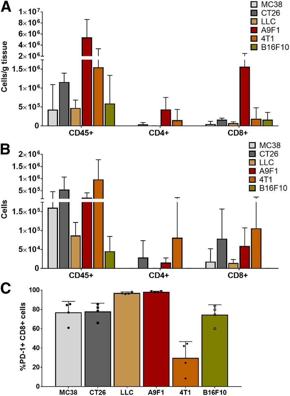
- FIGURE 2.
Evaluation of different syngeneic tumor models. (A) Highest density of total lymphocytes, CD4+, and CD8+ cells was found in smallest tumors, A9F1. (B) Highest number of lymphocytes was isolated from largest tumors, 4T1. (C) Percentage of CD8 cells that expressed PD-1 varied among different tumor types. In LLC and A9F1 tumors, over 97% of CD8 cells were found to be PD-1–positive, whereas in 4T1 tumors less than 30% of CD8 cells were positive for PD-1 (n = 4 for each tumor type; error bars represent SD); symbols within bars represent individual mice).
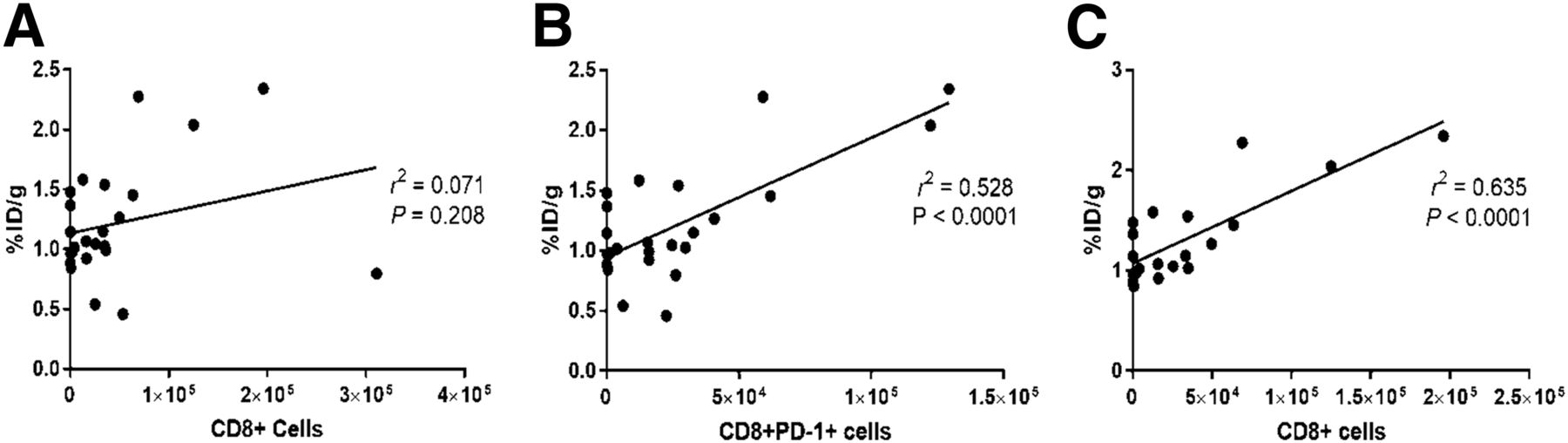
- FIGURE 3.
Correlation of 18F-AraG signal with number of CD8 cells present in TME. (A) 18F-AraG signal showed no correlation with number of intratumoral CD8+ cells. (B) 18F-AraG signal showed statistically significant correlation with number of CD8+PD-1+ cells. (C) Exclusion of 4T1 cells for which PD-1 expression indicated dysfunction led to statistically significant correlation between 18F-AraG signal and number of CD8+ cells. %ID = percentage injected dose.
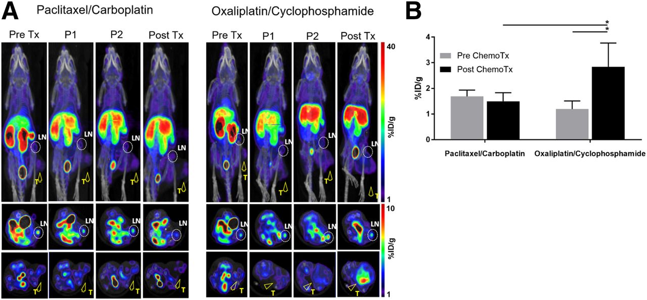
- FIGURE 4.
18F-AraG longitudinal imaging of MC38-bearing mice undergoing chemotherapy. Chemotherapy was administered once weekly for 2 wk. Mice were imaged 1 d before start of therapy (Pre Tx) and then 3 d (P1) and 6 d (P2) after first chemotherapy administration and 3 d after second chemotherapy administration (Post Tx). (A) Paclitaxel/carboplatin treatment did not lead to appreciable changes in signal intensity. Dramatic increase in signal intensity was detected after 2 oxaliplatin/cyclophosphamide injections. Encircled areas are TDLNs; arrowheads point to tumors. (B) 18F-AraG signal detected after oxaliplatin/cyclophosphamide treatment was significantly different from pretherapy signal as well as signal after paclitaxel-carboplatin treatment (n = 4 for each group; error bars represent SD). LN = lymph node; T = tumor; %ID = percentage injected dose. *P < 0.05.
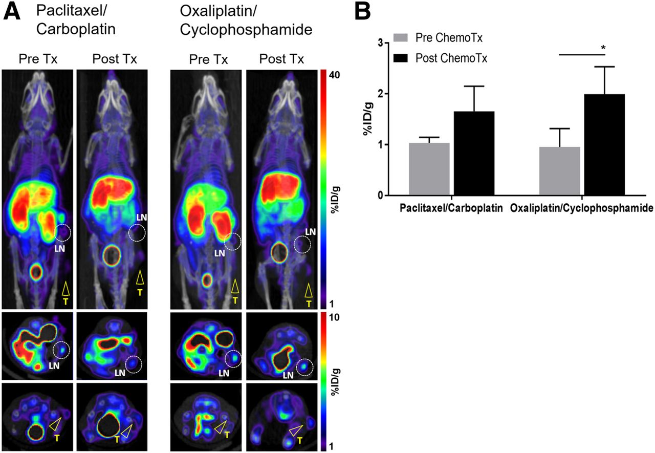
- FIGURE 5.
18F-AraG imaging of A9F1-bearing mice undergoing chemotherapy. (A) Paclitaxel/carboplatin treatment showed trend toward increase in signal. Oxaliplatin/cyclophosphamide treatment led to increase in intratumoral signal. Encircled areas are TDLNs; arrowheads point to tumors. (B) Signal detected after oxaliplatin/cyclophosphamide treatment was significantly different from pretherapy signal but not from signal after paclitaxel-carboplatin treatment (n = 4 for each group; error bars represent SD). LN = lymph node; T = tumor; %ID = percentage injected dose. *P < 0.05.
- FIGURE 6.
Lymphocyte profile of MC38 and A9F1 tumors after chemotherapy. (A) In MC38 tumors, oxaliplatin/cyclophosphamide treatment led to increase in total lymphocytes and number of CD8+ cells. In A9F1 tumors, oxaliplatin/cyclophosphamide treatment led to increase in total lymphocytes and number of CD4+ cells. (B) In MC38 tumors, ratio of CD8+ to CD4+FOXP3+ cells in oxaliplatin/cyclophosphamide group was 27 times higher than in paclitaxel/carboplatin-treated mice. (n = 4 for each group; error bars represent SD). *P < 0.05.


Additional Files
Supplemental Data
Files in this Data Supplement:


Jump to section
Related Articles
Cited By...
- Approaches to Imaging Immune Activation Using PET
- [18F]F-AraG Uptake in Vertebral Bone Marrow May Predict Survival in Patients with Non-Small Cell Lung Cancer Treated with Anti-PD-(L)1 Immunotherapy
- Total-Body Dynamic Imaging and Kinetic Modeling of [18F]F-AraG in Healthy Individuals and a Non-Small Cell Lung Cancer Patient Undergoing Anti-PD-1 Immunotherapy
- Total-body Dynamic Imaging and Kinetic Modeling of 18F-AraG in Healthy Individuals and a Non-Small Cell Lung Cancer Patient Undergoing Anti-PD-1 Immunotherapy
- Imaging of Activated T Cells
- Longitudinal Imaging of T Cells and Inflammatory Demyelination in a Preclinical Model of Multiple Sclerosis Using 18F-FAraG PET and MRI