


Article Figures & Data
Figures
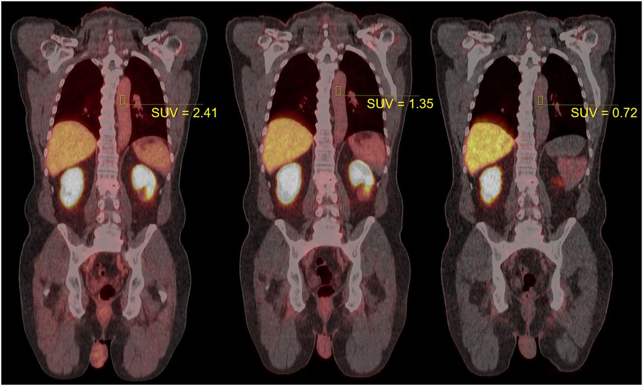
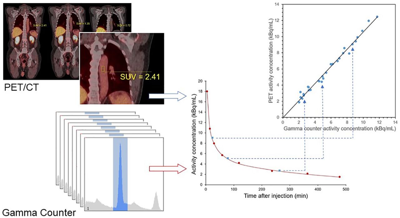
- FIGURE 1.
Example PET/CT images for typical patient. PET data were acquired 24, 79, and 264 min after injection (from left to right). Cylindric VOIs can be seen in descending aorta, with SUV as indicated. Fused display shows PET in hot-body color map, scaled from 0 to 20 SUV units in each image. Patient weighed 90.5 kg and was administered 339.7 MBq of 18F-DCFPyL.
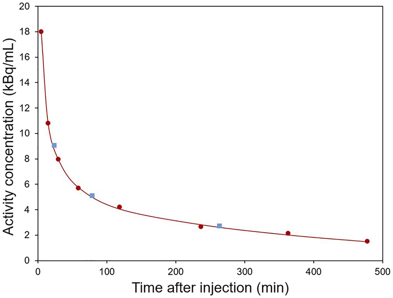
- FIGURE 2.
Example time–activity data for patient shown in Figure 1. Circles indicate whole blood samples counted on calibrated γ-counter. Squares indicate PET-derived data from VOIs in descending aorta. Line is triexponential model fit to γ-counter data. All data were decay-corrected to time of injection.
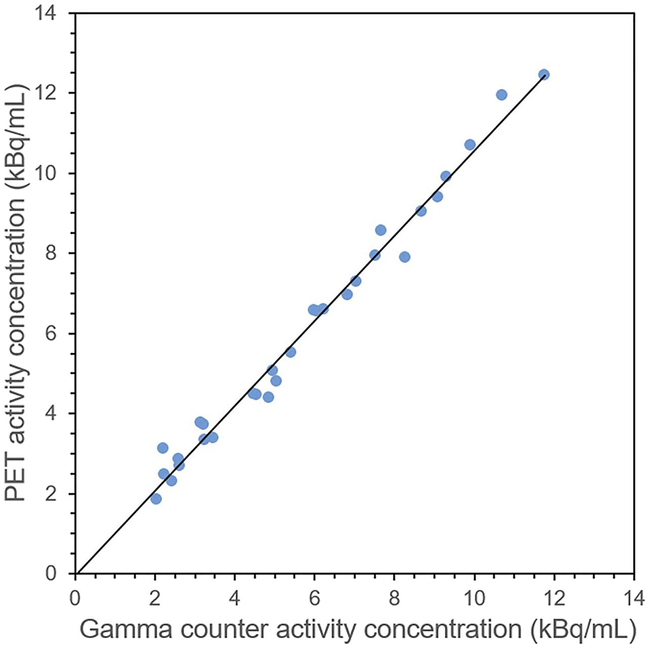
- FIGURE 3.
PET and γ-counter radioactivity concentration data for 10 patients, each with 3 corresponding measurements. Line indicates linear fit to data: y = 1.06x – 64, R 2 = 0.985.
- FIGURE 4.
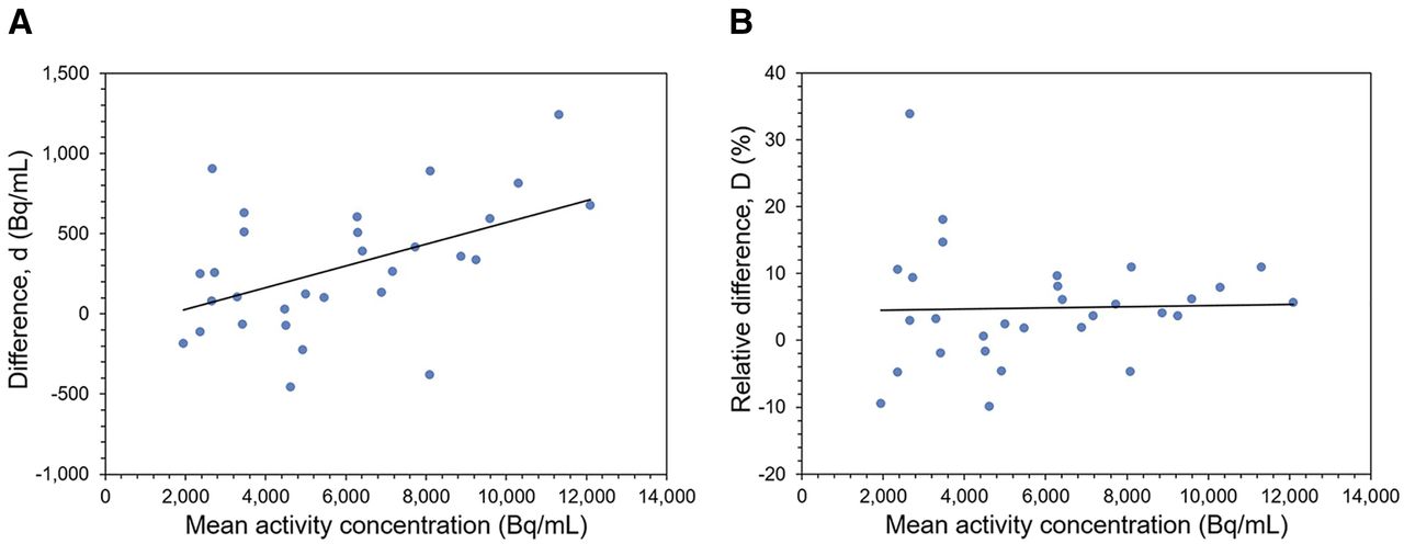
Bland–Altman plots showing difference between PET and γ-counter measurements in radioactivity concentration units (A) and relative units (B). Lines indicate linear regression. Relative difference was not proportional to radioactivity concentration and had mean value of 4.8% ± 8.6%.


