
Main menu

  • Home
  • Content
    • Current
    • Ahead of print
    • Past Issues
    • JNM Supplement
    • SNMMI Annual Meeting Abstracts
    • Continuing Education
    • JNM Podcasts
  • Subscriptions
    • Subscribers
    • Institutional and Non-member
    • Rates
    • Journal Claims
    • Corporate & Special Sales
  • Authors
    • Submit to JNM
    • Information for Authors
    • Assignment of Copyright
    • AQARA requirements
  • Info
    • Reviewers
    • Permissions
    • Advertisers
  • About
    • About Us
    • Editorial Board
    • Contact Information
  • More
    • Alerts
    • Feedback
    • Help
    • SNMMI Journals
  • SNMMI
    • JNM
    • JNMT
    • SNMMI Journals
    • SNMMI

User menu

  • Subscribe
  • My alerts
  • Log in
  • My Cart

Search

  • Advanced search
Journal of Nuclear Medicine
  • SNMMI
    • JNM
    • JNMT
    • SNMMI Journals
    • SNMMI
  • Subscribe
  • My alerts
  • Log in
  • My Cart
Journal of Nuclear Medicine

Advanced Search

  • Home
  • Content
    • Current
    • Ahead of print
    • Past Issues
    • JNM Supplement
    • SNMMI Annual Meeting Abstracts
    • Continuing Education
    • JNM Podcasts
  • Subscriptions
    • Subscribers
    • Institutional and Non-member
    • Rates
    • Journal Claims
    • Corporate & Special Sales
  • Authors
    • Submit to JNM
    • Information for Authors
    • Assignment of Copyright
    • AQARA requirements
  • Info
    • Reviewers
    • Permissions
    • Advertisers
  • About
    • About Us
    • Editorial Board
    • Contact Information
  • More
    • Alerts
    • Feedback
    • Help
    • SNMMI Journals
  • View or Listen to JNM Podcast
  • Visit JNM on Facebook
  • Join JNM on LinkedIn
  • Follow JNM on Twitter
  • Subscribe to our RSS feeds
Research ArticleClinical Investigation

Photoacoustic Molecular Imaging for the Identification of Lymph Node Metastasis in Head and Neck Cancer Using an Anti-EGFR Antibody–Dye Conjugate

Naoki Nishio, Nynke S. van den Berg, Brock A. Martin, Stan van Keulen, Shayan Fakurnejad, Eben L. Rosenthal and Katheryne E. Wilson
Journal of Nuclear Medicine May 2021, 62 (5) 648-655; DOI: https://doi.org/10.2967/jnumed.120.245241
Naoki Nishio
1Department of Otolaryngology–Head and Neck Surgery, Stanford University School of Medicine, Stanford, California
2Department of Otorhinolaryngology, Nagoya University Graduate School of Medicine, Nagoya, Aichi, Japan
  • Find this author on Google Scholar
  • Find this author on PubMed
  • Search for this author on this site
Nynke S. van den Berg
1Department of Otolaryngology–Head and Neck Surgery, Stanford University School of Medicine, Stanford, California
  • Find this author on Google Scholar
  • Find this author on PubMed
  • Search for this author on this site
Brock A. Martin
3Department of Pathology, Stanford University School of Medicine, Stanford, California; and
  • Find this author on Google Scholar
  • Find this author on PubMed
  • Search for this author on this site
Stan van Keulen
1Department of Otolaryngology–Head and Neck Surgery, Stanford University School of Medicine, Stanford, California
  • Find this author on Google Scholar
  • Find this author on PubMed
  • Search for this author on this site
Shayan Fakurnejad
1Department of Otolaryngology–Head and Neck Surgery, Stanford University School of Medicine, Stanford, California
  • Find this author on Google Scholar
  • Find this author on PubMed
  • Search for this author on this site
Eben L. Rosenthal
1Department of Otolaryngology–Head and Neck Surgery, Stanford University School of Medicine, Stanford, California
  • Find this author on Google Scholar
  • Find this author on PubMed
  • Search for this author on this site
Katheryne E. Wilson
4Department of Radiology, Stanford University School of Medicine, Stanford, California
  • Find this author on Google Scholar
  • Find this author on PubMed
  • Search for this author on this site
  • Article
  • Figures & Data
  • Supplemental
  • Info & Metrics
  • PDF
Loading

Article Figures & Data

Figures

  • Tables
  • Additional Files
  • FIGURE 1.
    • Download figure
    • Open in new tab
    • Download powerpoint
    FIGURE 1.

    Workflow of photoacoustic and fluorescence imaging of pan800 in HNSCC and LNs, including intraoperative, postoperative, and histologic imaging. All scale bars are 5 mm. H&E = hematoxylin and eosin.

  • FIGURE 2.
    • Download figure
    • Open in new tab
    • Download powerpoint
    FIGURE 2.

    Determining optical absorption spectra (A) and photoacoustic (B) and fluorescence (C) imaging sensitivity for pan800. deOxy hemo = deoxygenated hemoglobin; Oxy hemo = oxygenated hemoglobin.

  • FIGURE 3.
    • Download figure
    • Open in new tab
    • Download powerpoint
    FIGURE 3.

    Photoacoustic (A) and fluorescence (B) imaging of decreasing concentrations of pan800 in tissue-mimicking phantom. avg = average.

  • FIGURE 4.
    • Download figure
    • Open in new tab
    • Download powerpoint
    FIGURE 4.

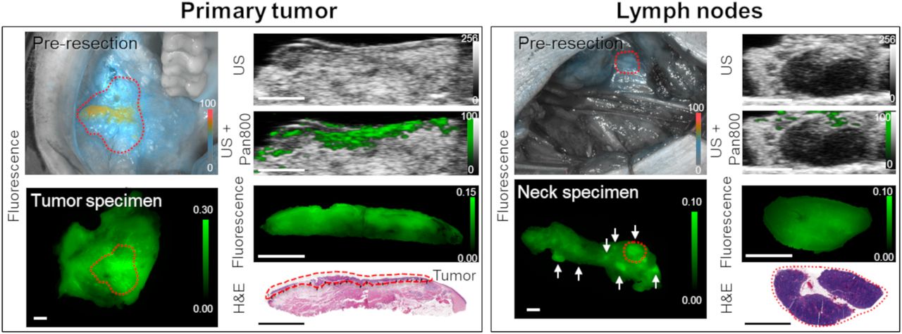
    Fluorescence, spectroscopic photoacoustic pan800 signal, and histology images from representative patient with primary tumor and benign LNs. Tumor is outlined in red dashed line. White arrows represent locations of several LNs within neck specimen; red dashed circle highlights the LN shown by histology at the bottom right. All scale bars are 5 mm. H&E = hematoxylin and eosin; US = ultrasound.

  • FIGURE 5.
    • Download figure
    • Open in new tab
    • Download powerpoint
    FIGURE 5.

    Discrimination of metastatic LNs from benign LNs from head and neck cancer patients using fluorescence imaging. (A) Bright-field and fluorescence images of metastatic and benign LNs corresponding to hematoxylin-and-eosin slides. (B) Quantitative analysis of mean fluorescence intensity for differentiating benign from metastatic LNs. All scale bars are 5 mm. H&E = hematoxylin and eosin.

  • FIGURE 6.
    • Download figure
    • Open in new tab
    • Download powerpoint
    FIGURE 6.

    Photoacoustic measurements for metastatic and benign LNs. (A) Representative ultrasound, spectroscopic photoacoustic, fluorescence, and histology images of benign and metastatic LNs. Photoacoustic signals corresponding to pan800 and oxy- and deoxyhemoglobin have been spectrally unmixed. All scale bars are 5 mm. (B) Quantitative analysis of PAMI signal using pan800 avg. (C) Quantitative analysis of PAMI signal using pan800 avg thresh. US = ultrasound.

  • FIGURE 7.
    • Download figure
    • Open in new tab
    • Download powerpoint
    FIGURE 7.

    Heterogeneity of pan800 in metastatic LNs. (A) Representative ultrasound, photoacoustic, fluorescence, and histology images of metastatic LN. All scale bars are 5 mm. (B) Quantitative and ROC analysis of ratio of PAMI signal using pan800 avg thresh to pan800 avg to differentiate benign from metastatic LNs. US = ultrasound.

Tables

  • Figures
  • Additional Files
    • View popup
    TABLE 1

    Patient Characteristics

    Patient no.Age (y)SexTumor locationcTN stage
    170FemaleBuccal mucosaT3N0
    268FemaleLateral tongueT3N2b
    371FemaleLateral tongueT1N0
    447FemaleRetromolar trigoneT4N2b
    556MaleBuccal mucosaT2N0
    660MaleEarT2N0
    770MaleLateral tongueT3N0

Additional Files

  • Figures
  • Tables
  • Supplemental Data

    Files in this Data Supplement:

    • Supplemental Data
PreviousNext
Back to top

In this issue

Journal of Nuclear Medicine: 62 (5)
Journal of Nuclear Medicine
Vol. 62, Issue 5
May 10, 2021
  • Table of Contents
  • Table of Contents (PDF)
  • About the Cover
  • Index by author
Print
Download PDF
Article Alerts
Sign In to Email Alerts with your Email Address
Email Article

Thank you for your interest in spreading the word on Journal of Nuclear Medicine.

NOTE: We only request your email address so that the person you are recommending the page to knows that you wanted them to see it, and that it is not junk mail. We do not capture any email address.

Enter multiple addresses on separate lines or separate them with commas.
Photoacoustic Molecular Imaging for the Identification of Lymph Node Metastasis in Head and Neck Cancer Using an Anti-EGFR Antibody–Dye Conjugate
(Your Name) has sent you a message from Journal of Nuclear Medicine
(Your Name) thought you would like to see the Journal of Nuclear Medicine web site.
Citation Tools
Photoacoustic Molecular Imaging for the Identification of Lymph Node Metastasis in Head and Neck Cancer Using an Anti-EGFR Antibody–Dye Conjugate
Naoki Nishio, Nynke S. van den Berg, Brock A. Martin, Stan van Keulen, Shayan Fakurnejad, Eben L. Rosenthal, Katheryne E. Wilson
Journal of Nuclear Medicine May 2021, 62 (5) 648-655; DOI: 10.2967/jnumed.120.245241

Citation Manager Formats

  • BibTeX
  • Bookends
  • EasyBib
  • EndNote (tagged)
  • EndNote 8 (xml)
  • Medlars
  • Mendeley
  • Papers
  • RefWorks Tagged
  • Ref Manager
  • RIS
  • Zotero
Share
Photoacoustic Molecular Imaging for the Identification of Lymph Node Metastasis in Head and Neck Cancer Using an Anti-EGFR Antibody–Dye Conjugate
Naoki Nishio, Nynke S. van den Berg, Brock A. Martin, Stan van Keulen, Shayan Fakurnejad, Eben L. Rosenthal, Katheryne E. Wilson
Journal of Nuclear Medicine May 2021, 62 (5) 648-655; DOI: 10.2967/jnumed.120.245241
Twitter logo Facebook logo LinkedIn logo Mendeley logo
  • Tweet Widget
  • Facebook Like
  • Google Plus One
Bookmark this article

Jump to section

  • Article
    • Abstract
    • MATERIALS AND METHODS
    • RESULTS
    • DISCUSSION
    • CONCLUSION
    • DISCLOSURE
    • Footnotes
    • REFERENCES
  • Figures & Data
  • Supplemental
  • Info & Metrics
  • PDF

Related Articles

  • This Month in JNM
  • PubMed
  • Google Scholar

Cited By...

  • Temporal dynamics of fluorescence and photoacoustic signals of a Cetuximab-IRDye800 conjugate in EGFR-overexpressing tumors
  • Precision Surgery Guided by Intraoperative Molecular Imaging
  • Google Scholar

More in this TOC Section

  • Feasibility of Ultra-Low-Activity 18F-FDG PET/CT Imaging Using a Long–Axial-Field-of-View PET/CT System
  • Is Long–Axial-Field-of-View PET/CT Cost-Effective? An International Health–Economic Analysis
  • Multicycle Dosimetric Behavior and Dose–Effect Relationships in [177Lu]Lu-DOTATATE Peptide Receptor Radionuclide Therapy
Show more Clinical Investigation

Similar Articles

Keywords

  • photoacoustic imaging
  • head and neck cancer
  • lymph node
  • antibody–dye
  • near-infrared
SNMMI

© 2025 SNMMI

Powered by HighWire