


Article Figures & Data
Figures
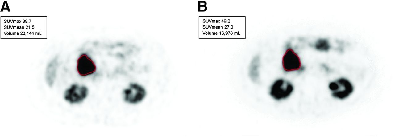
- FIGURE 1.
60-y-old woman with metastases of well-differentiated small bowel NET to liver and retroperitoneal lymph nodes (G1; Ki-67 index, 6%). Lesion-based assessment was done with bPET and iPET before cycle 2 of 177Lu-DOTATATE therapy. (A) Metastatic nodal mass chosen as marker lesion at bPET (SUVmax, 38.7) (outlined in red). (B) Same lesion before cycle 2 of therapy (SUVmax, 49.2) (outlined in red).
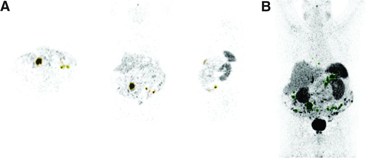
- FIGURE 2.
68Ga-DOTATATE tumor volume analysis using in-house automated segmentation software. (A) Multiplanar segmentation tool (to identify and confirm tumor sites). (B) Mask generated using tracer uptake in spleen as threshold (tumor lesions with SUVmax above that of spleen are outlined in green).
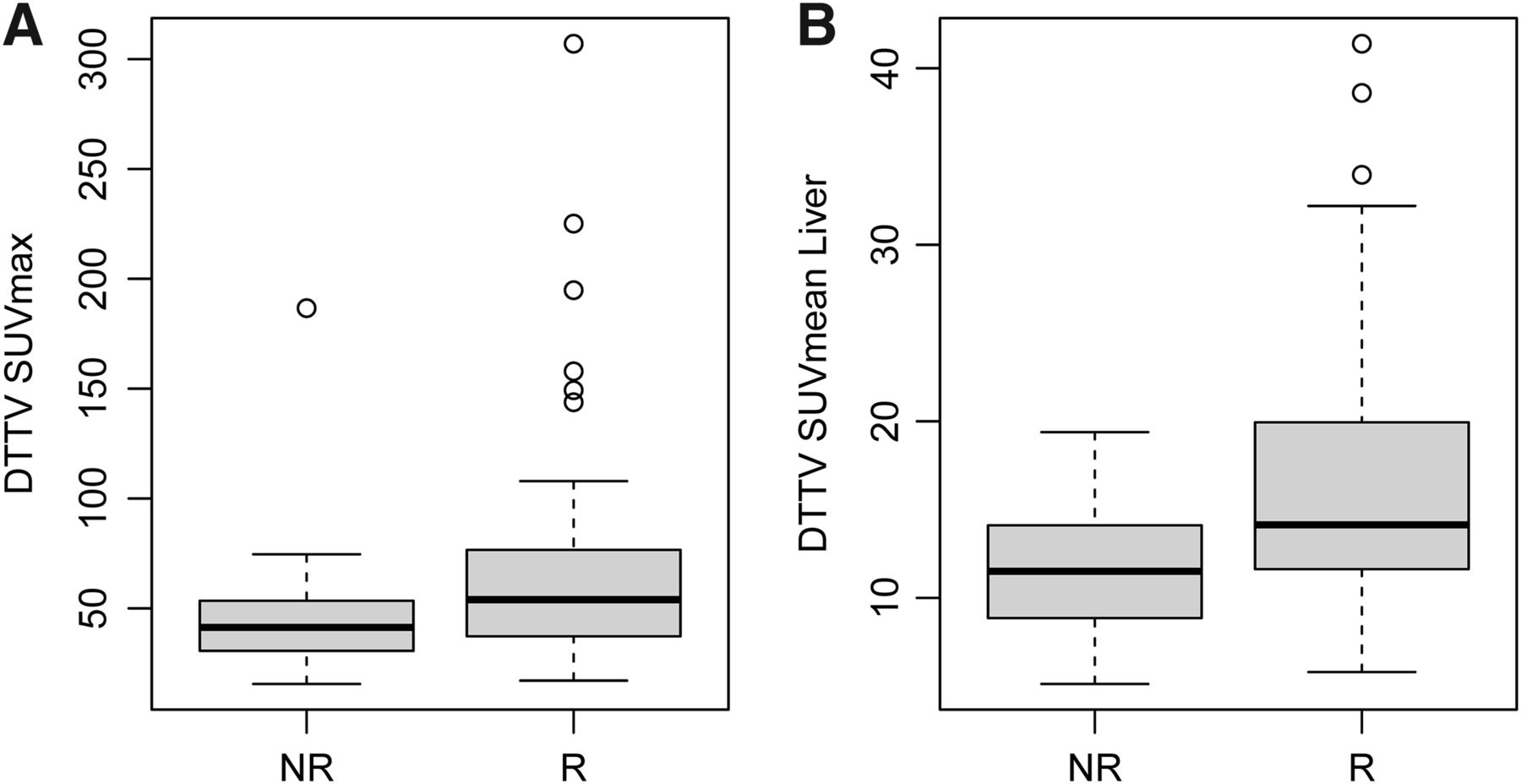
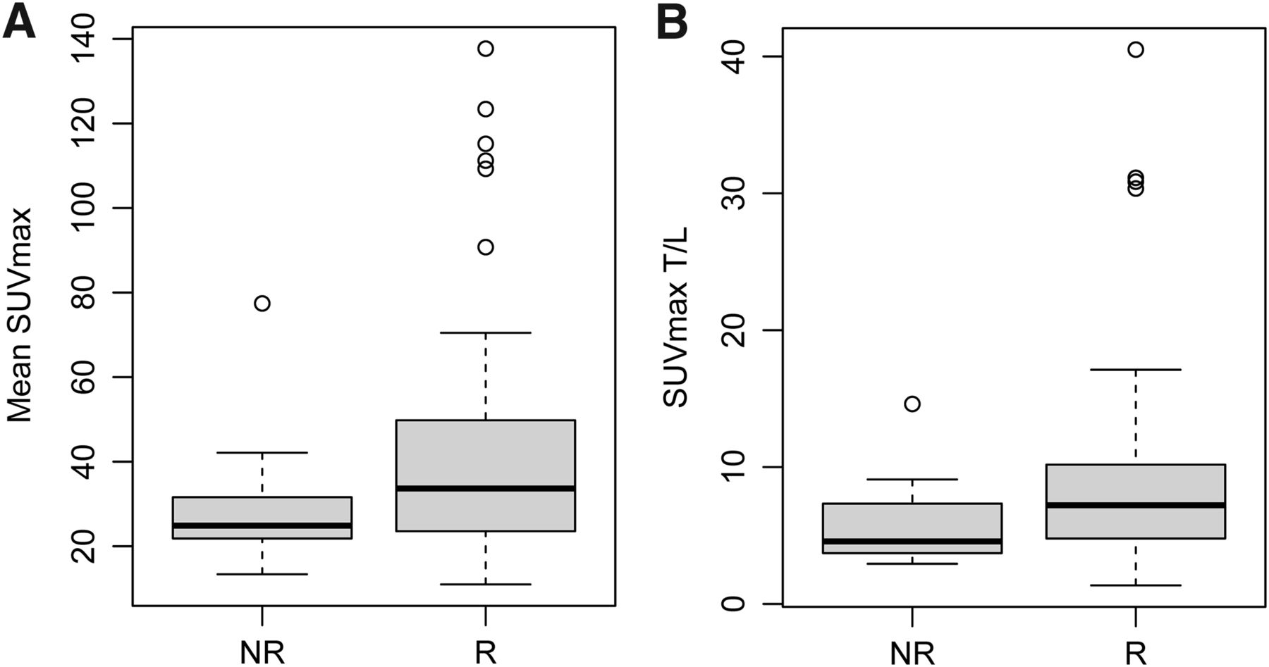
- FIGURE 3.
Lesion-based measures. Distributions of median SUVmax (P = 0.018) (A) and SUVmax T/L (P = 0.024) (B) are shown. Box plots represent median and upper and lower quartiles of each distribution, with whiskers showing limits of distribution (1.5 times interquartile range). NR = nonresponders; R = responders.
- FIGURE 4.
DTTV parameters. Distributions of mean DTTV SUVmax (P = 0.025) (A) and SUVmean obtained with liver as threshold (SUVmean Liver) (P = 0.0055) (B) are shown. Box plots represent median and upper and lower quartiles of each distribution, with whiskers showing limits of distribution (1.5 times interquartile range). NR = nonresponders; R = responders.
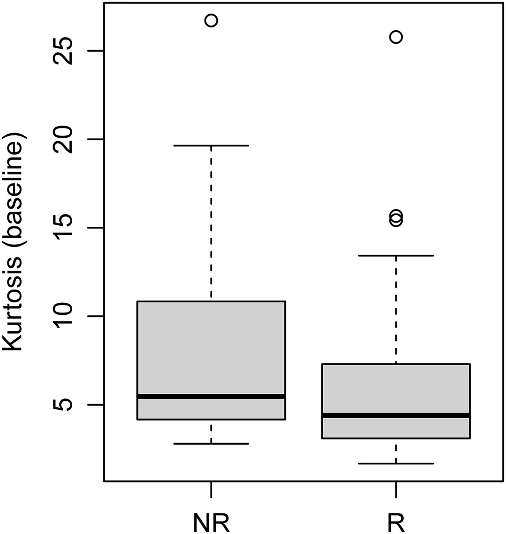
- FIGURE 5.
Kurtosis on baseline 68Ga-DOTATATE PET/CT. Distribution of kurtosis was estimated from 68Ga-DOTATATE tumor volumes (P = 0.031). Box plot represents median and upper and lower quartiles of each distribution, with whiskers showing limits of distribution (1.5 times interquartile range). NR = nonresponders; R = responders.


Tables
Characteristic Entire cohort iPET cohort No. of patients 91 36 Sex (M:F) 53:38 20:16 Mean age (y) 62.5 (SD = 12.9) 59.7 (SD = 14.2) Primary site (No. of patients) Gastrointestinal tract 48 19 Pancreas 19 8 Unknown primary 9 2 Bronchopulmonary 6 3 Adrenal 4 2 Other 5 2 Mean tumor Ki-67 index (%) 8.3 (SD = 7.8) 10.5 (SD = 8.3) Response status Nonresponders 20 6 Other 71 30 Mean follow-up (mo) 12.2 (SD = 7.2) 20.1 (SD = 9.7) - TABLE 2
bPET Reference Parameters, Lesion-Based Parameters, DTTV Parameters, and First-Order Heterogeneity Parameters
bPET Parameter All Patients (n = 91) Responders (n = 71) Nonresponders (n = 20) P* Reference tissue SUVmax liver 0.49 Mean (SD) 5.7 (2.1) 5.8 (2.3) 5.3 (1.6) Minimum–maximum 2–13.1 3.2–8.2 2–13.1 SUVmax spleen 0.30 Mean (SD) 13.8 (6.9) 14.1 (6.9) 12.6 (6.7) Minimum–maximum 4.4–45.8 4.4–26.7 5–45.8 Lesion based Mean SUVmax 0.018 Mean (SD) 38.7 (25.1) 41.7 (26.8) 28.2 (14.2) Minimum–maximum 11–137.7 13.4–77.4 11–137.7 Mean SUVmax T/L 0.024 Mean (SD) 8.3 (6.4) 9 (7) 5.8 (2.9) Minimum–maximum 1.4–40.5 1.4–40.5 2.9–14.6 Mean SUVmax T/S 0.13 Mean (SD) 3.5 (2.7) 3.7 (2.9) 2.8 (1.8) Minimum–maximum 0.2–14.6 0.2–14.6 1–8.3 DTTV DTTV liver† 0.12 Mean (SD) 554.1 (853.8) 611.5 (921.7) 350.4 (516.8) Minimum–maximum 4.7–4,891.8 4.7–4,891.8 8.1–2,200.9 DTTV spleen† 0.06 Mean (SD) 317.1 (616.3) 363.9 (677.6) 150.9 (265.4) Minimum–maximum 0–3,825.3 0–3,825.3 0–1,049.6 DTTV SUVmax 0.025 Mean (SD) 62.8 (46.4) 66.8 (48.4) 48.4 (36.2) Minimum–maximum 15.6–307) 17.1–307 15.6–186.6 DTTV SUVmean liver 0.0055 Mean (SD) 15.6 (7.3) 16.7 (7.7) 11.6 (3.6) Minimum–maximum 5.1–41.4 5.8–41.4 5.1–19.4 DTTV SUVmean spleen 0.06 Mean (SD) 23.5 (10.3) 24.5 (10.4) 20 (9.3) Minimum–maximum 0–50.4 0–50.4 7.7–45.3 Heterogeneity CoV 0.17 Mean (SD) 0.6 (0.2) 0.6 (0.2) 0.6 (0.3) Minimum–maximum 0.2–1.6 0.3–1.5 0.2–1.6 Skewness 0.055 Mean (SD) 1.5 (0.8) 1.4 (0.8) 1.9 (1.0) Minimum–maximum 0.1–4 0.1–4 0.6–4 Kurtosis 0.031 Mean (SD) 6.4 (4.8) 5.8 (4.1) 8.6 (6.4) Minimum–maximum 1.7–26.7 1.7–25.8 2.8–26.7 * P value.
↵† Measured in cubic centimeters.
DTTV SUVmean liver = SUVmean in segmented volume obtained with liver as threshold; DTTV SUVmean spleen = SUVmean in segmented volume obtained with spleen as threshold; DTTV liver = DTTV obtained with liver as threshold; DTTV spleen = DTTV obtained with spleen as threshold; DTTV SUVmean liver = DTTV SUVmean obtained with liver as threshold; DTTV SUVmean spleen = DTTV SUVmean obtained with spleen as threshold; CoV = coefficient of variation.
Wilcoxon rank sum test P values are shown.
- TABLE 3
iPET Reference Parameters, Lesion-Based Parameters, DTTV Parameters, and First-Order Heterogeneity Measures, Part 1
iPET All patients (n = 36) Responders (n = 30) Nonresponders (n = 6) P* Reference tissue SUVmax liver 0.011 Mean (SD) 5.8 (1.8) 6.1 (1.8) 4.2 (1.2) Minimum–maximum 2.8–11.2 3.6–11.2 2.8–6.4 SUVmax spleen 0.0085 Mean (SD) 19.4 (10.6) 21.2 (10.7) 10.6 (3.8) Minimum–maximum 4.3–49.1 7.3–49.1 4.3–14.6 Lesion based Mean SUVmax 0.048 Mean (SD) 34.3 (19.4) 37 (20.2) 21.2 (6) Minimum–maximum 6.8–93.1 6.8–93.1 12.4–29.9 Mean SUVmax T/L 0.57 Mean (SD) 6.1 (3.2) 6.3 (3.5) 5.2 (1.4) Minimum–maximum 0.9–17.4 0.9–17.4 2.9–6.8 Mean SUVmax T/S 0.92 Mean (SD) 2.1 (1.2) 2.1 (1.3) 2.2 (0.9) Minimum–maximum 0.1–6.1 0.1–6.1 1.5–3.9 * P value.
† Measured in cubic centimeters.
DTTV SUVmean liver = SUVmean in segmented volume obtained with liver as threshold; DTTV SUVmean spleen = SUVmean in segmented volume obtained with spleen as threshold; DTTV liver = DTTV obtained with liver as threshold; DTTV spleen = DTTV obtained with spleen as threshold; DTTV SUVmean liver = DTTV SUVmean obtained with liver as threshold; DTTV SUVmean spleen = DTTV SUVmean obtained with spleen as threshold; CoV = coefficient of variation.
Wilcoxon rank sum test P values are shown.
- TABLE 4
Univariable (UVA) and Multivariable (MVA) Analyses of Lesion-Based, Tumor Volume–Based, and Heterogeneity Parameters as Predictors of PFS
UVA MVA bPET covariate HR (95% CI) P HR (95% CI) P Lesion based Mean SUVmax 0.98 (0.97–1) 0.023 0.98 (0.96–1) 0.019 SUVmax T/L 0.92 (0.85–0.99) 0.028 0.89 (0.8–0.98) 0.018 SUVmax T/S 0.86 (0.75–1) 0.047 0.83 (0.69–0.99) 0.041 Tumor volume DTTV SUVmean liver 0.92 (0.87–0.98) 0.0053 0.9 (0.83–0.97) 0.0052 Heterogeneity Skewness 1.49 (1.07–2.07) 0.017 1.48 (1–2.18) 0.048 DTTV SUVmean liver = SUVmean from tumor volume obtained with liver or spleen as threshold.
Cox proportional hazards model P values are shown.
Supplemental Data
Files in this Data Supplement:
In this issue


Jump to section
Related Articles
Cited By...
- Dual Somatostatin Receptor/18F-FDG PET/CT Imaging in Patients with Well-Differentiated, Grade 2 and 3 Gastroenteropancreatic Neuroendocrine Tumors
- Interim Analysis of a Prospective Validation of 2 Blood-Based Genomic Assessments (PPQ and NETest) to Determine the Clinical Efficacy of 177Lu-DOTATATE in Neuroendocrine Tumors