


Article Figures & Data
Figures
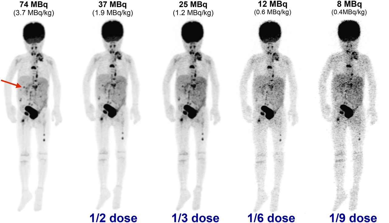
- FIGURE 1.
Maximum-intensity PET projections of 20-kg, 5-y-old patient with metastatic rhabdomyosarcoma; clinical full-dose image (far left, 74 MBq injected) and generated low-dose images are shown. Three liver lesions were identified in clinical image (arrow points to largest and highest-contrast lesion). Noise in generated images is visibly increased, as seen by pronounced heterogeneity of liver tissue. Confidence in lesion detection was compromised for doses lower than 1.2 MBq/kg, and no lesions could be identified at 0.4 MBq/kg. Window level was matched for all images.
- FIGURE 2.
(Top) Maximum-intensity PET projections of 2-y-old patient with Burkitt lymphoma. (Bottom) Axial slices through liver. Arrow indicates subtle 18F-FDG–avid lesion within liver that was not observed on MRI.
- FIGURE 3.
Measurements of liver background uptake for decreasing dosing regimens. Plots show mean (±SD) VOI measurements in background liver tissue from each patient. (A) Patients 2–5 y old (n = 4). (B) Patients 11–15 y old (n = 3).
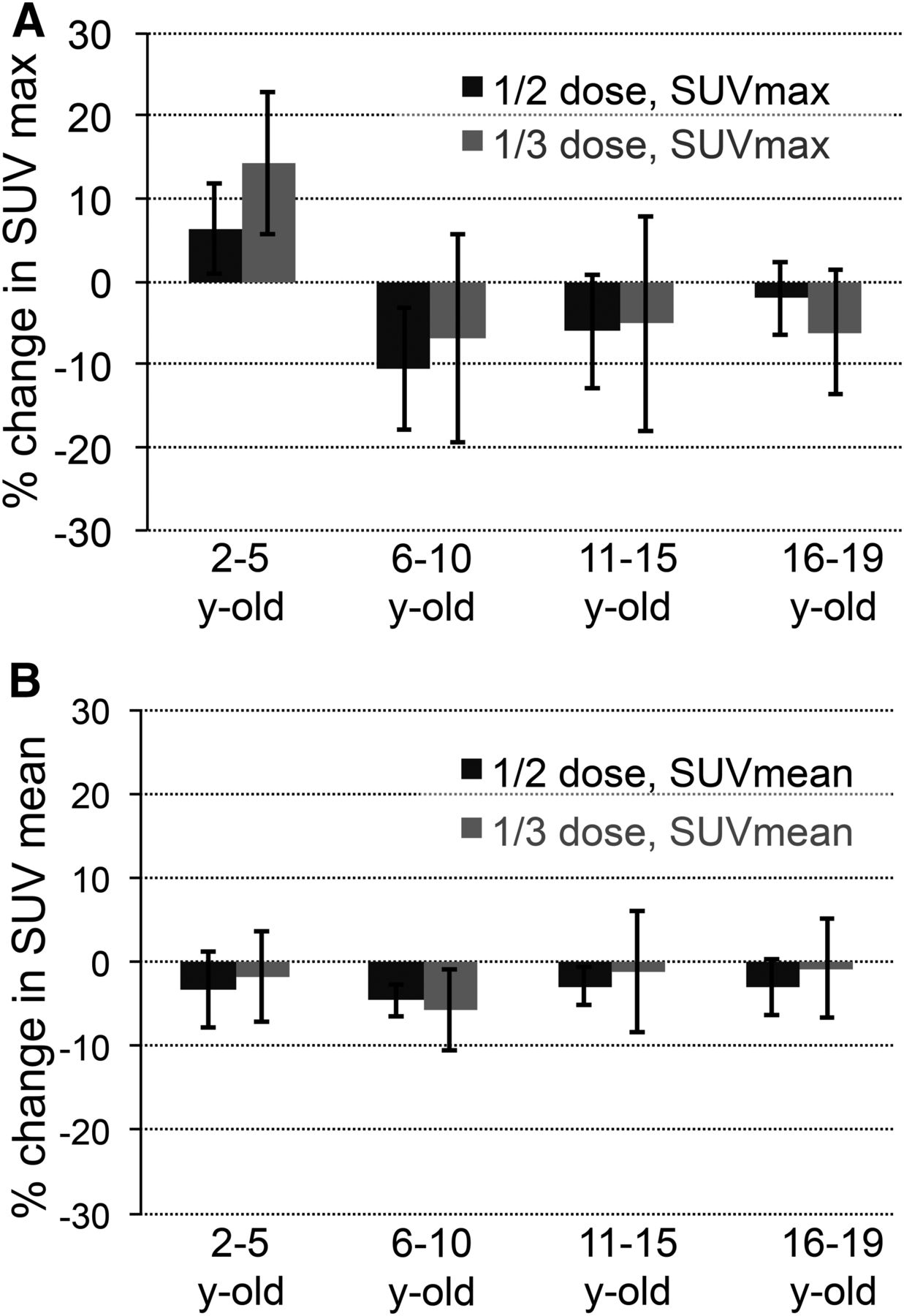
- FIGURE 4.
Bias and variability in lesion SUVmax (A) and SUVmean (B) at 2 dose levels in 4 age groups. Plot shows mean percentage difference ± SD compared with full-dose SUV measurement. Total lesion count was 28.
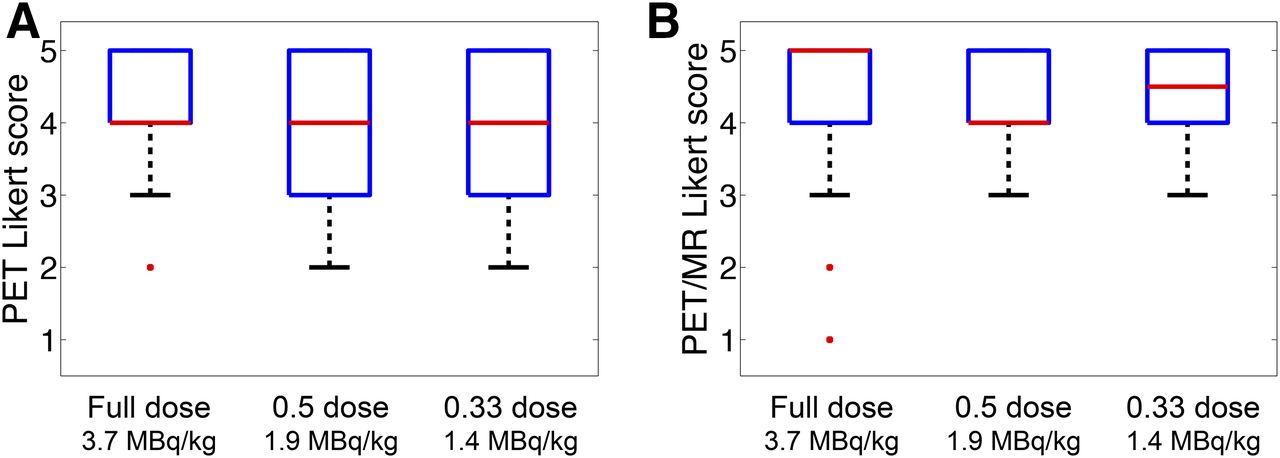
- FIGURE 5.
Summary of Likert scoring, averaged for all readers and grouped by dose level, for PET-only images (A) and PET/MR images (B). Plots show median (red lines), 25th–75th percentiles (box outlines), minimum (whisker), and outliers.
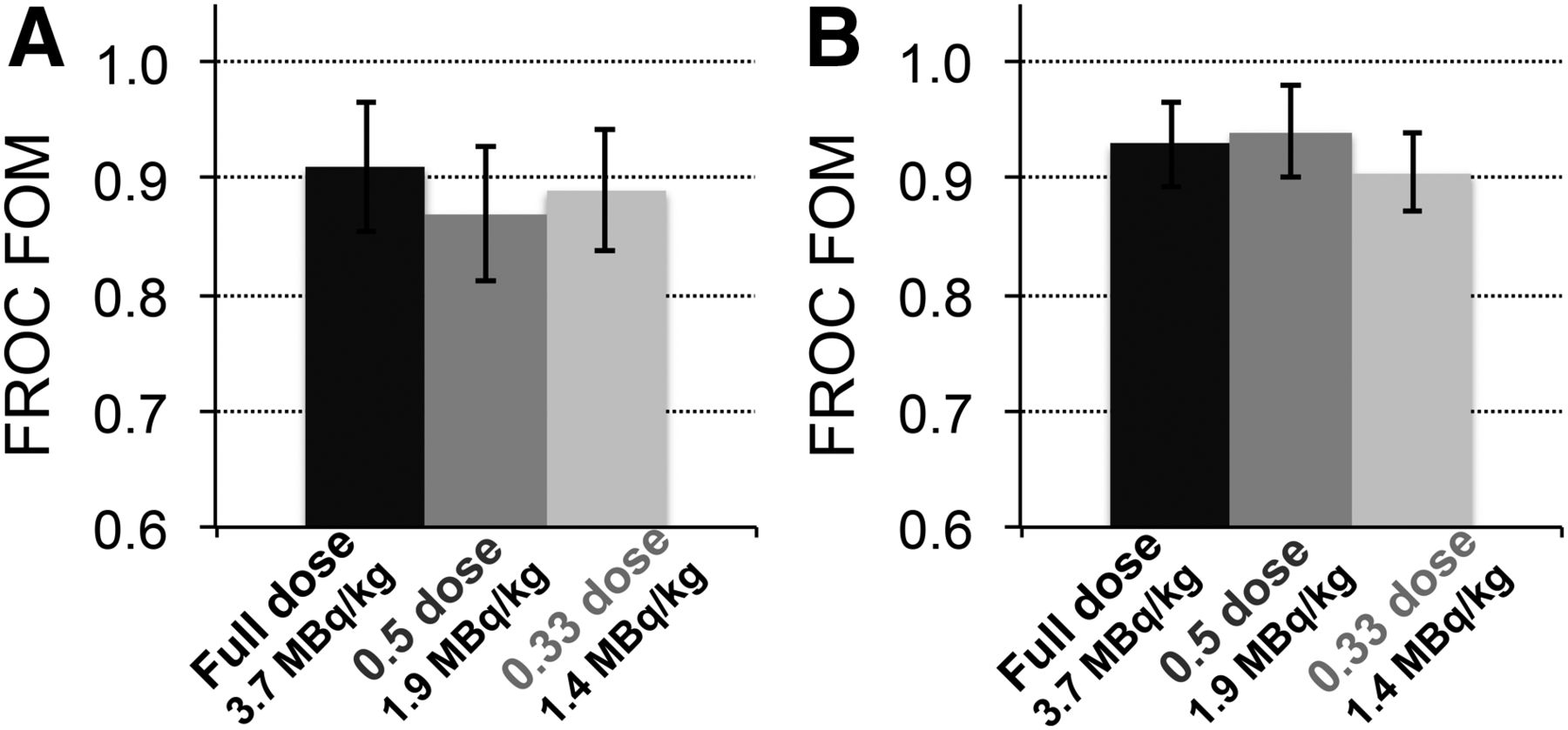
- FIGURE 6.
Estimated human observer performance quantified by JAFROC metric. Reader performance was measured at 3 dosing levels. (A) JAFROC values from readings of PET-only images. (B) JAFROC values from PET/MRI data using T1-weighted image.


Tables
Characteristic Data Average Median Patients (n) 11 Scans (n) 12 Age (y) 2–19 12 11 Weight (kg) 14–49 34 27 Lesions (n) n 28 Size (cm) 0.6–2.5 1.2 1.0 SUV 2.3–7.8 4.7 3.9 3.7 MBq/kg 1.9 MBq/kg 1.2 MBq/kg Index PET PET/MRI PET PET/MRI PET PET/MRI Mean 4.16 4.22 3.83 4.16 3.66 4.33 SD 0.83 1.13 1.01 1.03 1.20 0.75 % agreement 0.58 0.64 0.67 0.49 0.52 0.44 κ statistic 0.79 0.93 0.85 0.80 0.86 0.91 Scale: 5 = high diagnostic quality; 1 = poor diagnostic quality.
Reader 1 Reader 2 Reader 3 Parameter PET PET/MRI PET PET/MRI PET PET/MRI Full dose 19 18 23 23 17 19 ½ dose 16 21 27 28 16 17 ⅓ dose 17 18 21 25 14 14 False-positive* Full dose 2, 0 1, 1 4, 1 4, 1 1, 0 1, 0 ½ dose 0, 0 2, 2 9, 4 9, 4 1, 1 0, 0 ⅓ dose 0, 0 0, 0 4, 2 6, 4 0, 0 0, 0 False-negative Full dose 6 6 4 4 7 5 ½ dose 7 4 5 4 8 6 ⅓ dose 6 5 6 4 9 9 ↵* Presented as false-positive for all marked lesions, all lesions with confidence score ≥ 3.
Total lesions = 23.
PET PET/MRI Reader Full dose ½ dose ⅓ dose Full dose ½ dose ⅓ dose 1 0.91 0.89 0.95 0.93 0.96 0.93 2 0.93 0.88 0.83 0.93 0.92 0.89 3 0.89 0.83 0.89 0.94 0.92 0.89 Full-dose PET ½ dose PET ⅓ dose PET Full-dose PET/MRI ½ dose PET/MRI ⅓ dose PET/MRI Full-dose PET NA ½ dose PET 0.10 NA ⅓ dose PET 0.41 0.35 NA Full-dose PET/MRI 0.33 0.01 0.09 NA ½ dose PET/MRI 0.31 0.01 0.08 0.98 NA ⅓ dose PET/MRI 0.86 0.13 0.52 0.25 0.24 NA NA = not applicable.
95% confidence intervals for estimated combined reader performance were used.


