
Main menu

  • Home
  • Content
    • Current
    • Ahead of print
    • Past Issues
    • JNM Supplement
    • SNMMI Annual Meeting Abstracts
    • Continuing Education
    • JNM Podcasts
  • Subscriptions
    • Subscribers
    • Institutional and Non-member
    • Rates
    • Journal Claims
    • Corporate & Special Sales
  • Authors
    • Submit to JNM
    • Information for Authors
    • Assignment of Copyright
    • AQARA requirements
  • Info
    • Reviewers
    • Permissions
    • Advertisers
  • About
    • About Us
    • Editorial Board
    • Contact Information
  • More
    • Alerts
    • Feedback
    • Help
    • SNMMI Journals
  • SNMMI
    • JNM
    • JNMT
    • SNMMI Journals
    • SNMMI

User menu

  • Subscribe
  • My alerts
  • Log in
  • My Cart

Search

  • Advanced search
Journal of Nuclear Medicine
  • SNMMI
    • JNM
    • JNMT
    • SNMMI Journals
    • SNMMI
  • Subscribe
  • My alerts
  • Log in
  • My Cart
Journal of Nuclear Medicine

Advanced Search

  • Home
  • Content
    • Current
    • Ahead of print
    • Past Issues
    • JNM Supplement
    • SNMMI Annual Meeting Abstracts
    • Continuing Education
    • JNM Podcasts
  • Subscriptions
    • Subscribers
    • Institutional and Non-member
    • Rates
    • Journal Claims
    • Corporate & Special Sales
  • Authors
    • Submit to JNM
    • Information for Authors
    • Assignment of Copyright
    • AQARA requirements
  • Info
    • Reviewers
    • Permissions
    • Advertisers
  • About
    • About Us
    • Editorial Board
    • Contact Information
  • More
    • Alerts
    • Feedback
    • Help
    • SNMMI Journals
  • View or Listen to JNM Podcast
  • Visit JNM on Facebook
  • Join JNM on LinkedIn
  • Follow JNM on Twitter
  • Subscribe to our RSS feeds
Meeting ReportPhysics, Instrumentation & Data Sciences

Quantitative Assessment of Deep Learning-enhanced Actual Ultra-low-dose Amyloid PET/MR Imaging

Kevin Chen, Dawn Holley, Kim Halbert, Tyler Toueg, Athanasia Boumis, Gabriel Kennedy, Elizabeth Mormino, Mehdi Khalighi and Greg Zaharchuk
Journal of Nuclear Medicine May 2020, 61 (supplement 1) 521;
Kevin Chen
1Radiology Stanford University Stanford CA United States
  • Find this author on Google Scholar
  • Find this author on PubMed
  • Search for this author on this site
Dawn Holley
1Radiology Stanford University Stanford CA United States
  • Find this author on Google Scholar
  • Find this author on PubMed
  • Search for this author on this site
Kim Halbert
1Radiology Stanford University Stanford CA United States
  • Find this author on Google Scholar
  • Find this author on PubMed
  • Search for this author on this site
Tyler Toueg
2Neurology Stanford University Stanford CA United States
  • Find this author on Google Scholar
  • Find this author on PubMed
  • Search for this author on this site
Athanasia Boumis
1Radiology Stanford University Stanford CA United States
  • Find this author on Google Scholar
  • Find this author on PubMed
  • Search for this author on this site
Gabriel Kennedy
2Neurology Stanford University Stanford CA United States
  • Find this author on Google Scholar
  • Find this author on PubMed
  • Search for this author on this site
Elizabeth Mormino
2Neurology Stanford University Stanford CA United States
  • Find this author on Google Scholar
  • Find this author on PubMed
  • Search for this author on this site
Mehdi Khalighi
1Radiology Stanford University Stanford CA United States
  • Find this author on Google Scholar
  • Find this author on PubMed
  • Search for this author on this site
Greg Zaharchuk
1Radiology Stanford University Stanford CA United States
  • Find this author on Google Scholar
  • Find this author on PubMed
  • Search for this author on this site
  • Article
  • Figures & Data
  • Info & Metrics
Loading

Abstract

521

Objectives: Combining deep learning-based methods such as convolutional neural networks (CNNs) with the advantage of complementary information from simultaneous positron emission tomography (PET)/ magnetic resonance imaging (MRI), we have previously generated diagnostic quality amyloid PET images with simulated ultra-low (1%) radiotracer dose [1]. Here, we will demonstrate utility of this method with images acquired using actual ultra-low-dose radiotracer injections. Dramatically lowering injected dose will not only reduce radiation risk in subjects but also provide breakthroughs in PET/MRI scanning protocols, allowing for more frequent follow-ups of disease progression.

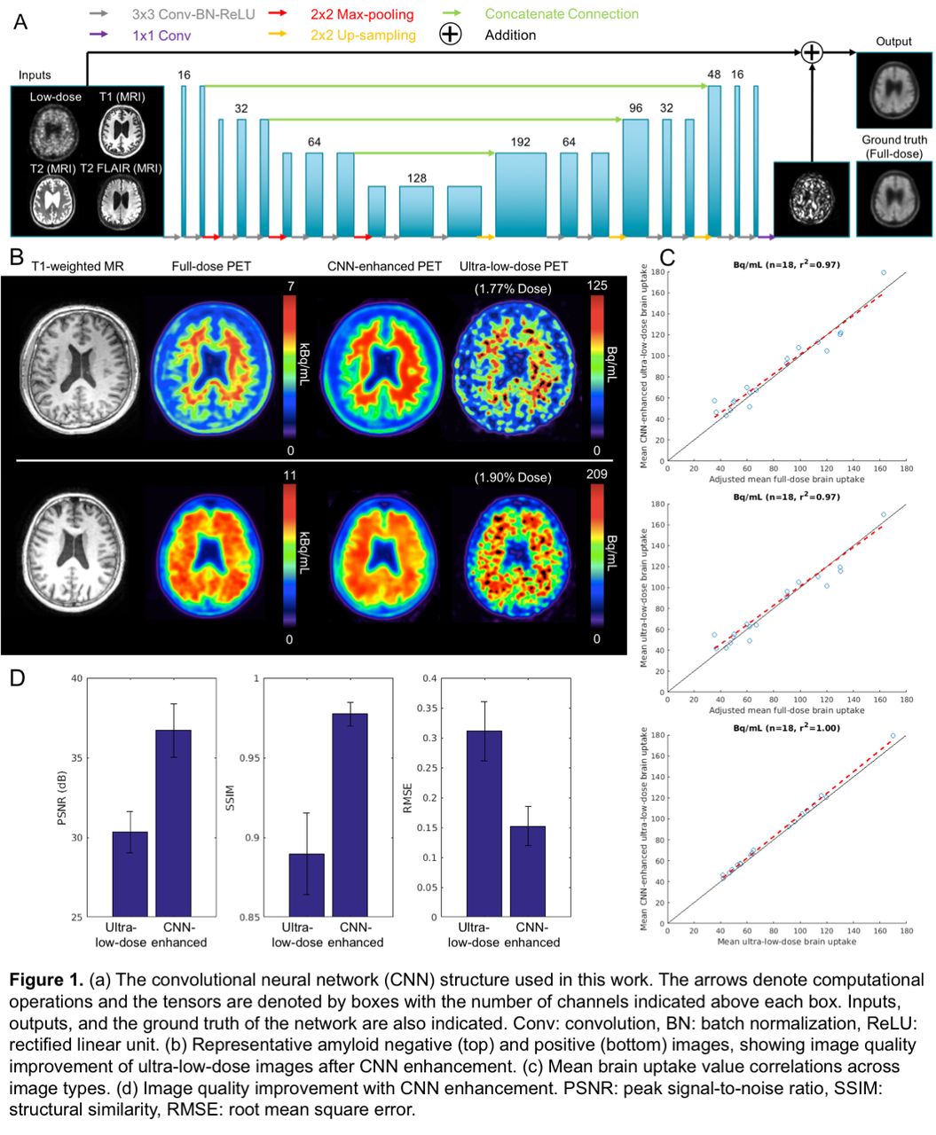
Methods: 50 total subjects were recruited for the study. 32 (19 female, mean±standard deviation [SD]: 68.2±7.1 years) were used for pre-training the network; 328±32 MBq of the amyloid radiotracer 18F-florbetaben were injected into the subject. 18 (8 female, 72.1±8.6 years) were scanned with the ultra-low-dose protocol. These subjects were scanned in two PET/MRI sessions (9 on same day: low-dose session followed by full dose; 9 on separate days: 1- to 42-day interval, mean 19.6 days), with 6.73±3.55 and 302±13 MBq 18F-florbetaben injections respectively (2.3%±1.3% dose for the ultra-low-dose sessions). For all scans, the T1-, T2-, and T2 FLAIR-weighted MR images were acquired simultaneously with PET (90-110 minutes after injection; 83-98 minutes for one subject) on an integrated PET/MR scanner with time-of-flight capabilities. A pre-trained low-dose CNN was trained based on Chen et al. (Figure 1a) [1]. The inputs of the network are the multi-contrast MR images and the simulated low-dose PET image (obtained from 1% random undersampling of the original list-mode data). The network was trained on the full-dose PET image as the ground truth. The last layer of the CNN was fine-tuned using the actual low-dose datasets, with actual low-dose images replacing simulations as inputs. 9-fold cross-validation was used to prevent tuning and testing on the same subjects (16 subjects for training, 2 subjects for testing per network trained). The mean uptake (full-dose images were multiplied by the low-dose percentage) within the brain was analyzed for correlation across image types. For each axial slice, the image quality of the synthesized and low-dose PET images within the brain were compared to the full-dose image using peak signal-to-noise ratio (PSNR), structural similarity (SSIM), and root mean square error (RMSE). Regional (from FreeSurfer) mean standard uptake value ratios (SUVRs, normalized to cerebellar cortex) were compared across image types.

Results: Qualitatively, the synthesized images show marked improvement in noise reduction to the ultra-low-dose image and resemble the ground truth image (Figure 1b). The mean radiotracer uptake within the brain highly correlated across image types, validating the amount of tracer injected for the ultra-low-dose portion (Figure 1c). All three metrics showed dramatic image quality improvement (Figure 1d) from the ultra-low-dose images to the synthesized images. Comparing the regional SUVRs of the synthesized and low-dose images to the full-dose images showed that the mean SUVR differences were close to zero for both image types, but the synthesized images had smaller SDs to their full-dose counterparts (SUVR difference, mean±SD: -0.03±0.16) than the low-dose images (SUVR difference: -0.02±0.17).

Conclusions: This work has shown that accurate amyloid PET images can be generated using trained CNNs with simultaneously-acquired MR images and PET images reconstructed from actual ultra-low-dose radiotracer injections. Acknowledgements: This work was made possible by the Michael J. Fox Foundation, the Stanford Alzheimer's Disease Research Center, the NIH Grant P41-EB015891, GE Healthcare, the Foundation of the ASNR, and Life Molecular Imaging.

Figure
  • Download figure
  • Open in new tab
  • Download powerpoint
Previous
Back to top

In this issue

Journal of Nuclear Medicine
Vol. 61, Issue supplement 1
May 1, 2020
  • Table of Contents
  • Index by author
Article Alerts
Sign In to Email Alerts with your Email Address
Email Article

Thank you for your interest in spreading the word on Journal of Nuclear Medicine.

NOTE: We only request your email address so that the person you are recommending the page to knows that you wanted them to see it, and that it is not junk mail. We do not capture any email address.

Enter multiple addresses on separate lines or separate them with commas.
Quantitative Assessment of Deep Learning-enhanced Actual Ultra-low-dose Amyloid PET/MR Imaging
(Your Name) has sent you a message from Journal of Nuclear Medicine
(Your Name) thought you would like to see the Journal of Nuclear Medicine web site.
Citation Tools
Quantitative Assessment of Deep Learning-enhanced Actual Ultra-low-dose Amyloid PET/MR Imaging
Kevin Chen, Dawn Holley, Kim Halbert, Tyler Toueg, Athanasia Boumis, Gabriel Kennedy, Elizabeth Mormino, Mehdi Khalighi, Greg Zaharchuk
Journal of Nuclear Medicine May 2020, 61 (supplement 1) 521;

Citation Manager Formats

  • BibTeX
  • Bookends
  • EasyBib
  • EndNote (tagged)
  • EndNote 8 (xml)
  • Medlars
  • Mendeley
  • Papers
  • RefWorks Tagged
  • Ref Manager
  • RIS
  • Zotero
Share
Quantitative Assessment of Deep Learning-enhanced Actual Ultra-low-dose Amyloid PET/MR Imaging
Kevin Chen, Dawn Holley, Kim Halbert, Tyler Toueg, Athanasia Boumis, Gabriel Kennedy, Elizabeth Mormino, Mehdi Khalighi, Greg Zaharchuk
Journal of Nuclear Medicine May 2020, 61 (supplement 1) 521;
Twitter logo Facebook logo LinkedIn logo Mendeley logo
  • Tweet Widget
  • Facebook Like
  • Google Plus One
Bookmark this article

Jump to section

  • Article
  • Figures & Data
  • Info & Metrics

Related Articles

  • No related articles found.
  • Google Scholar

Cited By...

  • No citing articles found.
  • Google Scholar

More in this TOC Section

Physics, Instrumentation & Data Sciences

  • AI-based methods for nuclear-medicine imaging: Need for objective task-specific evaluation
  • Keel-Edge Height Selection for Improved Multi-Pinhole 123I Brain SPECT Imaging
  • Ultra-Fast Reconstruction of Short List-Mode PET Data Frames for Real-Time Visualization and Processing
Show more Physics, Instrumentation & Data Sciences

Artificial Intelligence for Image Enhancement/Analysis and Disease Assessment

  • A fully unsupervised approach to create patient-like phantoms via Convolutional neural networks
  • A no-gold-standard technique for objective evaluation of quantitative nuclear-medicine imaging methods in the presence of correlated noise
Show more Artificial Intelligence for Image Enhancement/Analysis and Disease Assessment

Similar Articles

SNMMI

© 2025 SNMMI

Powered by HighWire