
Main menu

  • Home
  • Content
    • Current
    • Ahead of print
    • Past Issues
    • JNM Supplement
    • SNMMI Annual Meeting Abstracts
    • Continuing Education
    • JNM Podcasts
  • Subscriptions
    • Subscribers
    • Institutional and Non-member
    • Rates
    • Journal Claims
    • Corporate & Special Sales
  • Authors
    • Submit to JNM
    • Information for Authors
    • Assignment of Copyright
    • AQARA requirements
  • Info
    • Reviewers
    • Permissions
    • Advertisers
  • About
    • About Us
    • Editorial Board
    • Contact Information
  • More
    • Alerts
    • Feedback
    • Help
    • SNMMI Journals
  • SNMMI
    • JNM
    • JNMT
    • SNMMI Journals
    • SNMMI

User menu

  • Subscribe
  • My alerts
  • Log in
  • My Cart

Search

  • Advanced search
Journal of Nuclear Medicine
  • SNMMI
    • JNM
    • JNMT
    • SNMMI Journals
    • SNMMI
  • Subscribe
  • My alerts
  • Log in
  • My Cart
Journal of Nuclear Medicine

Advanced Search

  • Home
  • Content
    • Current
    • Ahead of print
    • Past Issues
    • JNM Supplement
    • SNMMI Annual Meeting Abstracts
    • Continuing Education
    • JNM Podcasts
  • Subscriptions
    • Subscribers
    • Institutional and Non-member
    • Rates
    • Journal Claims
    • Corporate & Special Sales
  • Authors
    • Submit to JNM
    • Information for Authors
    • Assignment of Copyright
    • AQARA requirements
  • Info
    • Reviewers
    • Permissions
    • Advertisers
  • About
    • About Us
    • Editorial Board
    • Contact Information
  • More
    • Alerts
    • Feedback
    • Help
    • SNMMI Journals
  • View or Listen to JNM Podcast
  • Visit JNM on Facebook
  • Join JNM on LinkedIn
  • Follow JNM on Twitter
  • Subscribe to our RSS feeds
Meeting ReportPhysics, Instrumentation & Data Sciences

AI-based methods for nuclear-medicine imaging: Need for objective task-specific evaluation

Zitong Yu, Md Ashequr Rahman, Thomas Schindler, Robert Gropler, Richard Laforest, Richard Wahl and Abhinav Jha
Journal of Nuclear Medicine May 2020, 61 (supplement 1) 575;
Zitong Yu
1Department of Biomedical Engineering Washington University in St. Louis Saint Louis MO United States
  • Find this author on Google Scholar
  • Find this author on PubMed
  • Search for this author on this site
Md Ashequr Rahman
1Department of Biomedical Engineering Washington University in St. Louis Saint Louis MO United States
  • Find this author on Google Scholar
  • Find this author on PubMed
  • Search for this author on this site
Thomas Schindler
2Mallinckrodt Institute of Radiology Washington University in St. Louis Saint Louis MO United States
  • Find this author on Google Scholar
  • Find this author on PubMed
  • Search for this author on this site
Robert Gropler
2Mallinckrodt Institute of Radiology Washington University in St. Louis Saint Louis MO United States
  • Find this author on Google Scholar
  • Find this author on PubMed
  • Search for this author on this site
Richard Laforest
2Mallinckrodt Institute of Radiology Washington University in St. Louis Saint Louis MO United States
  • Find this author on Google Scholar
  • Find this author on PubMed
  • Search for this author on this site
Richard Wahl
2Mallinckrodt Institute of Radiology Washington University in St. Louis Saint Louis MO United States
  • Find this author on Google Scholar
  • Find this author on PubMed
  • Search for this author on this site
Abhinav Jha
1Department of Biomedical Engineering Washington University in St. Louis Saint Louis MO United States
2Mallinckrodt Institute of Radiology Washington University in St. Louis Saint Louis MO United States
  • Find this author on Google Scholar
  • Find this author on PubMed
  • Search for this author on this site
  • Article
  • Figures & Data
  • Info & Metrics
Loading

Abstract

575

Objectives: Artificial intelligence (AI)-based methods are showing promise in reconstructing and processing nuclear-medicine images[1,2] Conventionally these methods are evaluated using figures of merit (FoMs) such as root mean squared error (RMSE)[3,4], structural similarity index (SSIM)[3,5], and peak signal-to-noise ratio (PSNR)[3,6]. However, nuclear-medicine images are acquired for clinical tasks, such as detection and quantification, and it is unclear if these metrics correlate with task performance. Our objective was to study whether evaluation of the AI-based methods using conventional FoMs yielded the same interpretation as objective evaluation on clinical tasks.

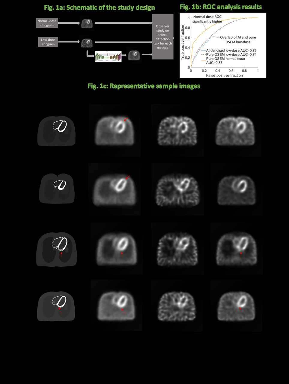
Methods: There is much interest in using AI-based methods to acquire images at lower dose/acquisition times. Given this interest, we conducted this study in the context of evaluating an AI-based method to denoise myocardial perfusion SPECT (MPS) images acquired at five-times lower dose (Fig. 1a). Using the XCAT phantom[7], a total of 14,000 digital phantoms with and without defects were generated. 18 types of myocardial defects, including 3 extent types, 3 severity types, and 2 locations, all based on existing clinical data[8,9], were evenly distributed in the defect-present population. Projection data for these phantoms at both normal and five-times lower dose were generated using highly realistic simulations that modeled various SPECT image-degrading processes. Both these sets of images were reconstructed using a 2D OSEM-based technique. A convolutional neural network (CNN)-based method was developed to predict the normal-dose image from the low-dose image (i.e. denoise the low-dose image). The CNN was optimized and trained using 12,000 low and normal-dose image pairs, and the rest 2000 were used for testing. The performance of this AI-based method on the clinical task of detecting myocardial perfusion defects was objectively evaluated using a previously validated Hotelling-observer-based technique designed for this task[10]. The detection performance was quantified using area under the ROC curve (AUC). Also, the RMSE, SSIM, and PSNR were computed for the CNN-denoised images. For comparison, we also computed these FoMs for the images reconstructed using the pure OSEM technique.

Results: The AI-based method significantly outperformed the pure OSEM method (p value < 0.001) yielding higher SSIM and PSNR values and lower RMSE values (Table 1). For e.g. the SSIM with the AI and OSEM-based methods were 0.869 (95 % CI: 0.868, 0.871) and 0.851 (95 % CI: 0.850, 0.853), respectively. Further, visually, the images look less noisy (Fig. 1c). However, in the observer study, the AI and OSEM-based methods yielded AUC values of 0.73 (95 % CI: 0.71,0.76) and 0.74 (95 % CI: 0.72, 0.77), respectively (no statistical difference, in fact mean AUC slightly lower with AI-based approach) with almost overlapping ROC curves (Fig. 1b). Thus, the results from the observer study were in conflict with evaluation using the conventional metrics.

Conclusions: The results motivate the need for objective evaluation of AI-based methods for nuclear-medicine imaging on clinically relevant tasks[11] as evaluation with conventional metrics may not capture that aspect of the performance. Further, the results motivate optimization of AI-based approaches on clinically relevant tasks.

View this table:
  • View inline
  • View popup

Table 1: Comparing AI and purely OSEM-based methods using conventional metrics and using AUC

Figure
  • Download figure
  • Open in new tab
  • Download powerpoint
Previous
Back to top

In this issue

Journal of Nuclear Medicine
Vol. 61, Issue supplement 1
May 1, 2020
  • Table of Contents
  • Index by author
Article Alerts
Sign In to Email Alerts with your Email Address
Email Article

Thank you for your interest in spreading the word on Journal of Nuclear Medicine.

NOTE: We only request your email address so that the person you are recommending the page to knows that you wanted them to see it, and that it is not junk mail. We do not capture any email address.

Enter multiple addresses on separate lines or separate them with commas.
AI-based methods for nuclear-medicine imaging: Need for objective task-specific evaluation
(Your Name) has sent you a message from Journal of Nuclear Medicine
(Your Name) thought you would like to see the Journal of Nuclear Medicine web site.
Citation Tools
AI-based methods for nuclear-medicine imaging: Need for objective task-specific evaluation
Zitong Yu, Md Ashequr Rahman, Thomas Schindler, Robert Gropler, Richard Laforest, Richard Wahl, Abhinav Jha
Journal of Nuclear Medicine May 2020, 61 (supplement 1) 575;

Citation Manager Formats

  • BibTeX
  • Bookends
  • EasyBib
  • EndNote (tagged)
  • EndNote 8 (xml)
  • Medlars
  • Mendeley
  • Papers
  • RefWorks Tagged
  • Ref Manager
  • RIS
  • Zotero
Share
AI-based methods for nuclear-medicine imaging: Need for objective task-specific evaluation
Zitong Yu, Md Ashequr Rahman, Thomas Schindler, Robert Gropler, Richard Laforest, Richard Wahl, Abhinav Jha
Journal of Nuclear Medicine May 2020, 61 (supplement 1) 575;
Twitter logo Facebook logo LinkedIn logo Mendeley logo
  • Tweet Widget
  • Facebook Like
  • Google Plus One
Bookmark this article

Jump to section

  • Article
  • Figures & Data
  • Info & Metrics

Related Articles

  • No related articles found.
  • Google Scholar

Cited By...

  • An Investigation of Lesion Detection Accuracy for Artificial Intelligence-Based Denoising of Low-Dose 64Cu-DOTATATE PET Imaging in Patients with Neuroendocrine Neoplasms
  • An Investigation of Lesion Detection Accuracy for Artificial Intelligence-Based Denoising of Low-Dose 64Cu-DOTATATE PET Imaging in Patients with Neuroendocrine Neoplasms
  • Artificial Intelligence in Nuclear Medicine: Opportunities, Challenges, and Responsibilities Toward a Trustworthy Ecosystem
  • Nuclear Medicine and Artificial Intelligence: Best Practices for Evaluation (the RELAINCE Guidelines)
  • Nuclear Medicine and Artificial Intelligence: Best Practices for Algorithm Development
  • Google Scholar

More in this TOC Section

Physics, Instrumentation & Data Sciences

  • Exploration of Multi-objective Optimization with Genetic Algorithms for PET Image Reconstruction
  • Ultra-Fast Reconstruction of Short List-Mode PET Data Frames for Real-Time Visualization and Processing
Show more Physics, Instrumentation & Data Sciences

SPECT, Image Quality, Therapy

  • Clinical feasibility of quantitative holmium-166 SPECT in presence of technetium-99m colloid
  • Data-driven respiratory signal estimation from finely sampled projection data in conventional cardiac perfusion SPECT imaging.
Show more SPECT, Image Quality, Therapy

Similar Articles

SNMMI

© 2025 SNMMI

Powered by HighWire