
Main menu

  • Home
  • Content
    • Current
    • Ahead of print
    • Past Issues
    • JNM Supplement
    • SNMMI Annual Meeting Abstracts
  • Subscriptions
    • Subscribers
    • Institutional and Non-member
    • Rates
    • Corporate & Special Sales
    • Journal Claims
  • Authors
    • Submit to JNM
    • Information for Authors
    • Assignment of Copyright
    • AQARA requirements
  • Info
    • Permissions
    • Advertisers
    • Continuing Education
  • About
    • About Us
    • Editorial Board
    • Contact Information
  • More
    • Alerts
    • Feedback
    • Help
    • SNMMI Journals
  • SNMMI
    • JNM
    • JNMT
    • SNMMI Journals
    • SNMMI

User menu

  • Subscribe
  • My alerts
  • Log in
  • My Cart

Search

  • Advanced search
Journal of Nuclear Medicine
  • SNMMI
    • JNM
    • JNMT
    • SNMMI Journals
    • SNMMI
  • Subscribe
  • My alerts
  • Log in
  • My Cart
Journal of Nuclear Medicine

Advanced Search

  • Home
  • Content
    • Current
    • Ahead of print
    • Past Issues
    • JNM Supplement
    • SNMMI Annual Meeting Abstracts
  • Subscriptions
    • Subscribers
    • Institutional and Non-member
    • Rates
    • Corporate & Special Sales
    • Journal Claims
  • Authors
    • Submit to JNM
    • Information for Authors
    • Assignment of Copyright
    • AQARA requirements
  • Info
    • Permissions
    • Advertisers
    • Continuing Education
  • About
    • About Us
    • Editorial Board
    • Contact Information
  • More
    • Alerts
    • Feedback
    • Help
    • SNMMI Journals
  • Follow JNM on Twitter
  • Visit JNM on Facebook
  • Join JNM on LinkedIn
  • Subscribe to our RSS feeds
Meeting ReportPhysics, Instrumentation & Data Sciences

Ultra-Fast Reconstruction of Short List-Mode PET Data Frames for Real-Time Visualization and Processing

Matthew Spangler-Bickell, Timothy Deller and Floris Jansen
Journal of Nuclear Medicine May 2020, 61 (supplement 1) 570;
Matthew Spangler-Bickell
1Nuclear Medicine Unit IRCCS Ospedale San Raffaele Milan Italy
  • Find this author on Google Scholar
  • Find this author on PubMed
  • Search for this author on this site
Timothy Deller
2General Electric Elm Grove WI United States
  • Find this author on Google Scholar
  • Find this author on PubMed
  • Search for this author on this site
Floris Jansen
3GE Healthcare Charlton NY United States
  • Find this author on Google Scholar
  • Find this author on PubMed
  • Search for this author on this site
  • Article
  • Figures & Data
  • Info & Metrics
Loading

Abstract

570

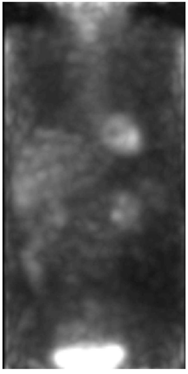
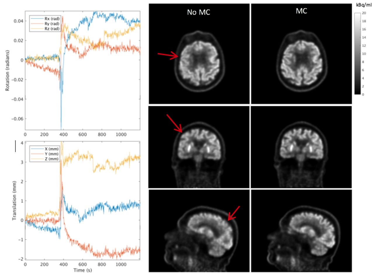
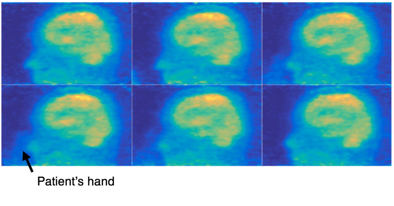
Introduction: Clinical PET reconstruction typically begins after the entire frame of data has been acquired. In this paper we present a framework for performing reconstructions of very short frames of list-mode data (∼1 sec) faster than real-time (i.e. < 1 sec), with the objective of using this framework during the scan to enable various applications:• The technologist can observe the tracer distribution in near real-time during the scan and verify the patient positioning, the injection success, any motion, etc., and intervene accordingly.• Perform data-driven motion estimation so that as soon as the scan is completed a fully motion corrected reconstruction can be performed.• Alert the technologist if motion exceeds a threshold.• Conduct a whole body survey over a few seconds to quickly see the tracer distribution, identify areas of interest, and adjust the subsequent bed durations accordingly.• For gated cardiac imaging, quick reconstructions of the gates can aid in determining which gate most closely matches the attenuation map.The developed framework runs on top of the PET research toolbox distributed by GE Healthcare (Chicago, IL). Method: A fast TOF-based ray-tracing projector is used within a list-mode (LM) reconstruction algorithm. Batches of LM data corresponding to a chosen frame duration are reconstructed using MLEM without subsets. 2-5 iterations are performed. Attenuation correction is applied, unless motion estimation is to be performed as it might bias the motion estimates. Randoms correction is calculated independently for each detected event, based on the per-second single events information. Scatter correction is not performed. For visualisation purposes, a 1 sec duration has been found to be adequate. To estimate head motion, frame durations as low as 0.3 sec provide sufficiently accurate motion estimates [1]. Clinical Results: A standard clinical 18F-FDG brain data set was acquired on a Discovery MI PET/CT (GE Healthcare). Frames of 1 sec duration were reconstructed, each taking approximately 0.2 sec on an 8-core CPU desktop system. In figure 1, the patient’s hand can be seen entering the field-of-view, which was not observed during the scan.In figure 2, a patient undergoing a brain scan on a SIGNA PET/MR (GE Healthcare) spoke to the technologist, resulting in head motion. Motion was estimated from the frames, and a fully motion corrected LM reconstruction was then performed (figure 3).Using data from a whole body 18F-FDG scan, very short frames of 0.3 sec each were reconstructed from several bed positions over the abdomen and thorax and stitched together (figure 4). This demonstrates the feasibility of an ultra-fast whole body survey.

Conclusions: A framework for performing ultra-fast reconstructions (< 1 sec) of very short frames (~ 1 sec) has been presented, allowing near real-time reconstruction of the tracer distribution during a PET scan. Such a framework opens many avenues, including real-time feedback to the technologist during the scan, data-driven motion correction in the head and abdomen, and rapid whole body surveys. Future work includes extracting non-rigid deformation parameters for data-driven motion correction with respiratory and cardiac motion. Fig. 1: Example reconstructions of 1 second frames. The patient’s hand can be seen entering the FOV. Fig. 2: Selected frames of 1 sec duration where the patient can be seen speaking during the scan. Fig. 3: Left: Plots of the motion estimates. The patient spoke at about 380 sec. Right: List-mode reconstructions of the full data set without and with motion correction (MC). Fig. 4: Very short frames of 0.3 sec each from several bed positions to demonstrate a whole body survey. Acknowledgement: We gratefully acknowledge patient data acquired at KU Leuven. References: [1] M. G. Spangler-Bickell, et al, “Effect of image noise on registration in PET brain imaging,” in IEEE MIC Proceeding, 2019

Figure
  • Download figure
  • Open in new tab
  • Download powerpoint
Figure
  • Download figure
  • Open in new tab
  • Download powerpoint
Figure
  • Download figure
  • Open in new tab
  • Download powerpoint
Figure
  • Download figure
  • Open in new tab
  • Download powerpoint
Previous
Back to top

In this issue

Journal of Nuclear Medicine
Vol. 61, Issue supplement 1
May 1, 2020
  • Table of Contents
  • Index by author
Article Alerts
Sign In to Email Alerts with your Email Address
Email Article

Thank you for your interest in spreading the word on Journal of Nuclear Medicine.

NOTE: We only request your email address so that the person you are recommending the page to knows that you wanted them to see it, and that it is not junk mail. We do not capture any email address.

Enter multiple addresses on separate lines or separate them with commas.
Ultra-Fast Reconstruction of Short List-Mode PET Data Frames for Real-Time Visualization and Processing
(Your Name) has sent you a message from Journal of Nuclear Medicine
(Your Name) thought you would like to see the Journal of Nuclear Medicine web site.
Citation Tools
Ultra-Fast Reconstruction of Short List-Mode PET Data Frames for Real-Time Visualization and Processing
Matthew Spangler-Bickell, Timothy Deller, Floris Jansen
Journal of Nuclear Medicine May 2020, 61 (supplement 1) 570;

Citation Manager Formats

  • BibTeX
  • Bookends
  • EasyBib
  • EndNote (tagged)
  • EndNote 8 (xml)
  • Medlars
  • Mendeley
  • Papers
  • RefWorks Tagged
  • Ref Manager
  • RIS
  • Zotero
Share
Ultra-Fast Reconstruction of Short List-Mode PET Data Frames for Real-Time Visualization and Processing
Matthew Spangler-Bickell, Timothy Deller, Floris Jansen
Journal of Nuclear Medicine May 2020, 61 (supplement 1) 570;
del.icio.us logo Digg logo Reddit logo Twitter logo Facebook logo Google logo Mendeley logo
  • Tweet Widget
  • Facebook Like
  • Google Plus One
Bookmark this article

Jump to section

  • Article
  • Figures & Data
  • Info & Metrics

Related Articles

  • No related articles found.
  • Google Scholar

Cited By...

  • No citing articles found.
  • Google Scholar

More in this TOC Section

Physics, Instrumentation & Data Sciences

  • Keel-Edge Height Selection for Improved Multi-Pinhole 123I Brain SPECT Imaging
  • Deep Learning Based 3D Dose Estimation from Prompt Gamma Distribution for Proton Therapy Monitoring
Show more Physics, Instrumentation & Data Sciences

SPECT, Image Quality, Therapy

  • Exploration of Multi-objective Optimization with Genetic Algorithms for PET Image Reconstruction
  • Keel-Edge Height Selection for Improved Multi-Pinhole 123I Brain SPECT Imaging
  • Clinical feasibility of quantitative holmium-166 SPECT in presence of technetium-99m colloid
Show more SPECT, Image Quality, Therapy

Similar Articles

SNMMI

© 2022 Journal of Nuclear Medicine

Powered by HighWire