


Article Figures & Data
Figures
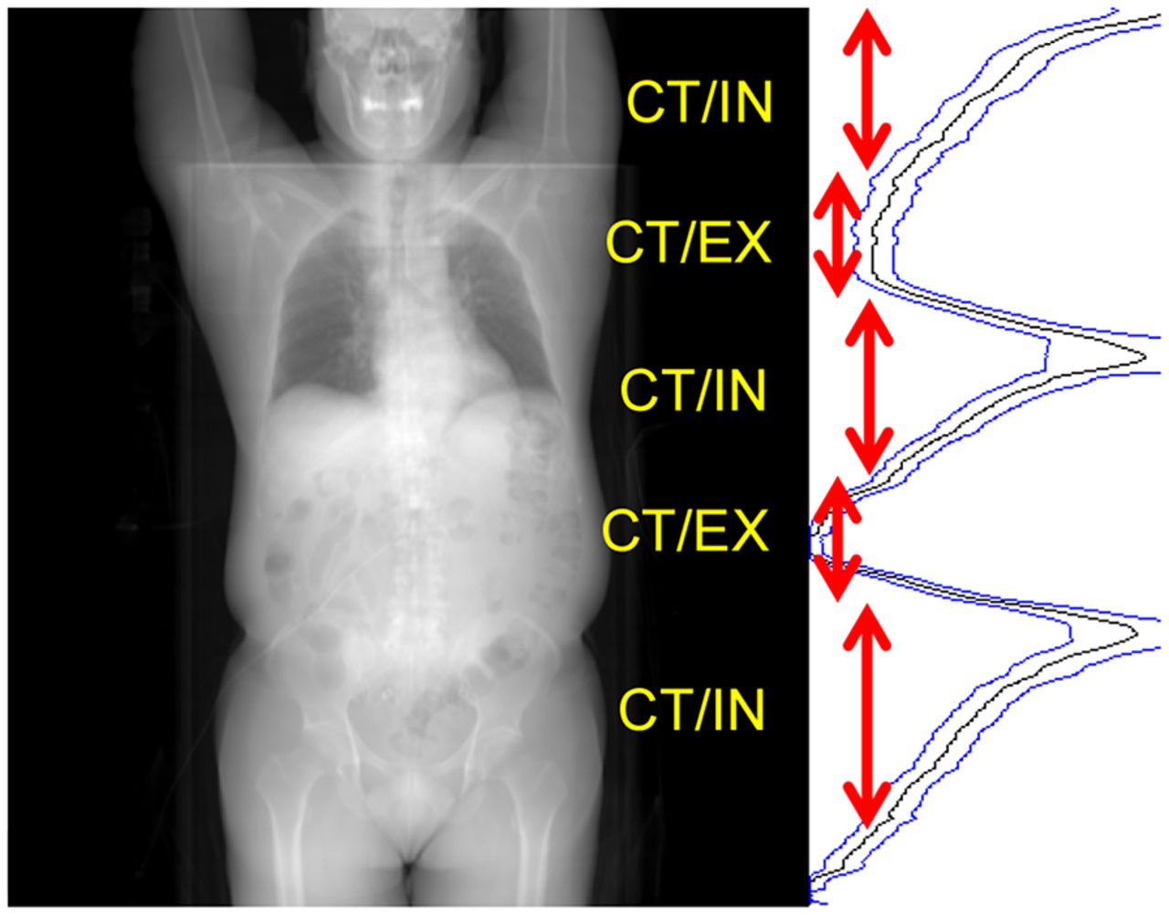
- FIGURE 1.
Anterior projection of patient’s whole-body CT image series, alongside a respiratory waveform aligned with the CT projection, with increasing amplitude toward right. Axial zones where CT/in and CT/ex occurred are indicated.
- FIGURE 2.
Respiratory waveform amplitudes plotted vs. time in PET scan. (A) Amplitude range used for OG. (B–F) Time and amplitude ranges for successive positions between upper lung and upper abdomen. a.u. = arbitrary units.
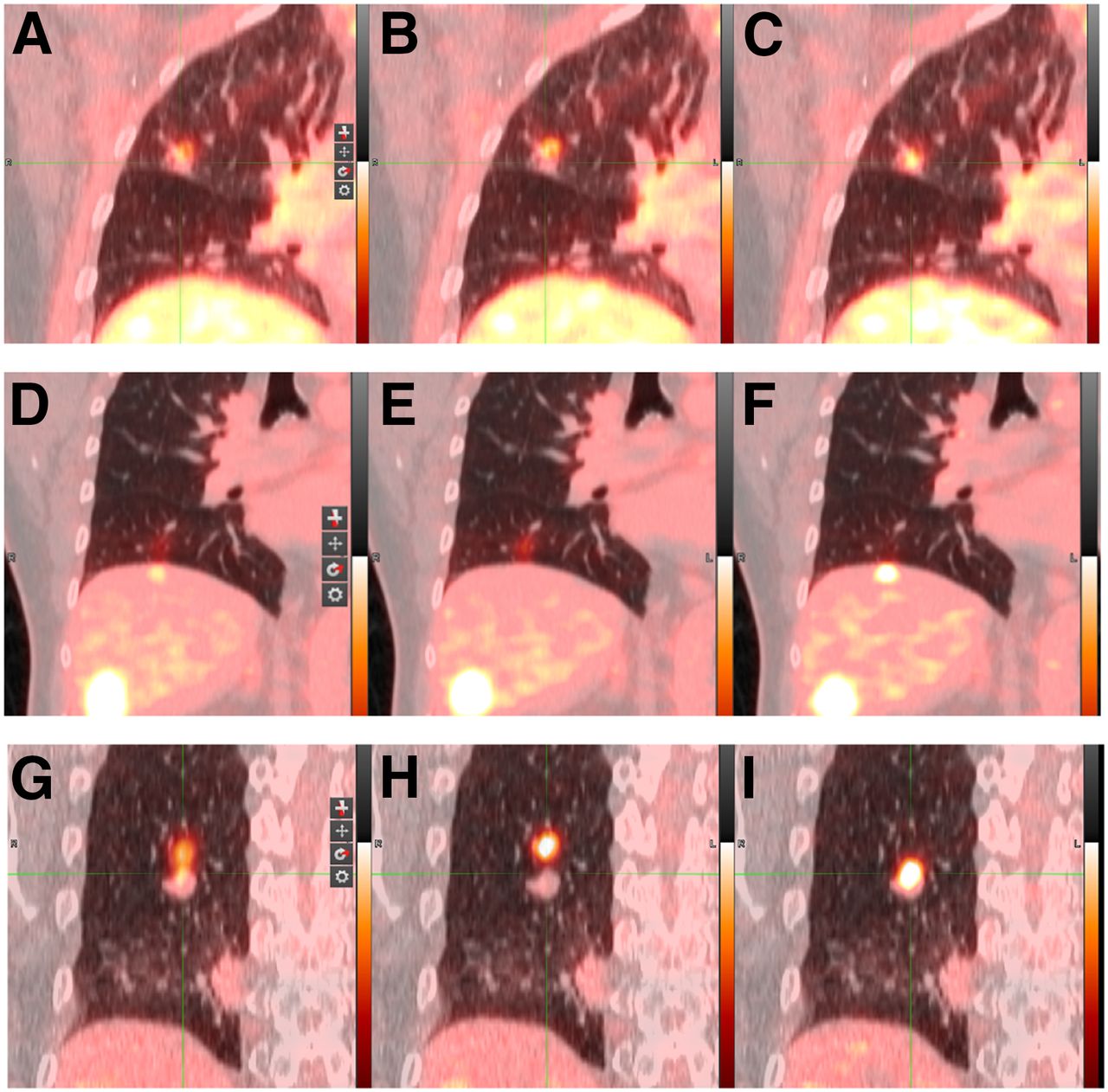
- FIGURE 3.
Coronal PET/CT images from 3 patient scans. Static (A, D, and G), PET/ex (B, E, and H), and PET/matched (C, F, and I) reconstructions are shown.


Tables
Parameter n PET/static PET/ex PET/matched PET/CT distance (mm) CT/ex 8 2.2 ± 3.8 (7.4) 2.9 ± 4.5 (9.0) 3.1 ± 3.9 (10.7) CT/in 18 3.5 ± 4.9 (11.1) 5.1 ± 5.2 (17.0) 2.8 ± 4.0 (8.3) Relative to static image SUVmax 40 — 1.21 ± 0.17 (1.56) 1.21 ± 0.26 (1.82) SUVpeak 40 — 1.12 ± 0.11 (1.45) 1.12 ± 0.22 (2.02) Liver noise 28 — 1.15 ± 0.15 (1.41) 1.40 ± 0.33 (2.39) Data are average ± SD, followed by maximum in parentheses.
Observation Rate PET/static was aligned with CT/in 13/21 PET/ex was aligned with CT/in 13/23 PET/matched was aligned with CT/in 18/21 PET/static was aligned with CT/in but PET/ex was not 1/10 PET/static was aligned with CT/in but PET/matched was not 0/3 PET/ex was aligned with CT/in but PET/static was not 0/8 PET/ex was aligned with CT/in but PET/matched was not 0/3 PET/matched was aligned with CT/in but PET/static was not 6/8 PET/matched was aligned with CT/in but PET/ex was not 7/10


Jump to section
Related Articles
Cited By...
- No citing articles found.