


Article Figures & Data
Figures
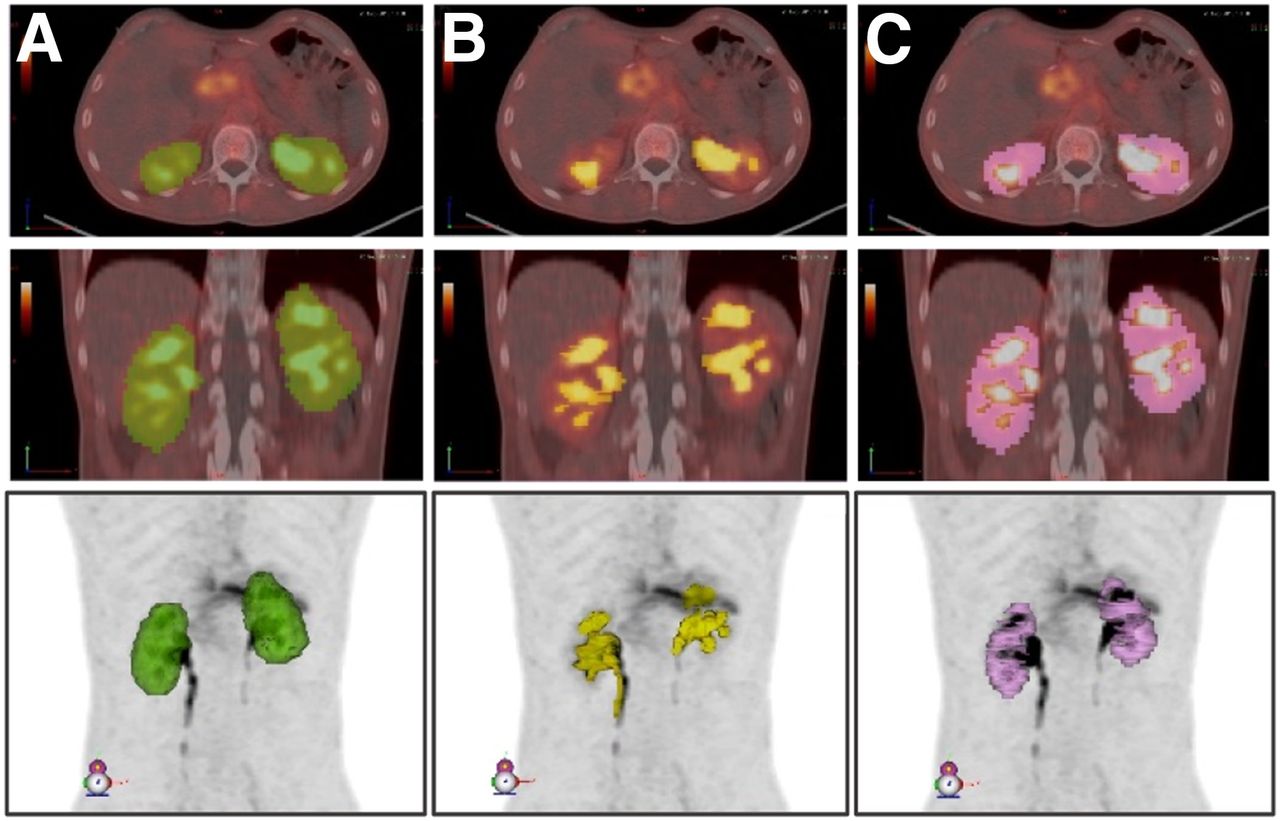
- FIGURE 1.
Delineated volumes used for determination of renal cortex volume: entire kidney volume (A) from which urine, including in renal calyces, is subtracted (B) to yield renal cortex volume (C). Images are shown for patient 6 and are representative of method applied for all patients. All volumes are shown in axial (top), coronal (middle), and maximum-intensity-projection views (bottom).
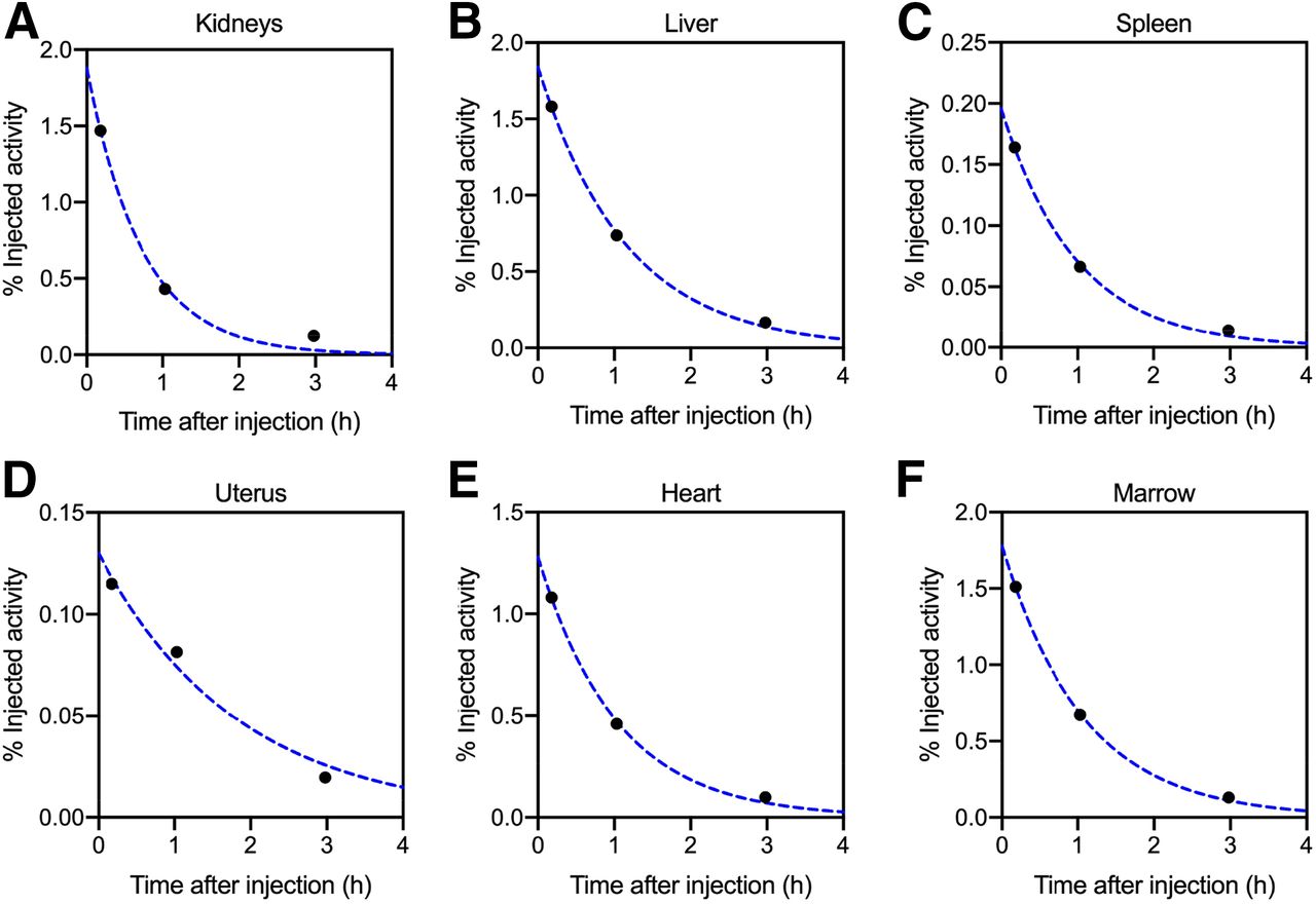
- FIGURE 2.
Percentage injected activity curves for patient 3 are shown for various source organs. Solid circles are measured values, and dotted lines are monoexponential functions fit to data.
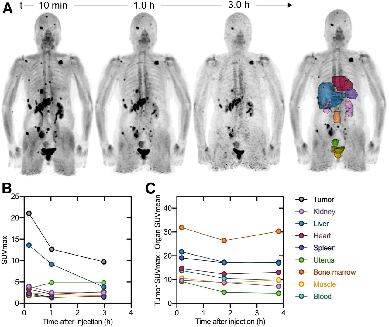
- FIGURE 3.
Patient 3 (female). (A) 68Ga-FAPI-46 maximum-intensity projections and delineated organs for dose calculations. (B) SUVmax at 3 time points after tracer injection. (C) TBR at 3 time points after tracer injection. SUVmax and TBR for bladder are excluded from plot. Data values are available in Supplemental Table 1.
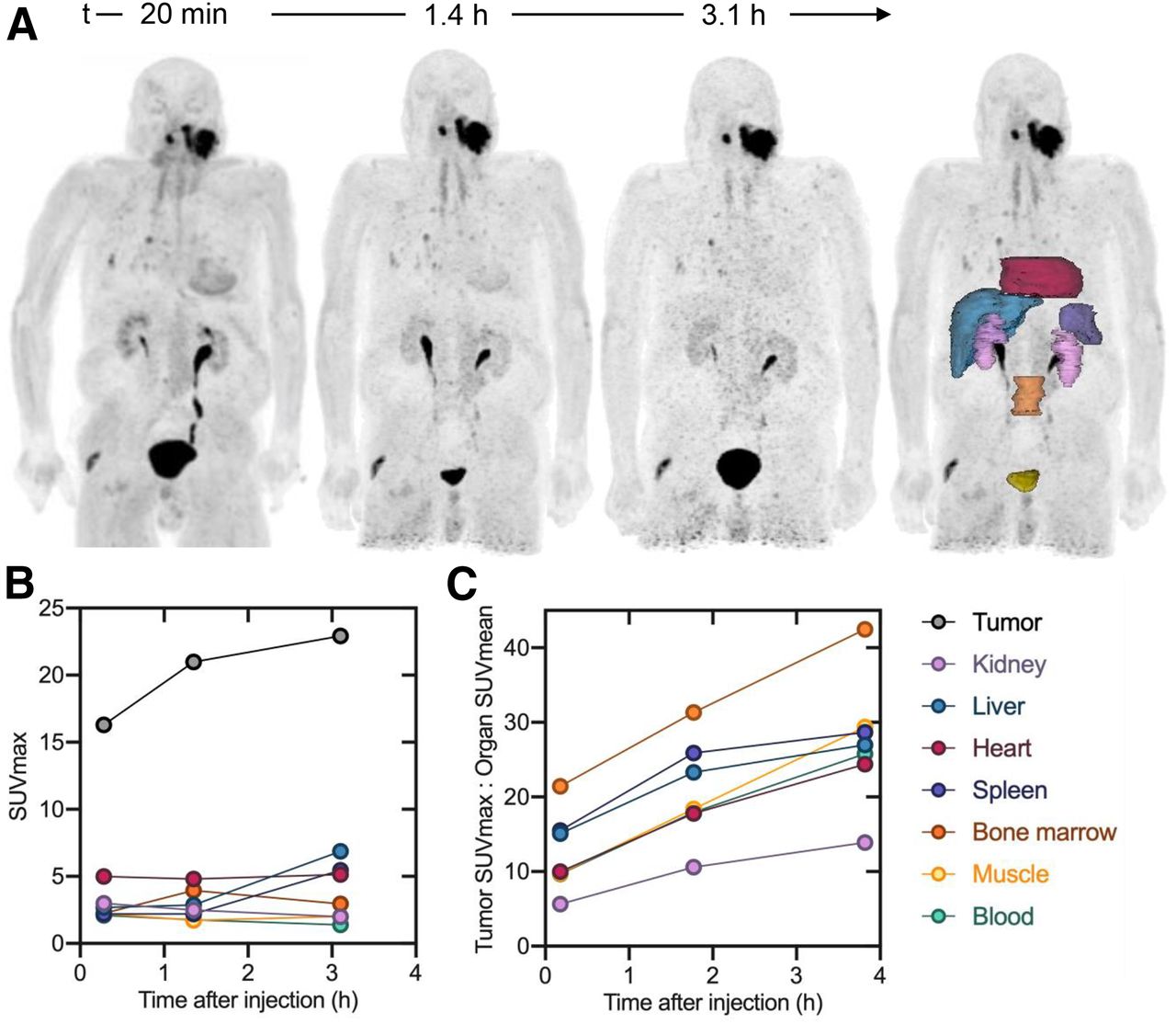
- FIGURE 4.
Patient 5 (male). (A) 68Ga-FAPI-46 maximum-intensity projection and delineated organs for dose calculations. (B) SUVmax at 3 time points after tracer injection. (C) TBR at 3 time points after tracer injection. SUVmax and TBR for bladder are excluded from plot. Data values are available in Supplemental Table 1.
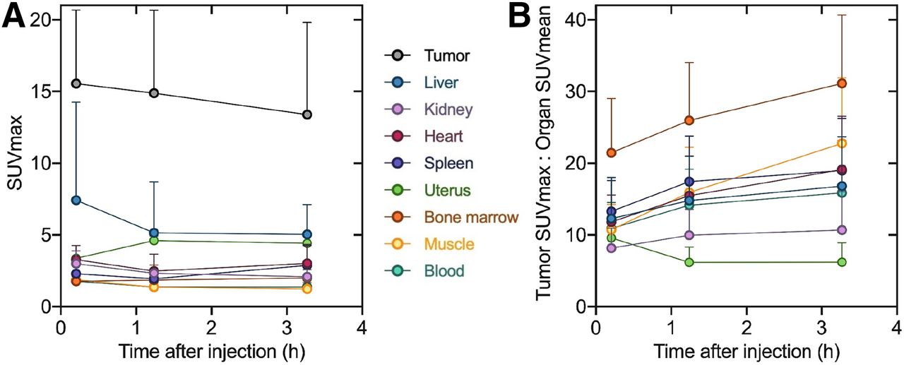
- FIGURE 5.
Pooled tumor and organ SUVmax (A) and TBR (B) at 3 time points after tracer injection (excluding bladder). Results are shown as mean and SD for 6 patients. Data values are available in Table 4.


Tables
Patient no. Sex Age (y) Diagnosis Injected activity (MBq) 1 F 63 Cholangiocellular carcinoma 246 2 M 81 Pancreatic cancer with peritonitis carcinomatosa 240 3 F 78 Breast cancer 234 4 M 56 Oropharynx carcinoma 239 5 M 78 Head and neck cancer 214 6 M 62 Gastric cancer 243 - TABLE 2
Monoexponential Function Fitting Parameters and Time-Integrated Activity Coefficients (Residence Times) for 68Ga-FAPI-46 in Various Organs
Organ A (%IA) λ (h−1) TIAC (h) Liver 3.49 (2.26) 0.88 (0.12) 0.0378 (0.0198) Kidney 2.07 (0.65) 1.08 (0.26) 0.0195 (0.0062) Bladder 6.82 (2.32) 1.47 (0.91) 0.0595 (0.0319) Heart 1.69 (0.30) 0.94 (0.06) 0.0182 (0.0035) Spleen 0.71 (0.62) 0.96 (0.12) 0.0074 (0.0066) Marrow 2.61 (0.63) 2.05 (2.97) 0.0250 (0.0114) Uterus (n = 2) 0.13 (0.004) 0.50 (0.07) 0.0027 (0.0005) A = activity, expressed as %IA = percentage injected activity;
 ; λ = rate constant; TIAC = time-integrated activity coefficient.
Data are mean followed by SD in parentheses for 6 patients. Representative percentage injected activity curves with monoexponential curve fits overlaid are available in Supplemental Figure 1. Per-patient coefficients and TIACs are available in Supplemental Table 2.
- TABLE 3
68Ga-FAPI-46 Dosimetry Summary of Mean Absorbed and Effective Doses Using OLINDA/EXM
Organ Dose per injected activity (mGy/MBq) Effective dose per injected activity (mSv/MBq) Adrenals 5.60E−03 (8.12E−04) 2.80E−05 (4.04E−06) Brain 4.59E−03 (6.12E−04) 2.29E−05 (3.06E−06) Breasts 4.55E−03 (6.47E−04) 2.28E−04 (3.23E−05) Gallbladder wall 5.62E−03 (8.53E−04) — Lower large intestine wall 5.72E−03 (6.96E−04) 6.86E−04 (8.33E−05) Small intestine 5.48E−03 (6.37E−04) 2.74E−05 (3.20E−06) Stomach wall 5.32E−03 (7.25E−04) 6.38E−04 (8.69E−05) Upper large intestine wall 5.47E−03 (6.97E−04) 2.74E−05 (3.50E−06) Heart wall 1.11E−02 (1.26E−03) — Kidneys 1.60E−02 (4.60E−03) 7.98E−05 (2.29E−05) Liver 1.01E−02 (7.96E−03) 5.05E−04 (4.00E−04) Lungs 5.02E−03 (7.09E−04) 6.02E−04 (8.48E−05) Muscle 4.96E−03 (6.54E−04) 2.48E−05 (3.27E−06) Ovaries 5.76E−03 (6.91E−04) 1.15E−03 (1.38E−04) Pancreas 5.69E−03 (8.49E−04) 2.84E−05 (4.24E−06) Red marrow 7.08E−03 (1.00E−03) 8.49E−04 (1.20E−04) Osteogenic cells 9.38E−03 (1.30E−03) 9.38E−05 (1.30E−05) Skin 4.41E−03 (6.33E−04) 4.41E−05 (6.33E−06) Spleen 6.96E−03 (2.76E−03) 3.48E−05 (1.39E−05) Testes 4.88E−03 (6.69E−04) 1.15E−03 (1.38E−04) Thymus 5.10E−03 (6.40E−04) 2.55E−05 (3.21E−06) Thyroid 4.84E−03 (5.72E−04) 2.42E−04 (2.85E−05) Urinary bladder wall 4.83E−02 (8.55E−03) 2.41E−03 (4.27E−04) Uterus 9.54E−03 (5.36E−03) 4.76E−05 (2.67E−05) Total body 5.82E−03 (1.18E−03) 7.80E−03 (1.31E−03) Total body dose for 200 MBq 1.16 mGy (0.24 mGy) 1.56 mSv (0.26 mSv) Data are mean followed by SD in parentheses for 6 patients. Effective doses in ovaries and testes are equivalent because of use of hermaphroditic adult phantom weighting. Gallbladder wall and heart wall effective doses are not available based on ICRP radiation weighting factors. Nonpooled OLINDA/EXM reports, including β and photon contribution to total dose, are available in Supplemental Table 3.
- TABLE 4
Pooled Tumor-to-Organ SUV Ratio, SUVmax, and SUVmean at 3 Time Points After 68Ga-FAPI-46 Administration
Parameter 10 min 1.2 h 3.3 h SUVmax-to-SUVmean ratio Tumor-to-liver 12.28 (5.75) 14.79 (6.22) 16.80 (6.90) Tumor-to-kidney 8.17 (2.76) 9.96 (3.64) 10.68 (5.34) Tumor-to-blood 10.89 (3.61) 14.15 (5.02) 15.87 (7.30) Tumor-to-muscle 10.71 (3.56) 15.91 (6.33) 22.77 (9.15) Tumor-to-heart 11.78 (3.76) 15.48 (5.49) 19.11 (7.44) Tumor-to-spleen 13.27 (4.31) 17.44 (6.36) 18.99 (7.27) Tumor-to-marrow 21.46 (7.56) 25.96 (8.09) 31.13 (9.52) Tumor-to-uterus (n = 2) 9.60 (0.46) 6.19 (2.12) 6.20 (2.70) SUVmax Tumor 15.54 (5.13) 14.89 (5.77) 13.39 (6.44) Liver 7.42 (6.84) 5.15 (3.56) 5.04 (2.07) Kidney 3.00 (0.89) 2.33 (0.26) 2.08 (0.49) Blood 1.77 (0.35) 1.38 (0.26) 1.37 (0.31) Muscle 1.87 (0.49) 1.37 (0.45) 1.24 (0.56) Heart 3.28 (1.00) 2.48 (1.17) 3.02 (1.31) Spleen 2.30 (0.23) 1.94 (0.42) 2.88 (1.38) Marrow 1.77 (0.34) 1.85 (1.05) 2.00 (0.59) Uterus (n = 2) 3.37 (0.23) 4.61 (0.32) 4.42 (0.58) SUVmean Tumor 3.87 (1.30) 3.37 (1.11) 2.81 (1.44) Liver 1.51 (0.96) 1.10 (0.58) 0.81 (0.34) Kidney 1.92 (0.34) 1.47 (0.17) 1.25 (0.22) Blood 1.43 (0.25) 1.03 (0.15) 0.83 (0.15) Muscle 1.48 (0.41) 0.96 (0.32) 0.61 (0.24) Heart 1.32 (0.21) 0.95 (0.15) 0.68 (0.15) Spleen 1.17 (0.20) 0.85 (0.17) 0.69 (0.16) Marrow 0.73 (0.12) 0.56 (0.11) 0.41 (0.10) Uterus (n = 2) 2.08 (0.27) 2.53 (0.24) 1.97 (0.42) Data are mean followed by SD in parentheses for 6 patients.

Supplemental Data
Files in this Data Supplement:


Jump to section
Related Articles
Cited By...
- Evaluation of Fibroblast Activation Protein Expression Using 68Ga-FAPI46 PET in Hypertension-Induced Tissue Changes
- Acquisition Duration Optimization Using Visual Grading Regression in [68Ga]FAPI-46 PET Imaging of Oncologic Patients
- Prognostic Implications of 68Ga-FAPI-46 PET/CT-Derived Parameters on Overall Survival in Various Types of Solid Tumors
- 68Ga-FAP-2286 PET of Solid Tumors: Biodistribution, Dosimetry, and Comparison with 18F-FDG
- Characterizing Normal Variant [68Ga]Ga-FAPI-46 Uptake in the Epididymis
- Incidental Focal 68Ga-FAPI-46 Uptake in a Urachal Remnant: A Potential Pitfall Mimicking a Malignant Peritoneal Lesion
- Could FAP-Targeted Molecular Imaging Replace 18F-FDG for Standard-of-Care Oncologic PET?
- Three-Time-Point PET Analysis of 68Ga-FAPI-46 in a Variety of Cancers
- PET Imaging of Fibroblast Activation Protein in Various Types of Cancer Using 68Ga-FAP-2286: Comparison with 18F-FDG and 68Ga-FAPI-46 in a Single-Center, Prospective Study
- A Dimeric FAP-Targeting Small-Molecule Radioconjugate with High and Prolonged Tumor Uptake
- Dual-Tracer PET/CT Protocol with [18F]-FDG and [68Ga]Ga-FAPI-46 for Cancer Imaging: A Proof of Concept
- Synthesis, Preclinical Evaluation, and a Pilot Clinical PET Imaging Study of 68Ga-Labeled FAPI Dimer
- Initial Clinical Experience with 90Y-FAPI-46 Radioligand Therapy for Advanced-Stage Solid Tumors: A Case Series of 9 Patients
- An ultra-high-affinity small organic ligand of fibroblast activation protein for tumor-targeting applications
- FAPI PET/CT: Will It End the Hegemony of 18F-FDG in Oncology?
- The Latest Developments in Imaging of Fibroblast Activation Protein
- Fibroblast Activation Protein-Targeted PET/CT with 68Ga-FAPI for Imaging IgG4-Related Disease: Comparison to 18F-FDG PET/CT
- FAPI-74 PET/CT Using Either 18F-AlF or Cold-Kit 68Ga Labeling: Biodistribution, Radiation Dosimetry, and Tumor Delineation in Lung Cancer Patients