
Main menu

  • Home
  • Content
    • Current
    • Ahead of print
    • Past Issues
    • JNM Supplement
    • SNMMI Annual Meeting Abstracts
    • Continuing Education
    • JNM Podcasts
  • Subscriptions
    • Subscribers
    • Institutional and Non-member
    • Rates
    • Journal Claims
    • Corporate & Special Sales
  • Authors
    • Submit to JNM
    • Information for Authors
    • Assignment of Copyright
    • AQARA requirements
  • Info
    • Reviewers
    • Permissions
    • Advertisers
  • About
    • About Us
    • Editorial Board
    • Contact Information
  • More
    • Alerts
    • Feedback
    • Help
    • SNMMI Journals
  • SNMMI
    • JNM
    • JNMT
    • SNMMI Journals
    • SNMMI

User menu

  • Subscribe
  • My alerts
  • Log in
  • My Cart

Search

  • Advanced search
Journal of Nuclear Medicine
  • SNMMI
    • JNM
    • JNMT
    • SNMMI Journals
    • SNMMI
  • Subscribe
  • My alerts
  • Log in
  • My Cart
Journal of Nuclear Medicine

Advanced Search

  • Home
  • Content
    • Current
    • Ahead of print
    • Past Issues
    • JNM Supplement
    • SNMMI Annual Meeting Abstracts
    • Continuing Education
    • JNM Podcasts
  • Subscriptions
    • Subscribers
    • Institutional and Non-member
    • Rates
    • Journal Claims
    • Corporate & Special Sales
  • Authors
    • Submit to JNM
    • Information for Authors
    • Assignment of Copyright
    • AQARA requirements
  • Info
    • Reviewers
    • Permissions
    • Advertisers
  • About
    • About Us
    • Editorial Board
    • Contact Information
  • More
    • Alerts
    • Feedback
    • Help
    • SNMMI Journals
  • View or Listen to JNM Podcast
  • Visit JNM on Facebook
  • Join JNM on LinkedIn
  • Follow JNM on Twitter
  • Subscribe to our RSS feeds
Research ArticleOncology

Can Intraoperative Fluorescence Imaging Identify All Lesions While the Road Map Created by Preoperative Nuclear Imaging Is Masked?

Phillipa Meershoek, Tessa Buckle, Matthias N. van Oosterom, Gijs H. KleinJan, Henk G. van der Poel and Fijs W.B. van Leeuwen
Journal of Nuclear Medicine June 2020, 61 (6) 834-841; DOI: https://doi.org/10.2967/jnumed.119.235234
Phillipa Meershoek
1Interventional Molecular Imaging Laboratory, Department of Radiology, Leiden University Medical Center, Leiden, The Netherlands; and
  • Find this author on Google Scholar
  • Find this author on PubMed
  • Search for this author on this site
Tessa Buckle
1Interventional Molecular Imaging Laboratory, Department of Radiology, Leiden University Medical Center, Leiden, The Netherlands; and
2Department of Urology, Netherlands Cancer Center and Antoni van Leeuwenhoek Hospital, Amsterdam, The Netherlands
  • Find this author on Google Scholar
  • Find this author on PubMed
  • Search for this author on this site
Matthias N. van Oosterom
1Interventional Molecular Imaging Laboratory, Department of Radiology, Leiden University Medical Center, Leiden, The Netherlands; and
2Department of Urology, Netherlands Cancer Center and Antoni van Leeuwenhoek Hospital, Amsterdam, The Netherlands
  • Find this author on Google Scholar
  • Find this author on PubMed
  • Search for this author on this site
Gijs H. KleinJan
1Interventional Molecular Imaging Laboratory, Department of Radiology, Leiden University Medical Center, Leiden, The Netherlands; and
2Department of Urology, Netherlands Cancer Center and Antoni van Leeuwenhoek Hospital, Amsterdam, The Netherlands
  • Find this author on Google Scholar
  • Find this author on PubMed
  • Search for this author on this site
Henk G. van der Poel
2Department of Urology, Netherlands Cancer Center and Antoni van Leeuwenhoek Hospital, Amsterdam, The Netherlands
  • Find this author on Google Scholar
  • Find this author on PubMed
  • Search for this author on this site
Fijs W.B. van Leeuwen
1Interventional Molecular Imaging Laboratory, Department of Radiology, Leiden University Medical Center, Leiden, The Netherlands; and
2Department of Urology, Netherlands Cancer Center and Antoni van Leeuwenhoek Hospital, Amsterdam, The Netherlands
  • Find this author on Google Scholar
  • Find this author on PubMed
  • Search for this author on this site
  • Article
  • Figures & Data
  • Info & Metrics
  • PDF
Loading

Article Figures & Data

Figures

  • Tables
  • FIGURE 1.
    • Download figure
    • Open in new tab
    • Download powerpoint
    FIGURE 1.

    Overview of intraoperative SN removal and tumor-positive SNs.
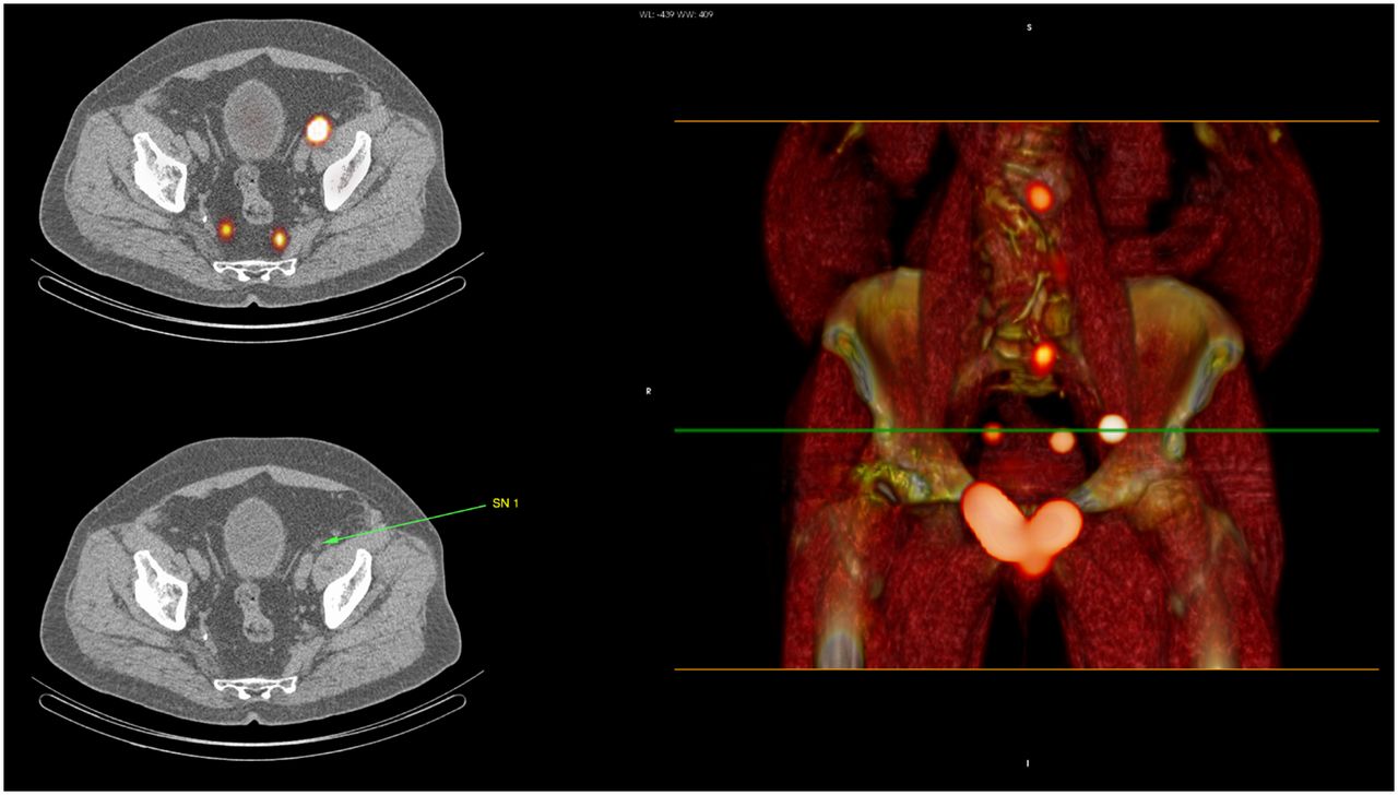
  • FIGURE 2.
    • Download figure
    • Open in new tab
    • Download powerpoint
    FIGURE 2.

    Location of iliac SN resected after unmasking of surgeon to SPECT/CT.

Tables

  • Figures
    • View popup
    TABLE 1

    Patient Characteristics

    CharacteristicAll patients (n = 25)Hybrid-tracer group (n = 15)Free-ICG group (n = 10)P
    Age at surgery (y)65.4 (6.1)64.3 (8.4)67.4 (4.4)0.247
    PSA at diagnosis8.6 (3.9)9.1 (4.6)7.7 (2.3)0.383
    PSA range at diagnosis (ng/mL)2.7–22.52.7–22.54.31–12.04
    Clinical stage0.870
     cT1c541
     cT2a422
     cT2b312
     cT2c743
     cT3a532
     cT3b110
    Biopsy Gleason sum1.000
     6211
     717107
     8321
     9321
    Prostate volume (mL)50.2 (21)48.1 (20.0)52.4 (25.8)0.641
    Briganti score16.6% (16.1)16.6 (15.6)16.6 (17.8)0.996
    Pathologic stage0.596
     pT2a413
     pT2c1495
     pT3a532
     pT3b110
     pT4110
    Pathologic Gleason sum0.194
     6330
     71578
     8330
     9222
    Nodal status1.00
     pN019118
     pN1642
    • Qualitative data are expressed as numbers; continuous data are expressed as mean followed by SD in parentheses.

    • View popup
    TABLE 2

    Results for Identified and Removed SNs and LNs: SPECT/CT and Ex Vivo Imaging

    ParameterAll patients (n = 25)Hybrid-tracer group (n = 15)Free-ICG group (n = 10)
    SPECT/CT
     Total identified SNs1115754
     Mean SNs per patient4.4 (SD, 2.5)3.8 (SD, 1.8)5.4 (SD, 3.0)
     SNs inside ePLND template82 (73.9%)50 (87.7%)32 (59.3%)
      External iliac261610
      Internal iliac21138
      Obturator fossa352114
     SNs outside ePLND template29 (26.1%)7 (12.3%)22 (40.7%)
      Common iliac1248
      Paravesical330
      Presacral505
      Pararectal606
      Inguinal303
    • Data are numbers.

    • View popup
    TABLE 3

    Results for Identified and Removed SNs and LNs: Surgery

    ParameterAll patients (n = 25)Hybrid-tracer group (n = 15)Free-ICG group (n = 10)
    Resected SNs1145856
    SNs removed under  fluorescence guidance73 (64.0%)44 (75.9%)29 (51.8%)
    SNs per patient4.6 (SD, 3.0)3.8 (SD, 2.0)5.6 (SD, 3.8)
    Fluorescent SNs removed  inside ePLND template56 (79.5%)34 (77.3%)22 (75.9%)
     Externa iliac20128
     Internal iliac1174
     Obturator fossa251510
    Fluorescent SNs removed  outside ePLND template17 (23.3%)10 (29.4%)7 (24.1%)
     Common iliac220
     Marcille000
     Cloquet000
     Pararectal505
     Presacral202
     Umbilical ligament880
     Aorta bifurcation000
     Undefined000
    SNs resected during eLNPD22 (19.3%)7 (12.1%)15 (26.8%)
    SNs identified after unmasking of SPECT18 (15.8%)6 (10.3%)12 (21.4%)
    SNs removed after unmasking1—1
    SNs not removed after unmasking17710
    Location
     Inside ePLND template (internal iliac)321
     Outside ePLND template
      Common iliac431
      Pararectal826
      Inguinal2—2
    Non-SNs removed under fluorescence  guidance21813
    Ex vivo imaging
     SNs that were fluorescent but  not radioactive14014
     Patients7 (28%)0 (0%)7 (70%)
    • Data are numbers.

    • View popup
    TABLE 4

    Results for Identified and Removed SNs and LNs: Pathology

    ParameterAll patients (n = 25)Hybrid-tracer group (n = 15)Free-ICG group (n = 10)
    Tumor-positive SNs
     Nodes1091
     Patients641
    Non-SNs removed320177143
    Tumor-positive nodes in non-SNs
     Nodes321
     Patients321
    Tumor-positive nodes inside ePLND template
     External iliac55*
     Internal iliac22
     Obturator fossa321†
    Tumor-positive nodes outside ePLND template
     Marcille11
     Cloquet11
     Pararectal11
    • ↵* 2 non-SNs.

    • ↵† 1 non-SN.

    • Data are numbers.

PreviousNext
Back to top

In this issue

Journal of Nuclear Medicine: 61 (6)
Journal of Nuclear Medicine
Vol. 61, Issue 6
June 1, 2020
  • Table of Contents
  • Table of Contents (PDF)
  • About the Cover
  • Index by author
Print
Download PDF
Article Alerts
Sign In to Email Alerts with your Email Address
Email Article

Thank you for your interest in spreading the word on Journal of Nuclear Medicine.

NOTE: We only request your email address so that the person you are recommending the page to knows that you wanted them to see it, and that it is not junk mail. We do not capture any email address.

Enter multiple addresses on separate lines or separate them with commas.
Can Intraoperative Fluorescence Imaging Identify All Lesions While the Road Map Created by Preoperative Nuclear Imaging Is Masked?
(Your Name) has sent you a message from Journal of Nuclear Medicine
(Your Name) thought you would like to see the Journal of Nuclear Medicine web site.
Citation Tools
Can Intraoperative Fluorescence Imaging Identify All Lesions While the Road Map Created by Preoperative Nuclear Imaging Is Masked?
Phillipa Meershoek, Tessa Buckle, Matthias N. van Oosterom, Gijs H. KleinJan, Henk G. van der Poel, Fijs W.B. van Leeuwen
Journal of Nuclear Medicine Jun 2020, 61 (6) 834-841; DOI: 10.2967/jnumed.119.235234

Citation Manager Formats

  • BibTeX
  • Bookends
  • EasyBib
  • EndNote (tagged)
  • EndNote 8 (xml)
  • Medlars
  • Mendeley
  • Papers
  • RefWorks Tagged
  • Ref Manager
  • RIS
  • Zotero
Share
Can Intraoperative Fluorescence Imaging Identify All Lesions While the Road Map Created by Preoperative Nuclear Imaging Is Masked?
Phillipa Meershoek, Tessa Buckle, Matthias N. van Oosterom, Gijs H. KleinJan, Henk G. van der Poel, Fijs W.B. van Leeuwen
Journal of Nuclear Medicine Jun 2020, 61 (6) 834-841; DOI: 10.2967/jnumed.119.235234
Twitter logo Facebook logo LinkedIn logo Mendeley logo
  • Tweet Widget
  • Facebook Like
  • Google Plus One
Bookmark this article

Jump to section

  • Article
    • Abstract
    • MATERIALS AND METHODS
    • RESULTS
    • DISCUSSION
    • CONCLUSION
    • DISCLOSURE
    • Acknowledgments
    • Footnotes
    • REFERENCES
  • Figures & Data
  • Info & Metrics
  • PDF

Related Articles

  • This Month in JNM
  • PubMed
  • Google Scholar

Cited By...

  • Optical Navigation of the Drop-In {gamma}-Probe as a Means to Strengthen the Connection Between Robot-Assisted and Radioguided Surgery
  • Google Scholar

More in this TOC Section

Oncology

  • Comparative PSMA expression at early (1hour) vs late (2hour) in primary and secondary sites of involvement in prostate cancer.
  • Imaging spectrum of peritoneal carcinomatosis associated with various etiologies on 18F-FDG PET/CT
  • Hybrid Imaging Features of Musculoskeletal Tumors
Show more Oncology

Clinical

  • Addition of 131I-MIBG to PRRT (90Y-DOTATOC) for Personalized Treatment of Selected Patients with Neuroendocrine Tumors
  • SUVs Are Adequate Measures of Lesional 18F-DCFPyL Uptake in Patients with Low Prostate Cancer Disease Burden
  • Hypermetabolism on Pediatric PET Scans of Brain Glucose Metabolism: What Does It Signify?
Show more Clinical

Similar Articles

Keywords

  • image-guided surgery
  • prostate cancer
  • sentinel node biopsy
  • SPECT/CT
  • robot-assisted surgery
  • fluorescence
SNMMI

© 2025 SNMMI

Powered by HighWire