
Main menu

  • Home
  • Content
    • Current
    • Ahead of print
    • Past Issues
    • JNM Supplement
    • SNMMI Annual Meeting Abstracts
    • Continuing Education
    • JNM Podcasts
  • Subscriptions
    • Subscribers
    • Institutional and Non-member
    • Rates
    • Journal Claims
    • Corporate & Special Sales
  • Authors
    • Submit to JNM
    • Information for Authors
    • Assignment of Copyright
    • AQARA requirements
  • Info
    • Reviewers
    • Permissions
    • Advertisers
  • About
    • About Us
    • Editorial Board
    • Contact Information
  • More
    • Alerts
    • Feedback
    • Help
    • SNMMI Journals
  • SNMMI
    • JNM
    • JNMT
    • SNMMI Journals
    • SNMMI

User menu

  • Subscribe
  • My alerts
  • Log in
  • Log out
  • My Cart

Search

  • Advanced search
Journal of Nuclear Medicine
  • SNMMI
    • JNM
    • JNMT
    • SNMMI Journals
    • SNMMI
  • Subscribe
  • My alerts
  • Log in
  • Log out
  • My Cart
Journal of Nuclear Medicine

Advanced Search

  • Home
  • Content
    • Current
    • Ahead of print
    • Past Issues
    • JNM Supplement
    • SNMMI Annual Meeting Abstracts
    • Continuing Education
    • JNM Podcasts
  • Subscriptions
    • Subscribers
    • Institutional and Non-member
    • Rates
    • Journal Claims
    • Corporate & Special Sales
  • Authors
    • Submit to JNM
    • Information for Authors
    • Assignment of Copyright
    • AQARA requirements
  • Info
    • Reviewers
    • Permissions
    • Advertisers
  • About
    • About Us
    • Editorial Board
    • Contact Information
  • More
    • Alerts
    • Feedback
    • Help
    • SNMMI Journals
  • View or Listen to JNM Podcast
  • Visit JNM on Facebook
  • Join JNM on LinkedIn
  • Follow JNM on Twitter
  • Subscribe to our RSS feeds
Research ArticleOncology

Prognostic Value of 18F-FDG PET/CT in a Large Cohort of Patients with Advanced Metastatic Neuroendocrine Neoplasms Treated with Peptide Receptor Radionuclide Therapy

Jingjing Zhang, Qingxing Liu, Aviral Singh, Christiane Schuchardt, Harshad R. Kulkarni and Richard P. Baum
Journal of Nuclear Medicine November 2020, 61 (11) 1560-1569; DOI: https://doi.org/10.2967/jnumed.119.241414
Jingjing Zhang
1Theranostics Center for Molecular Radiotherapy and Precision Oncology, ENETS Center of Excellence, Zentralklinik Bad Berka, Bad Berka, Germany
  • Find this author on Google Scholar
  • Find this author on PubMed
  • Search for this author on this site
Qingxing Liu
1Theranostics Center for Molecular Radiotherapy and Precision Oncology, ENETS Center of Excellence, Zentralklinik Bad Berka, Bad Berka, Germany
2Department of Nuclear Medicine, Peking Union Medical College Hospital, Chinese Academy of Medical Sciences and Peking Union Medical College, Beijing, China; and
  • Find this author on Google Scholar
  • Find this author on PubMed
  • Search for this author on this site
Aviral Singh
1Theranostics Center for Molecular Radiotherapy and Precision Oncology, ENETS Center of Excellence, Zentralklinik Bad Berka, Bad Berka, Germany
3GROW–School for Oncology and Developmental Biology, Maastricht University, Maastricht, the Netherlands
  • Find this author on Google Scholar
  • Find this author on PubMed
  • Search for this author on this site
Christiane Schuchardt
1Theranostics Center for Molecular Radiotherapy and Precision Oncology, ENETS Center of Excellence, Zentralklinik Bad Berka, Bad Berka, Germany
  • Find this author on Google Scholar
  • Find this author on PubMed
  • Search for this author on this site
Harshad R. Kulkarni
1Theranostics Center for Molecular Radiotherapy and Precision Oncology, ENETS Center of Excellence, Zentralklinik Bad Berka, Bad Berka, Germany
  • Find this author on Google Scholar
  • Find this author on PubMed
  • Search for this author on this site
Richard P. Baum
1Theranostics Center for Molecular Radiotherapy and Precision Oncology, ENETS Center of Excellence, Zentralklinik Bad Berka, Bad Berka, Germany
  • Find this author on Google Scholar
  • Find this author on PubMed
  • Search for this author on this site
  • Article
  • Figures & Data
  • Supplemental
  • Info & Metrics
  • PDF
Loading

Article Figures & Data

Figures

  • Tables
  • Additional Files
  • Figure1
    • Download figure
    • Open in new tab
    • Download powerpoint
  • FIGURE 1.
    • Download figure
    • Open in new tab
    • Download powerpoint
    FIGURE 1.

    Kaplan–Meier curves of OS and PFS for all patients (n = 495). (A) Median OS was 58.7 mo (95% CI, 52.8–64.6). (B) Median PFS was 19.6 mo (95% CI, 17.6–21.7).
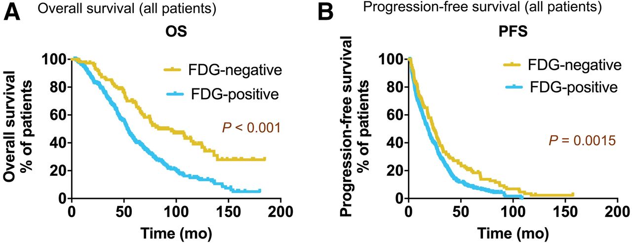
  • FIGURE 2.
    • Download figure
    • Open in new tab
    • Download powerpoint
    FIGURE 2.

    Kaplan–Meier survival analysis of all NEN patients (n = 495) stratified by baseline 18F-FDG status. Patients with 18F-FDG–negative lesions had significantly higher median OS (A) (83.1 mo vs. 53.2 mo, P < 0.001) and higher median PFS (B) (24.1 mo vs. 18.5 mo, P < 0.002) than patients with 18F-FDG–positive lesions.
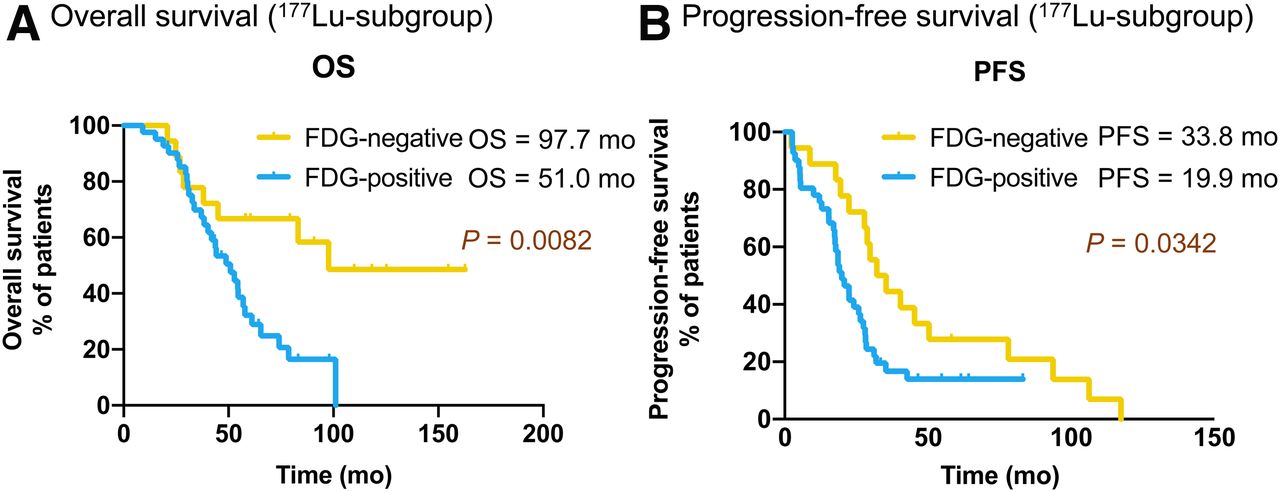
  • FIGURE 3.
    • Download figure
    • Open in new tab
    • Download powerpoint
    FIGURE 3.

    Kaplan–Meier curves of OS (A) and PFS (B) for 177Lu subgroup (n = 60) stratified by baseline 18F-FDG status.
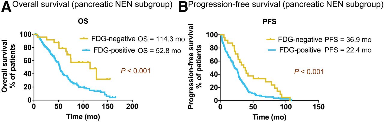
  • FIGURE 4.
    • Download figure
    • Open in new tab
    • Download powerpoint
    FIGURE 4.

    Kaplan–Meier survival of OS (A) and PFS (B) for pancreatic NEN subgroup (n = 199) stratified by baseline 18F-FDG status.

Tables

  • Figures
  • Additional Files
    • View popup
    TABLE 1

    Demographics of Patients with NENs (n = 495)

    DemographicData
    Sex
     Male299 (60.4)
     Female196 (39.6)
    Age (y)
     Median59.0 ± 10.7
     y ≤50111 (22.4)
     50< y ≤60146 (29.5)
     60< y ≤70165 (33.3)
     70< y ≤8073 (14.7)
    Primary tumor site
     Cancer of unknown primary49 (9.9)
     Lung38 (7.7)
     Midgut139 (28.1)
     Others42 (8.5)
     Pancreas199 (40.2)
     Rectum20 (4.0)
     Stomach8 (1.6)
    Ki-67 index grading
     G1 (Ki-67 < 3%)117 (23.6)
     G2 (Ki-67 = 3%–20%)245 (49.5)
     G3 (Ki-67 > 20%)29 (5.9)
     Not assessed104 (21.0)
    • Qualitative data are numbers followed by percentages in parentheses; continuous data are medians.

    • View popup
    TABLE 2

    Baseline 68Ga-SSTR and 18F-FDG PET Imaging of Patients with NENs

    ParameterData
    68Ga-SSTR uptake495 (100.0)
     SUVmax = liver (level 1)3 (0.6)
     Liver<SUVmax≤15 (level 2)161 (32.5)
     15<SUVmax≤20 (level 3)106 (21.4)
     SUVmax>20 (level 4)225 (45.5)
    18F-FDG uptake495 (100.0)
     Positive382 (77.2)
     Negative113 (22.8)
    Primary tumor uptake on 18F-FDG PET
     No uptake (level 1)376 (76.0)
     SUVmax≤10 (level 2)87 (17.6)
     10<SUVmax≤15 (level 3)16 (3.2)
     SUVmax>15 (level 4)16 (3.20)
    Primary tumor uptake on 68Ga-SSTR
     No uptake (level 1)262 (52.9)
     SUVmax≤15 (level 2)102 (20.6)
     15<SUVmax≤20 (level 3)37 (7.5)
     SUVmax>20 (level 4)94 (19.0)
    Liver tumor burden on 18F-FDG PET
     0 lesions (level 1)239 (48.3)
     1 lesion (level 2)49 (9.9)
     2 to ≤5 lesions (level 3)126 (25.5)
     >5 lesions (level 4)77 (15.6)
     Not assessed4 (0.8)
    Bone tumor burden on 18F-FDG PET
     0 lesions (level 1)409 (82.6)
     1 lesion (level 2)29 (5.9)
     2 to ≤5 lesions (level 3)36 (7.3)
     >5 lesions (level 4)19 (3.8)
     Not assessed2 (0.4)
    Lymph node tumor burden on 18F-FDG PET
     0 lesions (level 1)362 (73.1)
     1 lesion (level 2)55 (11.1)
     2 to ≤5 lesions (level 3)58 (11.7)
     >5 lesions (level 4)13 (2.6)
     Not assessed7 (1.4)
    Lung tumor burden on 18F-FDG PET
     0 lesions (level 1)463 (93.5)
     1 lesion (level 2)22 (4.4)
     2 to ≤5 lesions (level 3)6 (1.2)
     >5 lesions (level 4)4 (0.8)
    Liver tumor burden on 68Ga-SSTR PET
     0 lesions (level 1)76 (15.4)
     1 lesion (level 2)32 (6.5)
     2 to ≤5 lesions (level 3)153 (30.9)
     >5 lesions (level 4)234 (47.3)
    Liver tumor 68Ga-SSTR uptake
     No uptake (level 1)77 (15.6)
     SUVmax≤15 (level 2)117 (23.6)
     15<SUVmax≤20 (level 3)90 (18.2)
     SUVmax>20 (level 4)211 (42.6)
    • Data are numbers followed by percentages in parentheses.

    • View popup
    TABLE 3

    Univariate and Multivariate Analyses of Potential Factors Contributing to OS, Part 1

    OS (mo)Multivariate analysis
    FactorsMedian95% CIUnivariate analysis (P)HR and 95% CIP
    All patients58.752.8–64.6
    Sex
     Male53.747.6–59.80.040
     Female66.154.4–77.8
    Age (y)
     y ≤5069.861.7–77.80.024
     50< y ≤6061.651.5–71.7
     60< y ≤7053.049.8–56.3
     70< y ≤8049.039.6–58.5
    Grading
     Grade 178.566.2–90.8<0.0010.012
     Grade 255.446.9–63.91.4 (1.0–2.0)0.038
     Grade 333.218.8–7.62.5 (1.3–4.5)0.004
     Not assessed54.148.3–59.91.6 (1.1–2.2)0.009
    18F-FDG PET uptake
     Positive53.249.4–57.0<0.0010.002
     Negative83.157.0–109.20.5 (0.3–0.8)
    Primary tumor site
     Cancer of unknown primary65.147.4–82.70.0070.004
     Lung46.234.1–58.30.3 (0.1–0.7)0.004
     Midgut77.861.0–94.60.7 (0.3–1.6)0.344
     Others65.731.3–100.10.3 (0.1–0.7)0.008
     Pancreas54.449.3–59.60.4 (0.2–1.0)0.041
     Rectum55.450.3–60.40.5 (0.2–1.0)0.063
     Stomach46.933.3–60.50.6 (0.2–1.5)0.239
    • View popup
    TABLE 4

    Univariate and Multivariate Analyses of Potential Factors Contributing to OS, Part 2

    OS (mo)Multivariate analysis
    FactorsMedian95% CIUnivariate analysis (P)HR and 95% CIP
    Liver tumor burden on 18F-FDG PET
     0 lesions75.665.5–85.6<0.0010.034
     1 lesion55.436.9–73.91.7 (0.6–5.2)0.338
     2 to ≤5 lesions47.140.5–53.71.2 (0.4–3.8)0.712
     >5 lesions43.735.4–52.02.2 (0.7–6.4)0.157
     Not assessed54.633.7–75.52.3 (0.8–7.0)0.127
    Bone tumor burden on 18F-FDG PET
     0 lesions61.654.9–68.30.004
     1 lesion56.029.6–82.4
     2 to ≤5 lesions41.925.1–58.8
     >5 lesions43.419.2–67.7
     Not assessed32.6—
    Lymph node tumor burden on 18F-FDG PET
     0 lesions63.856.3–71.40.0060.035
     1 lesion51.634.6–68. 51.4 (0.5–4.1)0.540
     2 to ≤5 lesions46.237.2–55.31.7 (0.6–5.2)0.344
     >5 lesions37.410.0–64.71.8 (0.6–5.4)0.293
     Not assessed86.623.7–149.64.4 (1.3–15.3)0.018
    Grading of PRRT
     2 to ≤3 cycles33.2725.0–41.3<0.001<0.001
     4 to ≤5 cycles51.644.5–58.77.9 (3.9–15.9)<0.001
     6 to ≤7 cycles68.961.8–76.14.7 (2.6–8.4)<0.001
     8 to ≤10 cycles122.584.8–160.33.0 (1.8–5.0)<0.001
    Cumulative activity (GBq)
     Activity ≤1526.013.8–38.2<0.0010.038
     15< activity ≤2552.745.4–59.91.1 (0.6–2.0)0.745
     25< activity ≤3561.154.9–67.30.7 (0.4–1.1)0.084
     Activity >3577.866.0–89.60.8 (0.6–1.2)0.379
    • View popup
    TABLE 5

    Univariate and Multivariate Analyses of Potential Factors Contributing to PFS, Part 1

    PFS (mo)Multivariate analysis
    FactorsMedian95% CIUnivariate analysis (P)HR and 95% CIP
    All patients19.617.6–21.7
    Age
     y ≤5025.019.4–30.50.281
     50< y ≤6018.412.8–23.9
     60< y ≤7017.915.1–20.7
     70< y ≤8022.416.8–28.0
    Grading
     Grade 123.015.9–30.20.0030.039
     Grade 218.915.2–22.61.2 (0.9–1.5)0.150
     Grade 37.50.0–20.12.1 (1.3–3.4)0.003
     Not assessed19.813.8–25.71.1 (0.9–1.5)0.426
    18F-FDG PET uptake
     Positive18.515.9–21.10.0020.007
     Negative24.119.9–28.30.7 (0.5–0.9)
    Primary tumor site
     Cancer of unknown primary11.46.5–16.20.011
     Lung10.65.0–16.1
     Midgut22.617.2–28.0
     Others9.12.7–15.5
     Pancreas25.821.8–29.8
     Rectum19.911.8–27.9
     Stomach24.618.9–30.4
    • View popup
    TABLE 6

    Univariate and Multivariate Analyses of Potential Factors Contributing to PFS, Part 2

    PFS (mo)Multivariate analysis
    FactorsMedian95% CIUnivariate analysis (P)HR and 95% CIP
    Liver tumor burden on 18F-FDG PET
     0 lesions20.817.4–24.10.034
     1 lesion27.820.9–34.7
     2 to ≤5 lesions16.211.5–20.9
     >5 lesions17.914.1–21.7
     Not assessed28.20.0–58.7
    Bone tumor burden on 18F-FDG PET
     0 lesions22.419.3–25.5<0.0010.001
     1 lesion13.31.4–25.11.0 (0.2–4.1)0.982
     2 to ≤5 lesions10.58.4–12.51.7 (0.4–7.4)0.479
     >5 lesions11.50.0–24.92.2 (0.5–9.4)0.304
     Not assessed12.0—1.2 (0.3–5.2)0.848
    Lymph node tumor burden on 18F-FDG PET
     0 lesions21.618.6–24.7<0.0010.050
     1 lesion17.910.9–25.01.2 (0.5–3.1)0.587
     2 to ≤5 lesions15.412.3–18.61.3 (0.5–3.4)0.552
     >5 lesions6.54.5–8.51.3 (0.5–3.3)0.563
     Not assessed37.27.1–67.33.5 (1.2–10.1)0.019
    Lung tumor burden on 18F-FDG PET
     0 lesions19.917.5–22.3<0.001
     1 lesion18.416.5–20.3
     2 to ≤5 lesions3.70.0–21.3
     >5 lesions5.31.5–9.2
    Grading of PRRT
     2 to ≤3 cycles13.68.6–18.50.907
     4 to ≤5 cycles18.015.2–20.8
     6 to ≤7 cycles25.020.9–29.2
     8 to ≤10 cycles26.816.7–36.8

Additional Files

  • Figures
  • Tables
  • Supplemental Data

    Files in this Data Supplement:

    • Supplemental Data
PreviousNext
Back to top

In this issue

Journal of Nuclear Medicine: 61 (11)
Journal of Nuclear Medicine
Vol. 61, Issue 11
November 1, 2020
  • Table of Contents
  • Table of Contents (PDF)
  • About the Cover
  • Index by author
Print
Download PDF
Article Alerts
Sign In to Email Alerts with your Email Address
Email Article

Thank you for your interest in spreading the word on Journal of Nuclear Medicine.

NOTE: We only request your email address so that the person you are recommending the page to knows that you wanted them to see it, and that it is not junk mail. We do not capture any email address.

Enter multiple addresses on separate lines or separate them with commas.
Prognostic Value of 18F-FDG PET/CT in a Large Cohort of Patients with Advanced Metastatic Neuroendocrine Neoplasms Treated with Peptide Receptor Radionuclide Therapy
(Your Name) has sent you a message from Journal of Nuclear Medicine
(Your Name) thought you would like to see the Journal of Nuclear Medicine web site.
Citation Tools
Prognostic Value of 18F-FDG PET/CT in a Large Cohort of Patients with Advanced Metastatic Neuroendocrine Neoplasms Treated with Peptide Receptor Radionuclide Therapy
Jingjing Zhang, Qingxing Liu, Aviral Singh, Christiane Schuchardt, Harshad R. Kulkarni, Richard P. Baum
Journal of Nuclear Medicine Nov 2020, 61 (11) 1560-1569; DOI: 10.2967/jnumed.119.241414

Citation Manager Formats

  • BibTeX
  • Bookends
  • EasyBib
  • EndNote (tagged)
  • EndNote 8 (xml)
  • Medlars
  • Mendeley
  • Papers
  • RefWorks Tagged
  • Ref Manager
  • RIS
  • Zotero
Share
Prognostic Value of 18F-FDG PET/CT in a Large Cohort of Patients with Advanced Metastatic Neuroendocrine Neoplasms Treated with Peptide Receptor Radionuclide Therapy
Jingjing Zhang, Qingxing Liu, Aviral Singh, Christiane Schuchardt, Harshad R. Kulkarni, Richard P. Baum
Journal of Nuclear Medicine Nov 2020, 61 (11) 1560-1569; DOI: 10.2967/jnumed.119.241414
Twitter logo Facebook logo LinkedIn logo Mendeley logo
  • Tweet Widget
  • Facebook Like
  • Google Plus One
Bookmark this article

Jump to section

  • Article
    • Visual Abstract
    • Abstract
    • MATERIALS AND METHODS
    • RESULTS
    • DISCUSSION
    • CONCLUSION
    • DISCLOSURE
    • Acknowledgments
    • Footnotes
    • REFERENCES
  • Figures & Data
  • Supplemental
  • Info & Metrics
  • PDF

Related Articles

  • This Month in JNM
  • PubMed
  • Google Scholar

Cited By...

  • Dual Somatostatin Receptor/18F-FDG PET/CT Imaging in Patients with Well-Differentiated, Grade 2 and 3 Gastroenteropancreatic Neuroendocrine Tumors
  • Discordance Between Histopathologic Grading and Dual-Tracer PET/CT Findings in Metastatic NETs and Outcome of 177Lu-DOTATATE PRRT: Does In Vivo Molecular PET Perform Better from the Viewpoint of Prediction of Tumor Biology?
  • Dual PET Imaging in Bronchial Neuroendocrine Neoplasms: The NETPET Score as a Prognostic Biomarker
  • 18F-FDG PET is Superior to WHO Grading as a Prognostic Tool in Neuroendocrine Neoplasms and Useful in Guiding PRRT: A Prospective 10-Year Follow-up Study
  • Google Scholar

More in this TOC Section

Oncology

  • Rediscovering the indispensable role of FDG PET in kidney cancer imaging
  • The role of FDG PET/CT in clinical evaluation and management of nasopharyngeal carcinoma
  • Immune-related adverse events on 18F-FDG PET/CT in patients undergoing immune checkpoint inhibitor therapy
Show more Oncology

Clinical

  • TauIQ: A Canonical Image Based Algorithm to Quantify Tau PET Scans
  • Dual PET Imaging in Bronchial Neuroendocrine Neoplasms: The NETPET Score as a Prognostic Biomarker
  • Addition of 131I-MIBG to PRRT (90Y-DOTATOC) for Personalized Treatment of Selected Patients with Neuroendocrine Tumors
Show more Clinical

Similar Articles

Keywords

  • peptide receptor radionuclide therapy
  • 18F-FDG
  • neuroendocrine neoplasms
  • 177Lu
  • 90Y
  • prognostic factor
SNMMI

© 2025 SNMMI

Powered by HighWire