
Main menu

  • Home
  • Content
    • Current
    • Ahead of print
    • Past Issues
    • JNM Supplement
    • SNMMI Annual Meeting Abstracts
    • Continuing Education
    • JNM Podcasts
  • Subscriptions
    • Subscribers
    • Institutional and Non-member
    • Rates
    • Journal Claims
    • Corporate & Special Sales
  • Authors
    • Submit to JNM
    • Information for Authors
    • Assignment of Copyright
    • AQARA requirements
  • Info
    • Reviewers
    • Permissions
    • Advertisers
  • About
    • About Us
    • Editorial Board
    • Contact Information
  • More
    • Alerts
    • Feedback
    • Help
    • SNMMI Journals
  • SNMMI
    • JNM
    • JNMT
    • SNMMI Journals
    • SNMMI

User menu

  • Subscribe
  • My alerts
  • Log in
  • My Cart

Search

  • Advanced search
Journal of Nuclear Medicine
  • SNMMI
    • JNM
    • JNMT
    • SNMMI Journals
    • SNMMI
  • Subscribe
  • My alerts
  • Log in
  • My Cart
Journal of Nuclear Medicine

Advanced Search

  • Home
  • Content
    • Current
    • Ahead of print
    • Past Issues
    • JNM Supplement
    • SNMMI Annual Meeting Abstracts
    • Continuing Education
    • JNM Podcasts
  • Subscriptions
    • Subscribers
    • Institutional and Non-member
    • Rates
    • Journal Claims
    • Corporate & Special Sales
  • Authors
    • Submit to JNM
    • Information for Authors
    • Assignment of Copyright
    • AQARA requirements
  • Info
    • Reviewers
    • Permissions
    • Advertisers
  • About
    • About Us
    • Editorial Board
    • Contact Information
  • More
    • Alerts
    • Feedback
    • Help
    • SNMMI Journals
  • View or Listen to JNM Podcast
  • Visit JNM on Facebook
  • Join JNM on LinkedIn
  • Follow JNM on Twitter
  • Subscribe to our RSS feeds
Meeting ReportPhysics, Instrumentation & Data Sciences

A lesion insertion tool to study the impact of attenuation correction strategy in PET/MRI

Mahdjoub Hamdi, Dharshan Chandramohan, Peder Larson, Thomas Hope and Richard Laforest
Journal of Nuclear Medicine May 2019, 60 (supplement 1) 1354;
Mahdjoub Hamdi
4Washington University Saint Louis MO United States
5Washington University Saint Louis MO United States
  • Find this author on Google Scholar
  • Find this author on PubMed
  • Search for this author on this site
Dharshan Chandramohan
3University of California, San Francisco San Leandro CA United States
  • Find this author on Google Scholar
  • Find this author on PubMed
  • Search for this author on this site
Peder Larson
1UCSF School of Medicine San Francisco CA United States
  • Find this author on Google Scholar
  • Find this author on PubMed
  • Search for this author on this site
Thomas Hope
2University of California San Francisco San Francisco CA United States
  • Find this author on Google Scholar
  • Find this author on PubMed
  • Search for this author on this site
Richard Laforest
4Washington University Saint Louis MO United States
5Washington University Saint Louis MO United States
  • Find this author on Google Scholar
  • Find this author on PubMed
  • Search for this author on this site
  • Article
  • Figures & Data
  • Info & Metrics
Loading

Abstract

1354

Objectives: Attenuation correction in simultaneous PET/MRI was initially implemented using a two-point DIXON MR technique which neglected the bones. More recent implementations involve either using an ultra-short echo time based MR sequences (UTE or ZTE), a bone atlas or novel machine learning approaches which allows for inclusion of bones. Evaluation of the impact on the quantitative accuracy of PET uptake measurement of these various attenuation correction methods is much needed and this abstract presents the development of a lesion insertion tool to evaluate the accuracy of PET/MRI lesion SUV values resulting from the choice of attenuation correction strategy.

Methods: A lesion insertion tool has been developed to insert a spherical lesion into human body habitus from PET/MR images and simulate lesion sinogram data based on desired lesion size, location, activity and scan duration and accounting for attenuation, deadtime, scatter, normalization and Poisson statistics. Simulated sinograms are then added to patient sinograms and images are reconstructed with standard iterative image algorithm. The tool was evaluated using physical phantoms and FDG PET cancer patients imaged by PET/MRI. For those patients, a prior CT was registered to the MR based attenuation correction to provide a reference CT based attenuation correction.

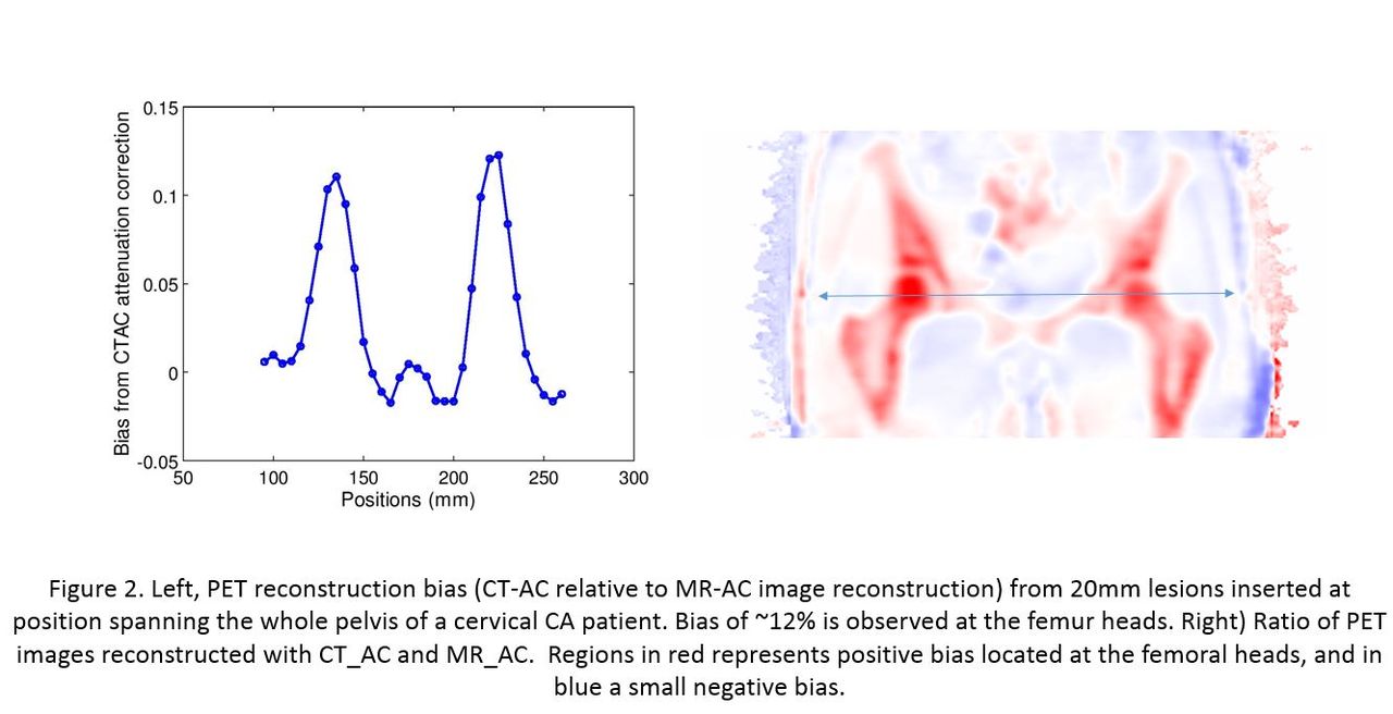
Results: The lesion insertion tool was tested using the NEMA-IEC phantom equipped with its standard set of 6 spheres (10 to 37 mm) filled with a 10:1 ratio with a total activity of 0.55 mCi and scanned for 30min. Simulated lesions of the same diameter were inserted and the contrast recovery coefficients (CRC) of the simulated lesion was compared to the measured values where an excellent agreement was found (Fig.1, left). In the same phantom, a 20mm lesion was placed at various position and images were generated with attenuation coefficient where the lung insert was assigned the air (in red) or bone (in blue) attenuation value. Lesion of 20mm were inserted at various position the pelvis area of cervical cancer patients injected with 18F-FDG and data acquired in PET/MR (fig.2). The simulated lesion data are shown to depict reliably the spatial information observed in patient data when comparing CT_AC reconstruction to MR_AC reconstruction, (DIXON attenuation correction is shown).

Conclusions: Excellent agreement of the inserted spherical lesions to the measured lesions CRC has been achieved and indicate that the proposed method can effectively be used to predict and study the quantitative bias on lesion uptake in patient data resulting from the choice of attenuation correction strategy. The evaluation of the methodology will be presented for the two major manufacturers of clinical PET/MRI cameras.

Figure
  • Download figure
  • Open in new tab
  • Download powerpoint
Figure
  • Download figure
  • Open in new tab
  • Download powerpoint
Previous
Back to top

In this issue

Journal of Nuclear Medicine
Vol. 60, Issue supplement 1
May 1, 2019
  • Table of Contents
  • Index by author
Article Alerts
Sign In to Email Alerts with your Email Address
Email Article

Thank you for your interest in spreading the word on Journal of Nuclear Medicine.

NOTE: We only request your email address so that the person you are recommending the page to knows that you wanted them to see it, and that it is not junk mail. We do not capture any email address.

Enter multiple addresses on separate lines or separate them with commas.
A lesion insertion tool to study the impact of attenuation correction strategy in PET/MRI
(Your Name) has sent you a message from Journal of Nuclear Medicine
(Your Name) thought you would like to see the Journal of Nuclear Medicine web site.
Citation Tools
A lesion insertion tool to study the impact of attenuation correction strategy in PET/MRI
Mahdjoub Hamdi, Dharshan Chandramohan, Peder Larson, Thomas Hope, Richard Laforest
Journal of Nuclear Medicine May 2019, 60 (supplement 1) 1354;

Citation Manager Formats

  • BibTeX
  • Bookends
  • EasyBib
  • EndNote (tagged)
  • EndNote 8 (xml)
  • Medlars
  • Mendeley
  • Papers
  • RefWorks Tagged
  • Ref Manager
  • RIS
  • Zotero
Share
A lesion insertion tool to study the impact of attenuation correction strategy in PET/MRI
Mahdjoub Hamdi, Dharshan Chandramohan, Peder Larson, Thomas Hope, Richard Laforest
Journal of Nuclear Medicine May 2019, 60 (supplement 1) 1354;
Twitter logo Facebook logo LinkedIn logo Mendeley logo
  • Tweet Widget
  • Facebook Like
  • Google Plus One
Bookmark this article

Jump to section

  • Article
  • Figures & Data
  • Info & Metrics

Related Articles

  • No related articles found.
  • Google Scholar

Cited By...

  • No citing articles found.
  • Google Scholar

More in this TOC Section

Physics, Instrumentation & Data Sciences

  • Exploration of Multi-objective Optimization with Genetic Algorithms for PET Image Reconstruction
  • AI-based methods for nuclear-medicine imaging: Need for objective task-specific evaluation
  • Ultra-Fast Reconstruction of Short List-Mode PET Data Frames for Real-Time Visualization and Processing
Show more Physics, Instrumentation & Data Sciences

Image Generation Posters

  • On Enhancing Monte-Carlo Scatter Correction for Y90 Bremsstrahlung SPECT using Guided Filtering
  • Data-driven rigid motion correction of PET brain images using list mode reconstruction
  • Simultaneous Attenuation Correction and Reconstruction of PET Images Using Deep Convolutional Encoder Decoder Networks from Emission Data
Show more Image Generation Posters

Similar Articles

SNMMI

© 2025 SNMMI

Powered by HighWire