
Main menu

  • Home
  • Content
    • Current
    • Ahead of print
    • Past Issues
    • JNM Supplement
    • SNMMI Annual Meeting Abstracts
    • Continuing Education
    • JNM Podcasts
  • Subscriptions
    • Subscribers
    • Institutional and Non-member
    • Rates
    • Journal Claims
    • Corporate & Special Sales
  • Authors
    • Submit to JNM
    • Information for Authors
    • Assignment of Copyright
    • AQARA requirements
  • Info
    • Reviewers
    • Permissions
    • Advertisers
  • About
    • About Us
    • Editorial Board
    • Contact Information
  • More
    • Alerts
    • Feedback
    • Help
    • SNMMI Journals
  • SNMMI
    • JNM
    • JNMT
    • SNMMI Journals
    • SNMMI

User menu

  • Subscribe
  • My alerts
  • Log in
  • My Cart

Search

  • Advanced search
Journal of Nuclear Medicine
  • SNMMI
    • JNM
    • JNMT
    • SNMMI Journals
    • SNMMI
  • Subscribe
  • My alerts
  • Log in
  • My Cart
Journal of Nuclear Medicine

Advanced Search

  • Home
  • Content
    • Current
    • Ahead of print
    • Past Issues
    • JNM Supplement
    • SNMMI Annual Meeting Abstracts
    • Continuing Education
    • JNM Podcasts
  • Subscriptions
    • Subscribers
    • Institutional and Non-member
    • Rates
    • Journal Claims
    • Corporate & Special Sales
  • Authors
    • Submit to JNM
    • Information for Authors
    • Assignment of Copyright
    • AQARA requirements
  • Info
    • Reviewers
    • Permissions
    • Advertisers
  • About
    • About Us
    • Editorial Board
    • Contact Information
  • More
    • Alerts
    • Feedback
    • Help
    • SNMMI Journals
  • View or Listen to JNM Podcast
  • Visit JNM on Facebook
  • Join JNM on LinkedIn
  • Follow JNM on Twitter
  • Subscribe to our RSS feeds
Meeting ReportPhysics, Instrumentation & Data Sciences

Data-driven rigid motion correction of PET brain images using list mode reconstruction

Samuel Hurley, Matthew Spangler-Bickell, Timothy Deller, Tyler Bradshaw, Floris Jansen and Alan McMillan
Journal of Nuclear Medicine May 2019, 60 (supplement 1) 1358;
Samuel Hurley
6Radiology University of Wisconsin - Madison Madison WI United States
  • Find this author on Google Scholar
  • Find this author on PubMed
  • Search for this author on this site
Matthew Spangler-Bickell
3Nuclear Medicine Unit IRCCS Ospedale San Raffaele Milano Italy
  • Find this author on Google Scholar
  • Find this author on PubMed
  • Search for this author on this site
Timothy Deller
2General Electric Elm Grove WI United States
  • Find this author on Google Scholar
  • Find this author on PubMed
  • Search for this author on this site
Tyler Bradshaw
5University of Wisconsin - Madison Madison WI United States
  • Find this author on Google Scholar
  • Find this author on PubMed
  • Search for this author on this site
Floris Jansen
1GE Healthcare Charlton NY United States
  • Find this author on Google Scholar
  • Find this author on PubMed
  • Search for this author on this site
Alan McMillan
4University of Wisconsin Madison WI United States
  • Find this author on Google Scholar
  • Find this author on PubMed
  • Search for this author on this site
  • Article
  • Figures & Data
  • Info & Metrics
Loading

Abstract

1358

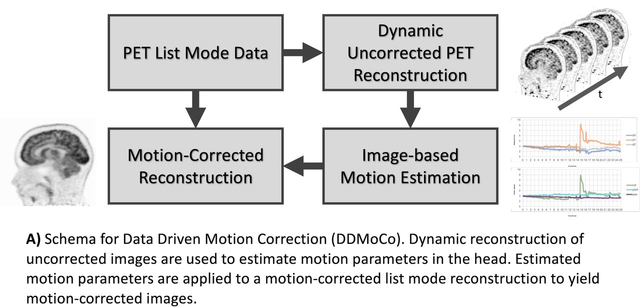
Introduction: Motion of the head during PET brain imaging studies results in burring and loss of spatial resolution. These movements are common and can be due to unintentional events, such as sneezing or coughing, or result from patients who have difficulty remaining still. Typically, head motion is corrected by splitting the acquisition into multiple frames, identifying the time that motion occurred, and throwing out data before or after the motion. Alternatively, one can register dynamic images from individual frames and average these images, but this suffers from suboptimal statistics and physics corrections. In this work, we propose a data-driven approach, using image-based motion estimation techniques on dynamic PET images and apply list-mode event-by-event reconstruction to compute a single data-driven motion-corrected (DDMoCo) image.

Methods: IRB-approved retrospective analysis was performed using clinical 18F-FDG PET/MR brain studies acquired on a Signa PET/MR scanner (GE Healthcare). Data used for this study included a 25-minute PET bed and vendor-provided MR-based attenuation correction scans. Dynamic PET images were generated without any corrections (e.g., scatter and attenuation) using a time-of-flight (TOF) list-mode reconstruction at varying time intervals (150s, 30s, and 5s) spanning the full 1500s PET scan. From the dynamic reconstructed images, image-based motion estimate was performed using the Analysis of Functional Images (AFNI) tool to compute rigid-body motion estimates. Motion parameters were then provided as inputs to an offline list-mode reconstruction tool [Spangler-Bickell, Khalighi, Hoo et al. (2018). IEEE TRPMS]. Images with and without MoCo were compared using peak signal-to-noise ratio (PSNR) and root mean squared error (RMSE).

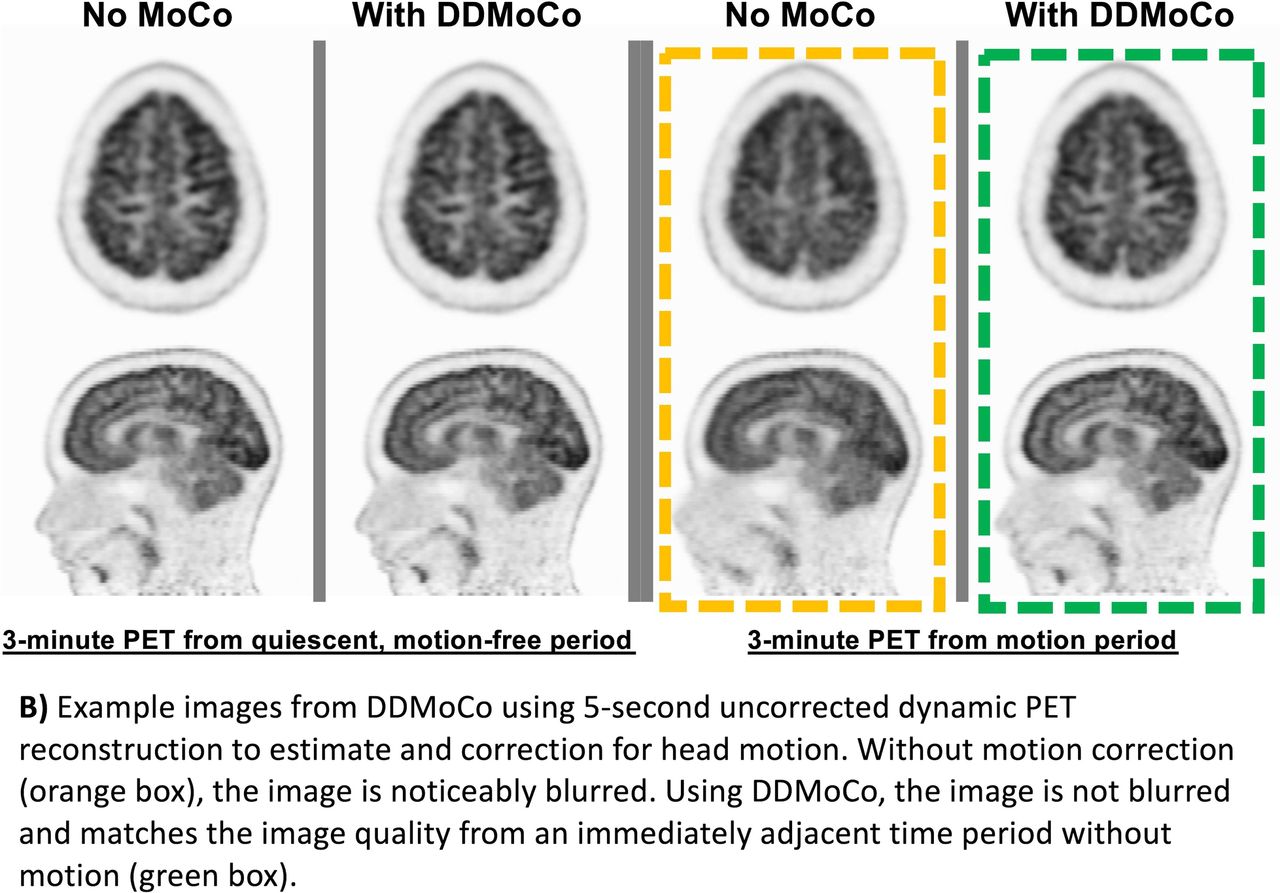
Results: The use of DDMoCo provided visually improved PET images in all cases. In subjects with minimal motion, use of DDMoCo provided subtle improvements in image quality. For such subjects there was little difference between motion estimates at 150s, 30s, or 5s time intervals. In one subject with a single abrupt motion period, a 3-minute PET scan was compared to the quiescent period immediately before the motion event to a 3-minute PET scan during which motion occurred. With the application of DDMoCo vs no correction, besides having greatly improved image sharpness and visual quality, PSNR was 42.1 vs. 39.9 (5.5% higher) and RMSE was 438 Bq/cc vs. 557 Bq/cc (22% lower) demonstrating better quantitative image performance. Discussion/Conclusions: DDMoCo provided robust estimation of motion using image-based estimation from uncorrected dynamic PET reconstructions. For subjects with minimal motion, dynamic images with 30-150s temporal resolution appeared to adequately sample the observed motion, which may be typical for routine clinical studies. Motion estimation at a temporal resolution of up to 5s was demonstrated in a subject with a large, abrupt motion, showing improved visual image quality and more accurate quantification compared to non-moving data. A major advantage of this approach over image-based registration and averaging is that image SNR can be maximized since all counts are used in a single list mode reconstruction. One limitation of DDMoCo is the additional computation time necessary to reconstruct many short duration frames; however, this process could be parallelized and performed offline at the desired temporal resolution. Compared to other methods of motion estimation, DDMoCo requires no additional hardware or integration with the scanner system and could be readily implemented into a clinical environment as a tool to compensate for minimal to moderate head motion.

Figure
  • Download figure
  • Open in new tab
  • Download powerpoint
Figure
  • Download figure
  • Open in new tab
  • Download powerpoint
Previous
Back to top

In this issue

Journal of Nuclear Medicine
Vol. 60, Issue supplement 1
May 1, 2019
  • Table of Contents
  • Index by author
Article Alerts
Sign In to Email Alerts with your Email Address
Email Article

Thank you for your interest in spreading the word on Journal of Nuclear Medicine.

NOTE: We only request your email address so that the person you are recommending the page to knows that you wanted them to see it, and that it is not junk mail. We do not capture any email address.

Enter multiple addresses on separate lines or separate them with commas.
Data-driven rigid motion correction of PET brain images using list mode reconstruction
(Your Name) has sent you a message from Journal of Nuclear Medicine
(Your Name) thought you would like to see the Journal of Nuclear Medicine web site.
Citation Tools
Data-driven rigid motion correction of PET brain images using list mode reconstruction
Samuel Hurley, Matthew Spangler-Bickell, Timothy Deller, Tyler Bradshaw, Floris Jansen, Alan McMillan
Journal of Nuclear Medicine May 2019, 60 (supplement 1) 1358;

Citation Manager Formats

  • BibTeX
  • Bookends
  • EasyBib
  • EndNote (tagged)
  • EndNote 8 (xml)
  • Medlars
  • Mendeley
  • Papers
  • RefWorks Tagged
  • Ref Manager
  • RIS
  • Zotero
Share
Data-driven rigid motion correction of PET brain images using list mode reconstruction
Samuel Hurley, Matthew Spangler-Bickell, Timothy Deller, Tyler Bradshaw, Floris Jansen, Alan McMillan
Journal of Nuclear Medicine May 2019, 60 (supplement 1) 1358;
Twitter logo Facebook logo LinkedIn logo Mendeley logo
  • Tweet Widget
  • Facebook Like
  • Google Plus One
Bookmark this article

Jump to section

  • Article
  • Figures & Data
  • Info & Metrics

Related Articles

  • No related articles found.
  • Google Scholar

Cited By...

  • No citing articles found.
  • Google Scholar

More in this TOC Section

Physics, Instrumentation & Data Sciences

  • AI-based methods for nuclear-medicine imaging: Need for objective task-specific evaluation
  • Keel-Edge Height Selection for Improved Multi-Pinhole 123I Brain SPECT Imaging
  • Ultra-Fast Reconstruction of Short List-Mode PET Data Frames for Real-Time Visualization and Processing
Show more Physics, Instrumentation & Data Sciences

Image Generation Posters

  • On Enhancing Monte-Carlo Scatter Correction for Y90 Bremsstrahlung SPECT using Guided Filtering
  • Simultaneous Attenuation Correction and Reconstruction of PET Images Using Deep Convolutional Encoder Decoder Networks from Emission Data
Show more Image Generation Posters

Similar Articles

SNMMI

© 2025 SNMMI

Powered by HighWire