
Main menu

  • Home
  • Content
    • Current
    • Ahead of print
    • Past Issues
    • JNM Supplement
    • SNMMI Annual Meeting Abstracts
    • Continuing Education
    • JNM Podcasts
  • Subscriptions
    • Subscribers
    • Institutional and Non-member
    • Rates
    • Journal Claims
    • Corporate & Special Sales
  • Authors
    • Submit to JNM
    • Information for Authors
    • Assignment of Copyright
    • AQARA requirements
  • Info
    • Reviewers
    • Permissions
    • Advertisers
  • About
    • About Us
    • Editorial Board
    • Contact Information
  • More
    • Alerts
    • Feedback
    • Help
    • SNMMI Journals
  • SNMMI
    • JNM
    • JNMT
    • SNMMI Journals
    • SNMMI

User menu

  • Subscribe
  • My alerts
  • Log in
  • Log out
  • My Cart

Search

  • Advanced search
Journal of Nuclear Medicine
  • SNMMI
    • JNM
    • JNMT
    • SNMMI Journals
    • SNMMI
  • Subscribe
  • My alerts
  • Log in
  • Log out
  • My Cart
Journal of Nuclear Medicine

Advanced Search

  • Home
  • Content
    • Current
    • Ahead of print
    • Past Issues
    • JNM Supplement
    • SNMMI Annual Meeting Abstracts
    • Continuing Education
    • JNM Podcasts
  • Subscriptions
    • Subscribers
    • Institutional and Non-member
    • Rates
    • Journal Claims
    • Corporate & Special Sales
  • Authors
    • Submit to JNM
    • Information for Authors
    • Assignment of Copyright
    • AQARA requirements
  • Info
    • Reviewers
    • Permissions
    • Advertisers
  • About
    • About Us
    • Editorial Board
    • Contact Information
  • More
    • Alerts
    • Feedback
    • Help
    • SNMMI Journals
  • View or Listen to JNM Podcast
  • Visit JNM on Facebook
  • Join JNM on LinkedIn
  • Follow JNM on Twitter
  • Subscribe to our RSS feeds
Meeting ReportPhysics, Instrumentation & Data Sciences

Whole-Body CT Plane Classification: Development and Comparison of Alternative Methods

Odai Salman and Ran Klein
Journal of Nuclear Medicine May 2019, 60 (supplement 1) 1184;
Odai Salman
1The Ottawa Hospital Ottawa ON Canada
2The Ottawa Hospital Ottawa ON Canada
  • Find this author on Google Scholar
  • Find this author on PubMed
  • Search for this author on this site
Ran Klein
1The Ottawa Hospital Ottawa ON Canada
2The Ottawa Hospital Ottawa ON Canada
  • Find this author on Google Scholar
  • Find this author on PubMed
  • Search for this author on this site
  • Article
  • Figures & Data
  • Info & Metrics
Loading

Abstract

1184

Objectives: Many medical image processing applications rely on targeted regions of interest within a larger volumetric image. In this work, we considered the application of classifying trans-axial CT image planes into 6 main anatomical body regions: head, neck, chest, abdomen, pelvis and legs, which can then, for example, be used to describe the location of tumors in automatically generated reports. We describe and compare three competing methods with regards to implementation complexity, performance and limitations.

Methods: We implemented three CT plane classification methods as follows: (1) An analytical approach which doesn’t require any training and relies solely on Hounsfield unit profiles to identify cut-planes between anatomical regions, (2) an approach using convolutional neural networks (CNNs) of varying configuration (322, 642 and 1282 input image matrix sizes) to classify each plane independently, requiring training using a labeled image set, and (3) CNN followed by a context based correction algorithm (CBCA) which improves the CNN classification using positional relationships between all CT planes. 140 whole-body CT datasets were semi-automatically labeled into anatomical regions using a custom viewer that was initialized with analytical approach cut-planes and were then manually adjusted. Of these, 90 CTs (32,562 planes) were used to train the CNNs. To evaluate performance, we used the remaining 50 CT image sets (18,278 planes) and compared automated plane classification to manual labels using bootstrapping (1000 samples, 30 image each) to estimate performance variability.

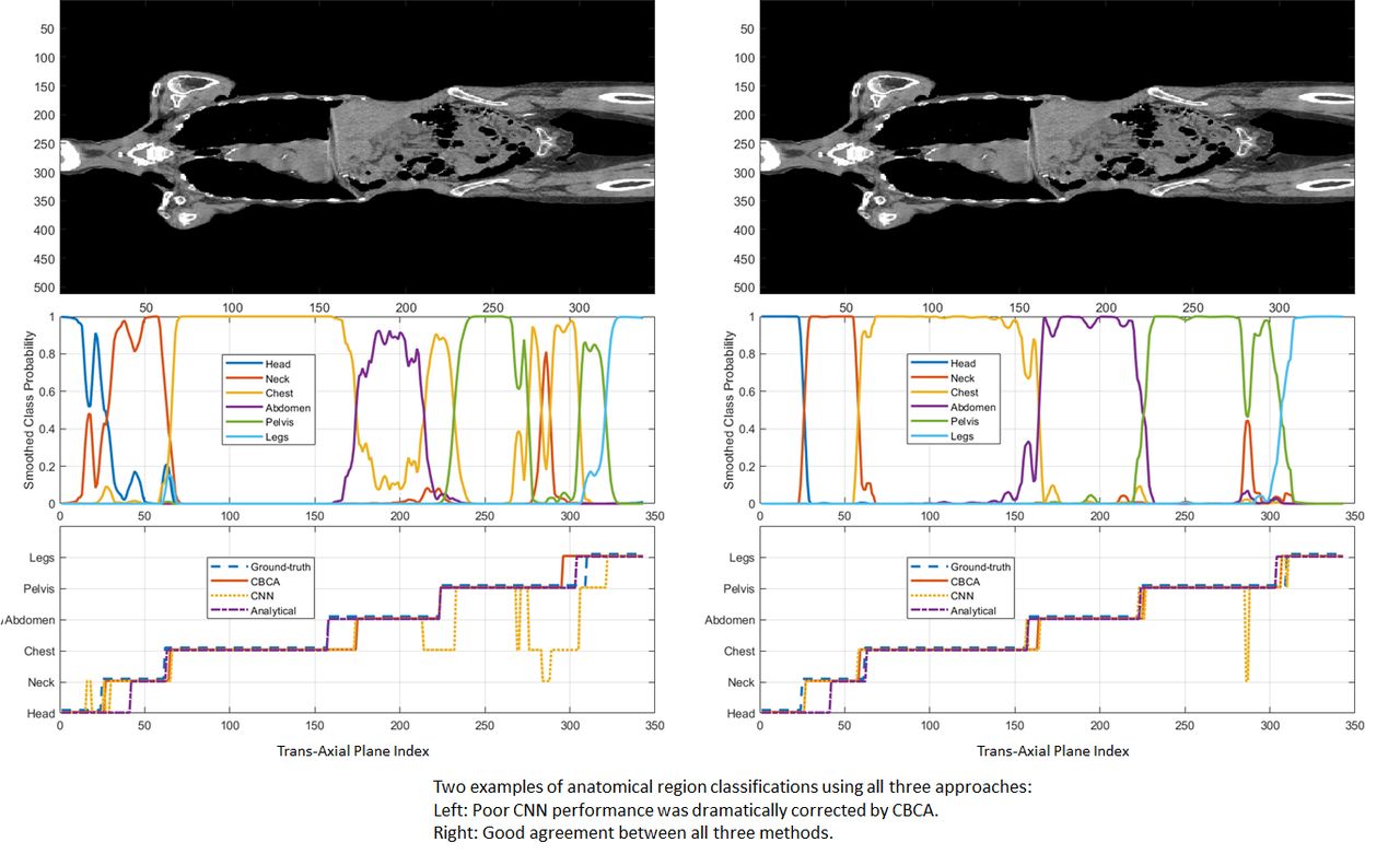
Results: Mean plane labeling accuracy ± standard-error of all 50 CTs with the analytical method was 85.91±0.00%. The three CNN variant accuracies were 95.11±0.09%, 95.85±0.05% and 96.05±0.02% and in all three cases were improved by CBCA to 95.50±0.06%, 96.20±0.07% and 96.32±0.01% (p<0.001 for all), eliminating out-of-order (e.g. pelvis proximal to neck) regions errors. Allowing for ±1 cm ambiguity in anatomical region boundary definition, plane classification accuracies further improved to 90.25±0.00% for analytical; 98.80±0.08%, 99.20±0.05%, 99.22±0.03% for CNNs; and 99.10±0.04%, 99.36±0.08%, 99.43±0.01% for CBCA (p<0.001). CBCA improvement was more dramatic with unoptimized CNNs. Execution times with all methods were <10 seconds per CT image set on a standard laptop.

Conclusions: The analytical approach achieved acceptable accuracy for anatomical region segmentation without the need for explicit data labelling and was effective for batch labelling CT data sets, greatly reducing manual labeling efforts. Convolutional neural networks achieved superior accuracy and allowed for rapid development and training but required labelled data. Post-hoc correction of CNN results using an analytical context-based correction further improved classification, achieving nearly perfect CT plane labeling and anatomical region segmentation.

Figure
  • Download figure
  • Open in new tab
  • Download powerpoint
Previous
Back to top

In this issue

Journal of Nuclear Medicine
Vol. 60, Issue supplement 1
May 1, 2019
  • Table of Contents
  • Index by author
Article Alerts
Sign In to Email Alerts with your Email Address
Email Article

Thank you for your interest in spreading the word on Journal of Nuclear Medicine.

NOTE: We only request your email address so that the person you are recommending the page to knows that you wanted them to see it, and that it is not junk mail. We do not capture any email address.

Enter multiple addresses on separate lines or separate them with commas.
Whole-Body CT Plane Classification: Development and Comparison of Alternative Methods
(Your Name) has sent you a message from Journal of Nuclear Medicine
(Your Name) thought you would like to see the Journal of Nuclear Medicine web site.
Citation Tools
Whole-Body CT Plane Classification: Development and Comparison of Alternative Methods
Odai Salman, Ran Klein
Journal of Nuclear Medicine May 2019, 60 (supplement 1) 1184;

Citation Manager Formats

  • BibTeX
  • Bookends
  • EasyBib
  • EndNote (tagged)
  • EndNote 8 (xml)
  • Medlars
  • Mendeley
  • Papers
  • RefWorks Tagged
  • Ref Manager
  • RIS
  • Zotero
Share
Whole-Body CT Plane Classification: Development and Comparison of Alternative Methods
Odai Salman, Ran Klein
Journal of Nuclear Medicine May 2019, 60 (supplement 1) 1184;
Twitter logo Facebook logo LinkedIn logo Mendeley logo
  • Tweet Widget
  • Facebook Like
  • Google Plus One
Bookmark this article

Jump to section

  • Article
  • Figures & Data
  • Info & Metrics

Related Articles

  • No related articles found.
  • Google Scholar

Cited By...

  • No citing articles found.
  • Google Scholar

More in this TOC Section

Physics, Instrumentation & Data Sciences

  • AI-based methods for nuclear-medicine imaging: Need for objective task-specific evaluation
  • Keel-Edge Height Selection for Improved Multi-Pinhole 123I Brain SPECT Imaging
  • Ultra-Fast Reconstruction of Short List-Mode PET Data Frames for Real-Time Visualization and Processing
Show more Physics, Instrumentation & Data Sciences

Data Analysis & Management Posters

  • Evaluation of Quantitative Whole Body Dynamic FDG-PET Using Block Sequence Regularized Expectation Maximization (BSREM) Reconstruction
  • Feasibility of 18F-THK5351 PET quantitation using the Centiloid scale
  • NaF-PET/CT for assessment of temporomandibular joint osseous activity in rheumatoid arthritis patients and healthy subjects
Show more Data Analysis & Management Posters

Similar Articles

SNMMI

© 2025 SNMMI

Powered by HighWire