


Article Figures & Data
Figures
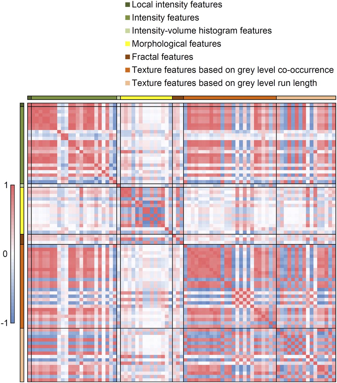
- FIGURE 1.
Correlation map for all radiomic features. Red = high positive correlation; blue = high negative correlation; white = no correlation.
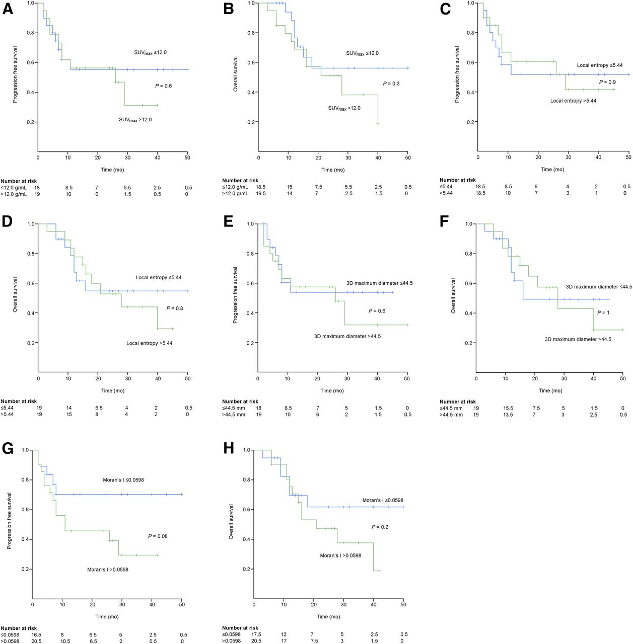
- FIGURE 2.
Estimated Kaplan–Meier curves for PFS of SUVmax (A), OS of SUVmax (B), PFS of local entropy (C), OS of local entropy (D), PFS of 3D maximum diameter (E), OS of 3D maximum diameter (F), PFS of Moran’s I (G), and OS Moran’s I (H).
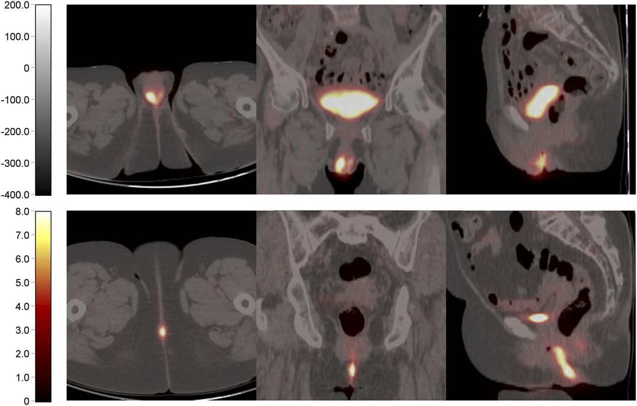
- FIGURE 3.
From left to right, transverse, coronal, and sagittal 18F-FDG PET/CT images of 2 women with vulvar carcinoma. (Top) An 81-y-old woman who had anterior midline 4-cm-diamter tumor with heterogeneous 18F-FDG uptake and relatively high Moran’s I (0.0924). Inguinal lymph node recurrence and death occurred 4 and 16 mo after surgery, respectively. (Bottom) A 74-y-old woman who had posterior midline 3-cm-diamter tumor with homogeneous 18F-FDG uptake and low Moan I (0.0213). At time of last follow-up, patient was alive and without recurrence.


Tables
Characteristic Data Mean age ± SD (y) 70 ± 10 Mean body mass index (kg/m2) 27 (range, 16–42) Mean time ± SD between PET/CT and surgery (d) 20 ± 17 (range, 1–74) Primary vulvar tumor site Anterior 20 (50) Lateral 17 (42.5) Posterior 3 (7.5) Tumor growth pattern Exophytic 29 (72.5) Ulcerative 7 (17.5) Infiltrative 4 (10) Neighboring tissues involved Extravulvar skin 4 (10) Urologic tract 4 (10) None 32 (80) Vulvar surgery Partial vulvectomy 6 (15) Radical vulvectomy 34 (85) Inguinofemoral lymph node surgery Unilateral 0 Bilateral 39 (97.5) None 1 (2.5) SLNB 3 (8) SLNB followed by IFL 16 (41) ILF 20 (51) Pelvic lymph node surgery Unilateral 8 (20) Bilateral 1 (2.5) None 31 (77.5) SLNB = sentinel lymph node biopsy; IFL 5 inguinofemoral lymphadenectomy.
Data are n followed by percentage in parentheses, unless otherwise specified.
- TABLE 2
Histopatologic Characteristics, Staging, and Postoperative Therapy of the 40 Patients
Characteristic Data Tumor diameter (mm) Median 50 (range, 30–90) 26–40 17 (42.5) >40 23 (57.5) Histologic grade G1 2 (5) G2 32 (80) G3 6 (15) Depth of stromal invasion (mm) Median 9 (range, 4–20) 1–4 3 >5 34 Unknown 3 Resection margin status R0 32 (80) R1 4 (10) VIN 4 (10) Lymph-vascular space invasion Yes 19 (47.5) No 21 (52.5) Metastatic lymph nodes Yes 23 (57.5) No 17 (42.5) Extracapsular invasion of metastatic lymph nodes Yes 12 (52) No 11 (48) Median no. of removed lymph nodes 17 (range, 0–28) Median no. of metastatic lymph nodes 1 (range, 0–7) Median size of lymph node metastases (mm) 13 (range, 1–27) FIGO stage IB 5 (12.5) II 12 (30) III 18 (45) IVA 5 (12.5) Adjuvant therapy Yes 27 (67.5) No 13 (32.5) G1 = well differentiated; G2 = moderately differentiated; G3 = poorly differentiated; R0 = no tumor; R1 = microscopic tumor; VIN = vulvar intraepithelial neoplasia; FIGO = International Federation of Gynecology and Obstetrics.
Data are n followed by percentage in parentheses, unless otherwise specified.
Parameter Data Follow-up for entire study (mo) Median 15 (range, 2–50) Mean ± SD 19 ± 13 Follow-up for surviving patients (mo) Median 26 (range, 6–50) Mean ± SD 25 ± 13 PFS Median 10 (range, 2–50) Mean ± SD 17 ± 14 OS Median 16 (range, 3–50) Mean ± SD 20 ± 13 Local or distant recurrence (n) 18 (45%)* Death (n) 18 (45%)† - TABLE 4
Association Between Histopathologic Characteristics and Imaging Features Derived from 18F-FDG PET Images
Histologic grade Lymph-vascular space invasion Metastatic lymph nodes Radiomic feature 1–2 3 P Depth of invasion (mm) No Yes P No Yes P SUVmax 12.1 ± 4.2 13.9 ± 3.0 0.3 R2 = 0.07 11.2 ± 3.2 13.7 ± 4.48 0.05 11.5 ± 4.2 13.0 ± 3.9 0.2 Local entropy 3.9 ± 0.11 3.9 ± 0.09 1 R2 = 0.10 3.9 ± 0.12 3.9 ± 0.10 0.6 3.9 ± 0.10 3.9 ± 0.11 0.4 Maximum 3D diameter 48.5 ± 19.4 44.6 ± 16.1 0.8 R2 = 0.08 47.6 ± 22.0 48.3 ± 15.0 0.7 46.9 ± 20.2 48.6 ± 18.0 0.9 Moran’s I 0.062 ± 0.018 0.057 ± 0.021 0.8 R2 = 0.013 0.059 ± 0.018 0.064 ± 0.020 0.6 0.062 ± 0.017 0.061 ± 0.020 0.9 R2 indicates the coefficient of determination.
PFS OS Parameter Hazard ratio P Hazard ratio P Univariate Cox regression analysis Age 1.1 (1.0–1.1) 0.06 1.1 (1.0–1.1) 0.006 Maximum diameter of primary tumor 1.0 (0.8–1.4) 0.9 1.1 (1.0–1.1) 0.6 Histologic grade 0.8 0.3 I–II 1 1 III 0.9 (0.3–3.0) 0.6 (0.2–1.7) Lymph-vascular space invasion 2.3 (0.9–6.1) 0.08 3.0 (1.1–7.9) 0.03 Depth of stromal invasion 1.0 (0.9–1.2) 0.4 1.1 (0.9–1.2) 0.4 Resection margin status 0.6 0.4 R0 1 1 R1 2.5 (0.3–18.7) 3.3 (0.4–25.3) VIN 1.9 (0.2–21.4) 2.5 (0.2–27.4) Metastatic lymph nodes* 2.1 (0.7–5.9) 0.2 3.0 (1.0–9.2) 0.05 Bilateral metastatic lymph nodes 3.0 (1.2–7.6) 0.02 2.7 (1.0–6.8) 0.04 Number of metastatic lymph nodes* 1.3 (1.0–1.5) 0.02 1.3 (1.0–1.5) 0.03 Extracapsular invasion of metastatic lymph nodes 5.3 (2.1–13.7) <0.0001 4.2 (1.6–10.6) 0.003 FIGO stage 0.4 0.1 IB 1 1 II 0.6 (0.1–3.9) 0.7 (0.01–5.0) III 0.3 (0.06–1.5) 0.4 (0.05–2.6) IVA 0.8 (0.2–3.0) 1.7 (0.4–7.6) Radiomic feature SUVmax 1.0 (0.9–1.2) 0.6 1.0 (0.9–1.1) 0.6 Local entropy 1.7 (0.03–99.1) 0.8 0.6 (0–33.2) 0.8 3D maximum diameter 1.0 (1.0–1.0) 0.5 1.0 (1.0–1.0) 0.8 Moran’s I 1.3 (1.0–1.6) 0.06 1.3 (1.0–1.7) 0.03 Multivariate Cox regression analysis Iterative forward selection Extracapsular invasion 9.5 (3.1–28.6) <0.0001 6.5 (2.3–18.4) <0.0001 Moran’s I 1.6 (1.2–2.3) 0.03 1.5 (1.1–2.1) 0.009 Iterative backward selection Extracapsular invasion 9.5 (3.1–28.6) 0.0001 4.3 (1.4–12.6) 0.009 Moran’s I 1.6 (1.2–2.3) 0.03 1.4 (1.1–2.0) 0.02 ↵* Metastatic lymph nodes indicates patients with metastatic lymph nodes; number of metastatic lymph nodes is the number of metastatic lymph nodes per patient.
R0 = no tumor; R1 = microscopic tumor; VIN = vulvar intraepithelial neoplasia; FIGO = International Federation of Gynecology and Obstetrics.
Data in parentheses are 95% confidence intervals.
Supplemental Data
Files in this Data Supplement:


