
Main menu

  • Home
  • Content
    • Current
    • Ahead of print
    • Past Issues
    • JNM Supplement
    • SNMMI Annual Meeting Abstracts
    • Continuing Education
    • JNM Podcasts
  • Subscriptions
    • Subscribers
    • Institutional and Non-member
    • Rates
    • Journal Claims
    • Corporate & Special Sales
  • Authors
    • Submit to JNM
    • Information for Authors
    • Assignment of Copyright
    • AQARA requirements
  • Info
    • Reviewers
    • Permissions
    • Advertisers
  • About
    • About Us
    • Editorial Board
    • Contact Information
  • More
    • Alerts
    • Feedback
    • Help
    • SNMMI Journals
  • SNMMI
    • JNM
    • JNMT
    • SNMMI Journals
    • SNMMI

User menu

  • Subscribe
  • My alerts
  • Log in
  • My Cart

Search

  • Advanced search
Journal of Nuclear Medicine
  • SNMMI
    • JNM
    • JNMT
    • SNMMI Journals
    • SNMMI
  • Subscribe
  • My alerts
  • Log in
  • My Cart
Journal of Nuclear Medicine

Advanced Search

  • Home
  • Content
    • Current
    • Ahead of print
    • Past Issues
    • JNM Supplement
    • SNMMI Annual Meeting Abstracts
    • Continuing Education
    • JNM Podcasts
  • Subscriptions
    • Subscribers
    • Institutional and Non-member
    • Rates
    • Journal Claims
    • Corporate & Special Sales
  • Authors
    • Submit to JNM
    • Information for Authors
    • Assignment of Copyright
    • AQARA requirements
  • Info
    • Reviewers
    • Permissions
    • Advertisers
  • About
    • About Us
    • Editorial Board
    • Contact Information
  • More
    • Alerts
    • Feedback
    • Help
    • SNMMI Journals
  • View or Listen to JNM Podcast
  • Visit JNM on Facebook
  • Join JNM on LinkedIn
  • Follow JNM on Twitter
  • Subscribe to our RSS feeds
Research ArticlePhysics and Instrumentation

Development and Evaluation of mini-EXPLORER: A Long Axial Field-of-View PET Scanner for Nonhuman Primate Imaging

Eric Berg, Xuezhu Zhang, Julien Bec, Martin S. Judenhofer, Brijesh Patel, Qiyu Peng, Maciej Kapusta, Matthias Schmand, Michael E. Casey, Alice F. Tarantal, Jinyi Qi, Ramsey D. Badawi and Simon R. Cherry
Journal of Nuclear Medicine June 2018, 59 (6) 993-998; DOI: https://doi.org/10.2967/jnumed.117.200519
Eric Berg
1Department of Biomedical Engineering, University of California–Davis, Davis, California
  • Find this author on Google Scholar
  • Find this author on PubMed
  • Search for this author on this site
Xuezhu Zhang
1Department of Biomedical Engineering, University of California–Davis, Davis, California
  • Find this author on Google Scholar
  • Find this author on PubMed
  • Search for this author on this site
Julien Bec
1Department of Biomedical Engineering, University of California–Davis, Davis, California
  • Find this author on Google Scholar
  • Find this author on PubMed
  • Search for this author on this site
Martin S. Judenhofer
1Department of Biomedical Engineering, University of California–Davis, Davis, California
  • Find this author on Google Scholar
  • Find this author on PubMed
  • Search for this author on this site
Brijesh Patel
1Department of Biomedical Engineering, University of California–Davis, Davis, California
  • Find this author on Google Scholar
  • Find this author on PubMed
  • Search for this author on this site
Qiyu Peng
1Department of Biomedical Engineering, University of California–Davis, Davis, California
2Cell and Tissue Imaging Department, Molecular Biophysics and Integrated Bioimaging Division, Lawrence Berkeley National Laboratory, Berkeley, California
  • Find this author on Google Scholar
  • Find this author on PubMed
  • Search for this author on this site
Maciej Kapusta
3Siemens Medical Solutions, Knoxville, Tennessee
  • Find this author on Google Scholar
  • Find this author on PubMed
  • Search for this author on this site
Matthias Schmand
3Siemens Medical Solutions, Knoxville, Tennessee
  • Find this author on Google Scholar
  • Find this author on PubMed
  • Search for this author on this site
Michael E. Casey
3Siemens Medical Solutions, Knoxville, Tennessee
  • Find this author on Google Scholar
  • Find this author on PubMed
  • Search for this author on this site
Alice F. Tarantal
4Departments of Pediatrics and Cell Biology and Human Anatomy, School of Medicine, and California National Primate Research Center, University of California–Davis, Davis, California; and
  • Find this author on Google Scholar
  • Find this author on PubMed
  • Search for this author on this site
Jinyi Qi
1Department of Biomedical Engineering, University of California–Davis, Davis, California
  • Find this author on Google Scholar
  • Find this author on PubMed
  • Search for this author on this site
Ramsey D. Badawi
1Department of Biomedical Engineering, University of California–Davis, Davis, California
5Department of Radiology, University of California–Davis, Sacramento, California
  • Find this author on Google Scholar
  • Find this author on PubMed
  • Search for this author on this site
Simon R. Cherry
1Department of Biomedical Engineering, University of California–Davis, Davis, California
5Department of Radiology, University of California–Davis, Sacramento, California
  • Find this author on Google Scholar
  • Find this author on PubMed
  • Search for this author on this site
  • Article
  • Figures & Data
  • Supplemental
  • Info & Metrics
  • PDF
Loading

Article Figures & Data

Figures

  • Tables
  • Additional Files
  • FIGURE 1.
    • Download figure
    • Open in new tab
    • Download powerpoint
    FIGURE 1.

    (A) Side view of scanner with covers removed showing gantry frame, detector rings, and scanner electronics. (B) View inside detector rings showing detectors mounted between tapered aluminum rails. (C) View from front of scanner with covers on and scanning bed installed.
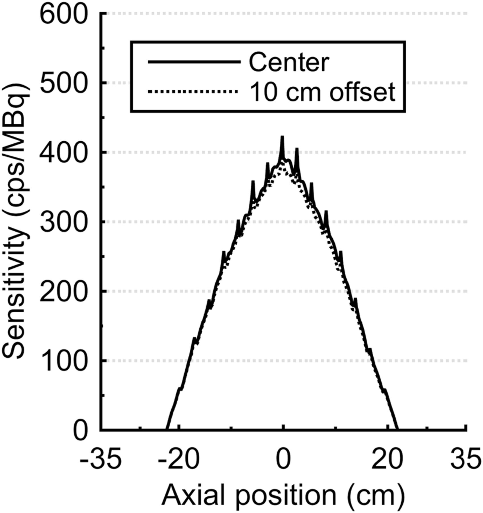
  • FIGURE 2.
    • Download figure
    • Open in new tab
    • Download powerpoint
    FIGURE 2.

    Axial NEMA NU-2 sensitivity profiles (2-mm axial slices) obtained with 46° acceptance angle.
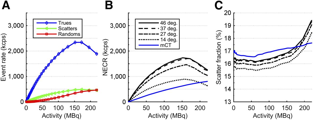
  • FIGURE 3.
    • Download figure
    • Open in new tab
    • Download powerpoint
    FIGURE 3.

    NEMA NU-4 monkey scatter phantom count rate performance. (A) True-, scatter-, and random-coincidence rates measured with 46° acceptance angle. (B) NECR vs. activity for each acceptance angle. (C) Scatter fraction vs. activity for each acceptance angle.
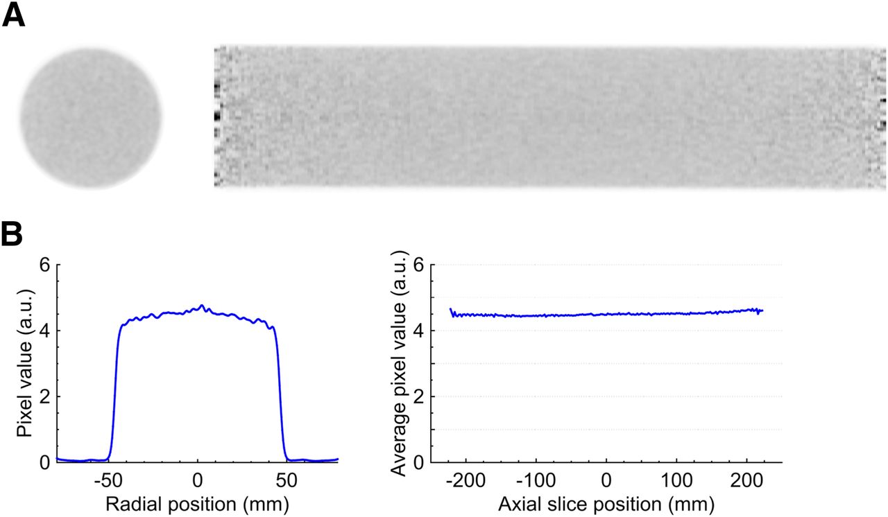
  • FIGURE 4.
    • Download figure
    • Open in new tab
    • Download powerpoint
    FIGURE 4.

    (A) Transaxial (left) and sagittal (right) image slices of reconstructed uniform cylinder. (B) Radial line profile through average of all transaxial image slices (left) and average image pixel value inside uniform cylinder for each axial slice (right).
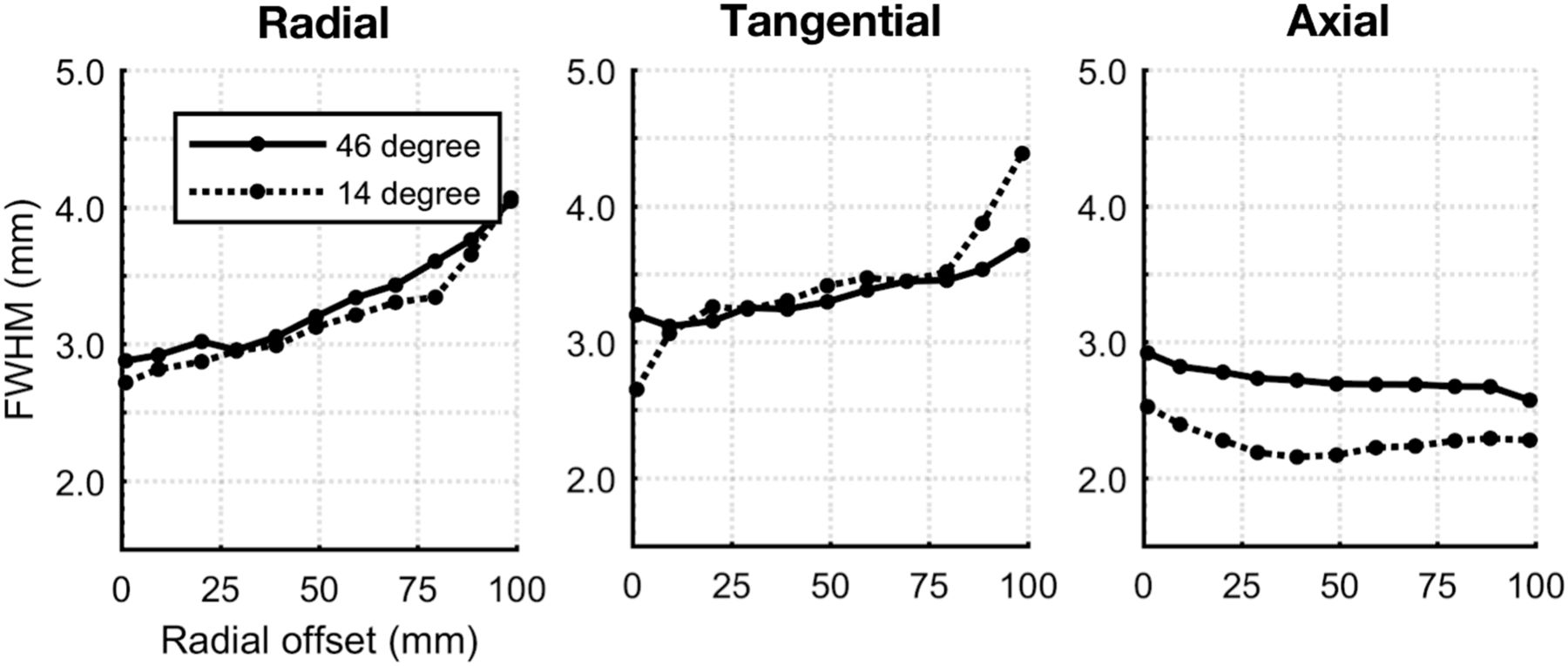
  • FIGURE 5.
    • Download figure
    • Open in new tab
    • Download powerpoint
    FIGURE 5.

    Reconstructed point-source spatial resolution vs. radial offset.
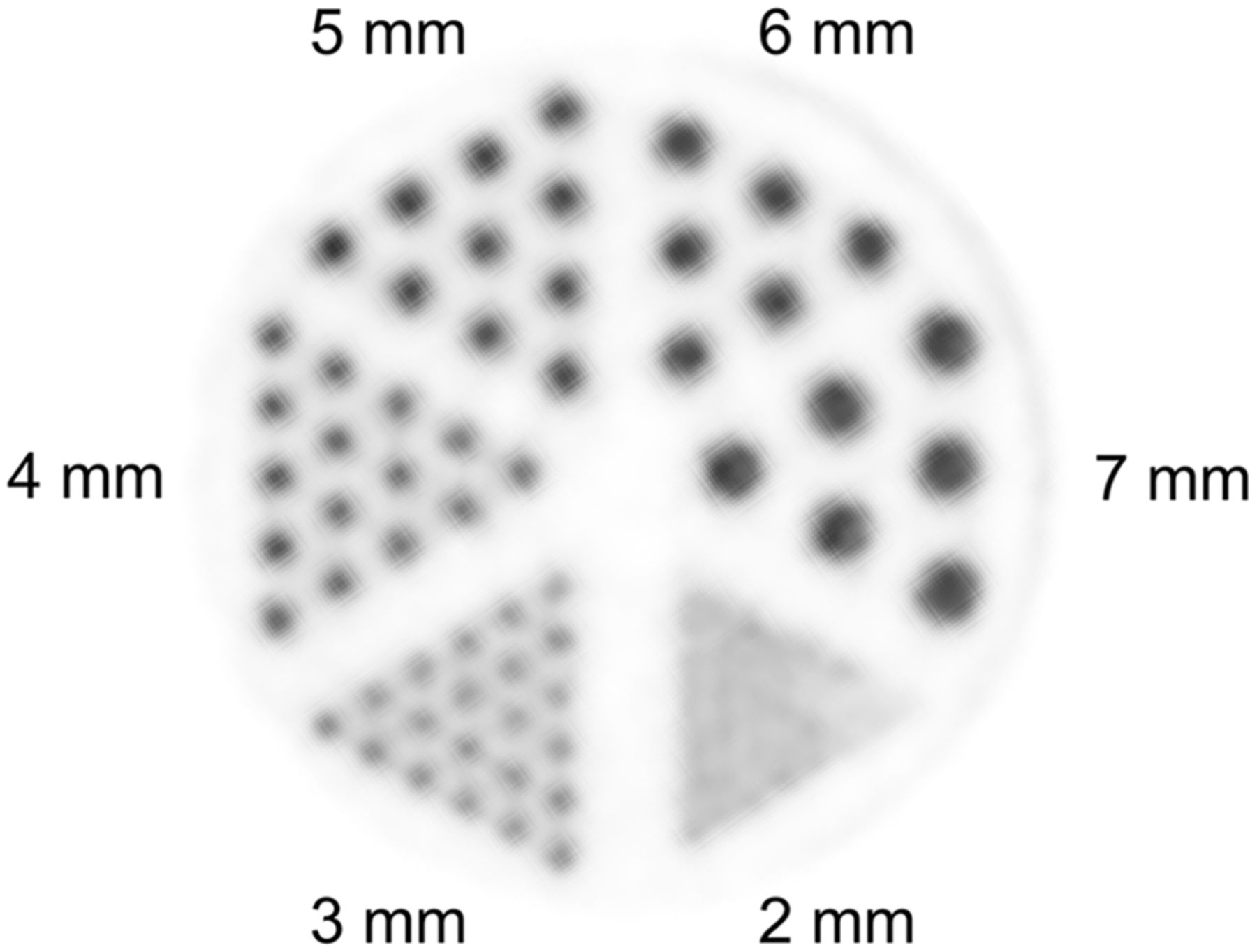
  • FIGURE 6.
    • Download figure
    • Open in new tab
    • Download powerpoint
    FIGURE 6.

    Transaxial slice of Derenzo phantom reconstructed image.
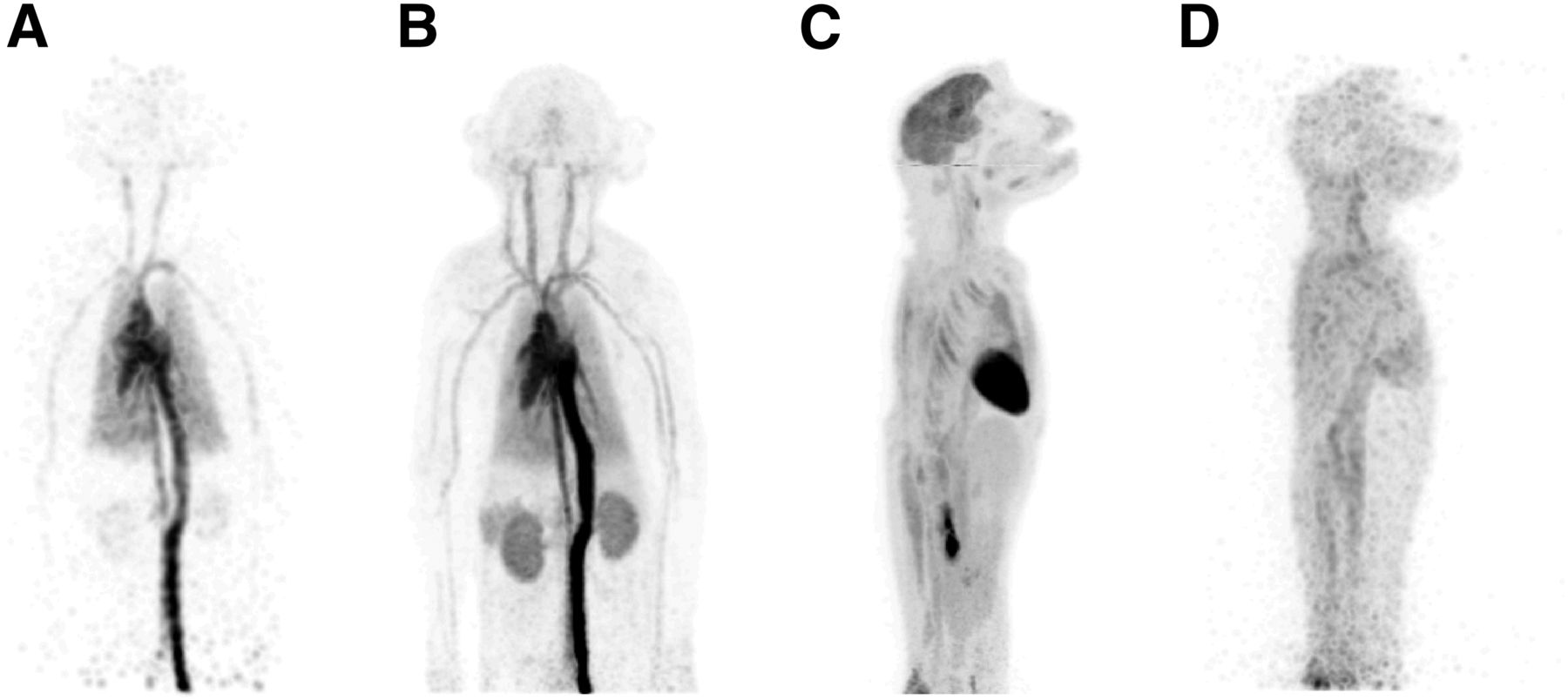
  • FIGURE 7.
    • Download figure
    • Open in new tab
    • Download powerpoint
    FIGURE 7.

    Maximum-intensity-projection images from 18F-FDG rhesus monkey study: 1-s frame at 5 s after injection (reconstructed using kernel method (37)) (A), 0–30 s after injection (B), 55–60 min after injection (C), and 18 h after injection (40-min scan) (D). Further images are provided as Supplemental Figure 4 and Supplemental Videos 1–6.

Tables

  • Figures
  • Additional Files
    • View popup
    TABLE 1

    Physical Characteristics of mini-EXPLORER

    ParameterDescription
    PhotodetectorPhotomultiplier tube
    ScintillatorLutetium oxyorthosilicate
    Crystal pitch4 × 4 × 20 mm
    Total number of crystals32,448
    Detector ring diameter43.5 cm
    Transaxial FOV32.0 cm
    Axial FOV45.7 cm
    • View popup
    TABLE 2

    Physical Performance of mini-EXPLORER

    ParameterAcceptance angle
    14°27°37°46°
    NU-2 sensitivity (%)2.44.04.85.0
    NU-4 peak monkey NECR (kcps)8951,4661,7071,741
    NU-4 scatter fraction (%)15.616.116.416.5

Additional Files

  • Figures
  • Tables
  • Supplemental Data

    Files in this Data Supplement:

    • Supplemental Data
    • Supplemental Video
    • Supplemental Video
    • Supplemental Video
    • Supplemental Video
    • Supplemental Video
    • Supplemental Video
PreviousNext
Back to top

In this issue

Journal of Nuclear Medicine: 59 (6)
Journal of Nuclear Medicine
Vol. 59, Issue 6
June 1, 2018
  • Table of Contents
  • Table of Contents (PDF)
  • About the Cover
  • Index by author
Print
Download PDF
Article Alerts
Sign In to Email Alerts with your Email Address
Email Article

Thank you for your interest in spreading the word on Journal of Nuclear Medicine.

NOTE: We only request your email address so that the person you are recommending the page to knows that you wanted them to see it, and that it is not junk mail. We do not capture any email address.

Enter multiple addresses on separate lines or separate them with commas.
Development and Evaluation of mini-EXPLORER: A Long Axial Field-of-View PET Scanner for Nonhuman Primate Imaging
(Your Name) has sent you a message from Journal of Nuclear Medicine
(Your Name) thought you would like to see the Journal of Nuclear Medicine web site.
Citation Tools
Development and Evaluation of mini-EXPLORER: A Long Axial Field-of-View PET Scanner for Nonhuman Primate Imaging
Eric Berg, Xuezhu Zhang, Julien Bec, Martin S. Judenhofer, Brijesh Patel, Qiyu Peng, Maciej Kapusta, Matthias Schmand, Michael E. Casey, Alice F. Tarantal, Jinyi Qi, Ramsey D. Badawi, Simon R. Cherry
Journal of Nuclear Medicine Jun 2018, 59 (6) 993-998; DOI: 10.2967/jnumed.117.200519

Citation Manager Formats

  • BibTeX
  • Bookends
  • EasyBib
  • EndNote (tagged)
  • EndNote 8 (xml)
  • Medlars
  • Mendeley
  • Papers
  • RefWorks Tagged
  • Ref Manager
  • RIS
  • Zotero
Share
Development and Evaluation of mini-EXPLORER: A Long Axial Field-of-View PET Scanner for Nonhuman Primate Imaging
Eric Berg, Xuezhu Zhang, Julien Bec, Martin S. Judenhofer, Brijesh Patel, Qiyu Peng, Maciej Kapusta, Matthias Schmand, Michael E. Casey, Alice F. Tarantal, Jinyi Qi, Ramsey D. Badawi, Simon R. Cherry
Journal of Nuclear Medicine Jun 2018, 59 (6) 993-998; DOI: 10.2967/jnumed.117.200519
Twitter logo Facebook logo LinkedIn logo Mendeley logo
  • Tweet Widget
  • Facebook Like
  • Google Plus One
Bookmark this article

Jump to section

  • Article
    • Abstract
    • MATERIALS AND METHODS
    • RESULTS
    • DISCUSSION
    • CONCLUSION
    • DISCLOSURE
    • Acknowledgments
    • Footnotes
    • REFERENCES
  • Figures & Data
  • Supplemental
  • Info & Metrics
  • PDF

Related Articles

  • This Month in JNM
  • PubMed
  • Google Scholar

Cited By...

  • Performance Evaluation of the uEXPLORER Total-Body PET/CT Scanner Based on NEMA NU 2-2018 with Additional Tests to Characterize PET Scanners with a Long Axial Field of View
  • Total-Body PET Imaging for up to 30 Days After Injection of 89Zr-Labeled Antibodies
  • Total-Body PET and Highly Stable Chelators Together Enable Meaningful 89Zr-Antibody PET Studies up to 30 Days After Injection
  • Total-Body Dynamic Reconstruction and Parametric Imaging on the uEXPLORER
  • PennPET Explorer: Human Imaging on a Whole-Body Imager
  • Google Scholar

More in this TOC Section

  • Performance Evaluation of the uMI Panorama PET/CT System in Accordance with the National Electrical Manufacturers Association NU 2-2018 Standard
  • A Multicenter Study on Observed Discrepancies Between Vendor-Stated and PET-Measured 90Y Activities for Both Glass and Resin Microsphere Devices
  • Ultra-Fast List-Mode Reconstruction of Short PET Frames and Example Applications
Show more Physics and Instrumentation

Similar Articles

Keywords

  • positron emission tomography
  • high sensitivity
  • long axial field of view
  • total-body imaging
  • Rhesus monkey
SNMMI

© 2025 SNMMI

Powered by HighWire