
Main menu

  • Home
  • Content
    • Current
    • Ahead of print
    • Past Issues
    • JNM Supplement
    • SNMMI Annual Meeting Abstracts
    • Continuing Education
    • JNM Podcasts
  • Subscriptions
    • Subscribers
    • Institutional and Non-member
    • Rates
    • Journal Claims
    • Corporate & Special Sales
  • Authors
    • Submit to JNM
    • Information for Authors
    • Assignment of Copyright
    • AQARA requirements
  • Info
    • Reviewers
    • Permissions
    • Advertisers
  • About
    • About Us
    • Editorial Board
    • Contact Information
  • More
    • Alerts
    • Feedback
    • Help
    • SNMMI Journals
  • SNMMI
    • JNM
    • JNMT
    • SNMMI Journals
    • SNMMI

User menu

  • Subscribe
  • My alerts
  • Log in
  • Log out
  • My Cart

Search

  • Advanced search
Journal of Nuclear Medicine
  • SNMMI
    • JNM
    • JNMT
    • SNMMI Journals
    • SNMMI
  • Subscribe
  • My alerts
  • Log in
  • Log out
  • My Cart
Journal of Nuclear Medicine

Advanced Search

  • Home
  • Content
    • Current
    • Ahead of print
    • Past Issues
    • JNM Supplement
    • SNMMI Annual Meeting Abstracts
    • Continuing Education
    • JNM Podcasts
  • Subscriptions
    • Subscribers
    • Institutional and Non-member
    • Rates
    • Journal Claims
    • Corporate & Special Sales
  • Authors
    • Submit to JNM
    • Information for Authors
    • Assignment of Copyright
    • AQARA requirements
  • Info
    • Reviewers
    • Permissions
    • Advertisers
  • About
    • About Us
    • Editorial Board
    • Contact Information
  • More
    • Alerts
    • Feedback
    • Help
    • SNMMI Journals
  • View or Listen to JNM Podcast
  • Visit JNM on Facebook
  • Join JNM on LinkedIn
  • Follow JNM on Twitter
  • Subscribe to our RSS feeds
Research ArticleTheranostics

Biochemical Recurrence of Prostate Cancer: Initial Results with [18F]PSMA-1007 PET/CT

Frederik L. Giesel, Leon Will, Claudia Kesch, Martin Freitag, Christophe Kremer, Jonas Merkle, Oliver C. Neels, Jens Cardinale, Boris Hadaschik, Markus Hohenfellner, Klaus Kopka, Uwe Haberkorn and Clemens Kratochwil
Journal of Nuclear Medicine April 2018, 59 (4) 632-635; DOI: https://doi.org/10.2967/jnumed.117.196329
Frederik L. Giesel
1Department of Nuclear Medicine, University Hospital Heidelberg, Heidelberg, Germany
2Cooperation Unit Nuclear Medicine, German Cancer Research Center (DKFZ), Heidelberg, Germany
3German Cancer Consortium (DKTK), Heidelberg, Germany
  • Find this author on Google Scholar
  • Find this author on PubMed
  • Search for this author on this site
Leon Will
1Department of Nuclear Medicine, University Hospital Heidelberg, Heidelberg, Germany
  • Find this author on Google Scholar
  • Find this author on PubMed
  • Search for this author on this site
Claudia Kesch
4Department of Urology, University Hospital Heidelberg, Heidelberg, Germany
  • Find this author on Google Scholar
  • Find this author on PubMed
  • Search for this author on this site
Martin Freitag
2Cooperation Unit Nuclear Medicine, German Cancer Research Center (DKFZ), Heidelberg, Germany
5Department of Radiology, German Cancer Research Center (DKFZ), Heidelberg, Germany; and
  • Find this author on Google Scholar
  • Find this author on PubMed
  • Search for this author on this site
Christophe Kremer
1Department of Nuclear Medicine, University Hospital Heidelberg, Heidelberg, Germany
  • Find this author on Google Scholar
  • Find this author on PubMed
  • Search for this author on this site
Jonas Merkle
1Department of Nuclear Medicine, University Hospital Heidelberg, Heidelberg, Germany
  • Find this author on Google Scholar
  • Find this author on PubMed
  • Search for this author on this site
Oliver C. Neels
6Division of Radiopharmaceutical Chemistry, German Cancer Research Center (DKFZ), Heidelberg, Germany
  • Find this author on Google Scholar
  • Find this author on PubMed
  • Search for this author on this site
Jens Cardinale
6Division of Radiopharmaceutical Chemistry, German Cancer Research Center (DKFZ), Heidelberg, Germany
  • Find this author on Google Scholar
  • Find this author on PubMed
  • Search for this author on this site
Boris Hadaschik
4Department of Urology, University Hospital Heidelberg, Heidelberg, Germany
  • Find this author on Google Scholar
  • Find this author on PubMed
  • Search for this author on this site
Markus Hohenfellner
4Department of Urology, University Hospital Heidelberg, Heidelberg, Germany
  • Find this author on Google Scholar
  • Find this author on PubMed
  • Search for this author on this site
Klaus Kopka
6Division of Radiopharmaceutical Chemistry, German Cancer Research Center (DKFZ), Heidelberg, Germany
  • Find this author on Google Scholar
  • Find this author on PubMed
  • Search for this author on this site
Uwe Haberkorn
1Department of Nuclear Medicine, University Hospital Heidelberg, Heidelberg, Germany
2Cooperation Unit Nuclear Medicine, German Cancer Research Center (DKFZ), Heidelberg, Germany
  • Find this author on Google Scholar
  • Find this author on PubMed
  • Search for this author on this site
Clemens Kratochwil
1Department of Nuclear Medicine, University Hospital Heidelberg, Heidelberg, Germany
  • Find this author on Google Scholar
  • Find this author on PubMed
  • Search for this author on this site
  • Article
  • Figures & Data
  • Info & Metrics
  • PDF
Loading

Abstract

Biochemical recurrence (BCR) is a concern for prostate cancer patients after local treatment. 68Ga-labeled prostate-specific membrane antigen (PSMA) ligands have significantly improved prostate cancer imaging. However, several 18F-labeled ligands that were developed as fluorinated tracers might present advantages. In this study, we analyzed the potential of 18F-PSMA-1007 in patients with BCR. Methods: Twelve patients with BCR after local treatment underwent PET/CT scans 1 and 3 h after injection of 18F-PSMA-1007. Results: 18F-PSMA-1007 PET/CT detected lesions in 9 of 12 patients (75%). A significant difference was observed when comparing the tracer uptake in 18F-PSMA-1007–positive lesions 1 and 3 h after injection (median SUVmax, 7.00 vs. 11.34; P < 0.001; n = 76). Forty-four (88%) of 50 18F-PSMA-1007–positive lymph nodes had a short-axis diameter of less than 8 mm. Conclusion: In this pilot study, 18F-PSMA-1007 PET/CT presented high potential for localization of recurrent disease in prostate cancer patients with BCR.

  • peptides
  • PET/CT
  • biochemical recurrence
  • 18F-PSMA-1007
  • PSMA-PET
  • prostate cancer

Biochemical recurrence (BCR) represents a concern for patients after initial curative treatment of prostate cancer. Early detection of recurrent disease is important, to be able to offer a salvage therapy with curative intent. However, in the setting of BCR, full-body bone scans and cross-sectional abdominopelvic CT imaging offer only limited detection rates for metastatic or recurrent prostate cancer lesions (1). Introduction of 68Ga-labeled prostate-specific membrane antigen (PSMA)–11 significantly improved prostate cancer imaging. With data from more than 3,000 evaluated patients so far (2–6), this tracer has already demonstrated impressive results in the setting of BCR and is most likely also superior to 18F-choline PET/CT (7,8).

Fluorinated tracers, however, might present practical advantages, as the longer half-life of 18F compared with 68Ga (110 min vs. 68 min) might enable centralized production and distribution to satellite centers. Furthermore, there are fewer limitations caused by generator capacity. Therefore, several 18F-labeled ligands were developed, and new tracers have already been evaluated in preliminary clinical investigations (9,10).

One candidate for introduction to clinical routine is 18F-PSMA-1007, which benefits from a low background activity in the urinary tract (10). Unspecific bladder activity can challenge delineation of primary tumors (11) or local recurrence (12,13) and can even hamper evaluation of the pelvic lymph nodes (LNs) by introducing halo artifacts in PET/MR systems (12,13). 18F-PSMA-1007 has already demonstrated its potential for primary tumor staging (10). In the present study, we report the results for 18F-PSMA-1007 PET/CT in the setting of BCR.

MATERIALS AND METHODS

Patients

From May 2016 to July 2017, 12 patients presenting with a rising level of serum prostate-specific antigen (PSA) (median, 0.60 ng/mL; range, 0.08–6.50 ng/mL) after previous local treatment underwent 18F-PSMA-1007 PET/CT. Detailed patient characteristics are provided in Table 1. The institutional review board approved the study (permit S-321), and all subjects gave written informed consent. The data of one study patient (patient 4) have also been published in a case report (14).

View this table:
  • View inline
  • View popup
TABLE 1

Patient Characteristics

PET/CT Imaging

All imaging was performed on a Biograph mCT Flow scanner (Siemens). Images were acquired 63 ± 6 min (1 h) and 180 ± 5 min (3 h) after injection of 18F-PSMA-1007. Median injected activity was 251.5 MBq (range, 154–326 MBq). Tracer synthesis, examination protocol, and image reconstruction were performed as previously described (10).

Tumor Uptake

Tumor lesions were evaluated by a dedicated, board-certified nuclear medicine physician. Lesions were counted and tracer uptake was quantified by SUVmax at 1 and 3 h after injection. Volumes of interest were automatically defined by an isocontour of 40% with e.soft/TrueD software (Siemens). Furthermore, ratios of tumor to blood pool (the volume of interest in the thoracic aorta) were calculated for each lesion.

CT Volumetric LN Assessment

Using semiautomated software (Fraunhofer MEVIS), volumetric analysis on CT correlates of PET-positive LNs was performed as previously described (15) and validated visually by a radiologist. After semiautomated segmentation, short-axis diameter, long-axis diameter, and volume were determined automatically.

Statistical Analysis

Statistical analysis was performed using SPSS software, version 23.0 (IBM Corp.). For comparison of tracer uptake, as well as the ratio of SUVmax to blood pool, at 1 and 3 h after injection, the Wilcoxon signed-rank test was used.

RESULTS

Patient Findings

18F-PSMA-1007–positive lesions were detected in 9 of 12 patients (75%). A single local recurrence was found in 3 of 12 patients. Patient 1, initially treated with low-dose brachytherapy, presented with focal uptake between the radioactive seeds (Fig. 1). LN metastases (n = 50) were found in 5 of 12 patients (patients 5–7, with a single 18F-PSMA-1007–positive pelvic LN; patient 5, with 17 positive LNs; and patient 8, with 30 positive LNs) (Fig. 2). Bone metastases (n = 23) were found in 3 of 12 patients, one of whom had no LN metastases. 18F-PSMA-1007 PET/CT was unable to detect any tumor-suggestive lesions in patients 10–12, all of whom had PSA levels of 0.5 ng/mL or less. Table 1 illustrates the distribution of 18F-PSMA-1007–positive lesions among the patients.

FIGURE 1.
  • Download figure
  • Open in new tab
  • Download powerpoint
FIGURE 1.

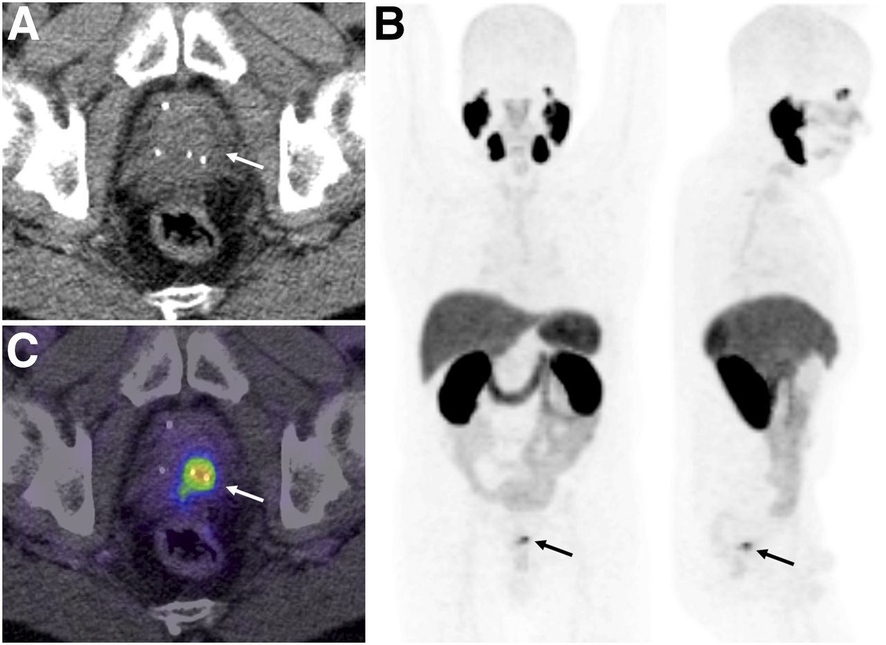
Images of patient 1. (A) After initial brachytherapy, no tumor detection within prostate is achievable with CT alone. (C) With 18F-PSMA-1007 PET/CT, good delineation of local recurrence is possible. (B) Maximum-intensity projection highlights advantage of low urinary clearance, because no PET signal derives from urinary bladder. Arrows indicate location of tumor.

FIGURE 2.
  • Download figure
  • Open in new tab
  • Download powerpoint
FIGURE 2.

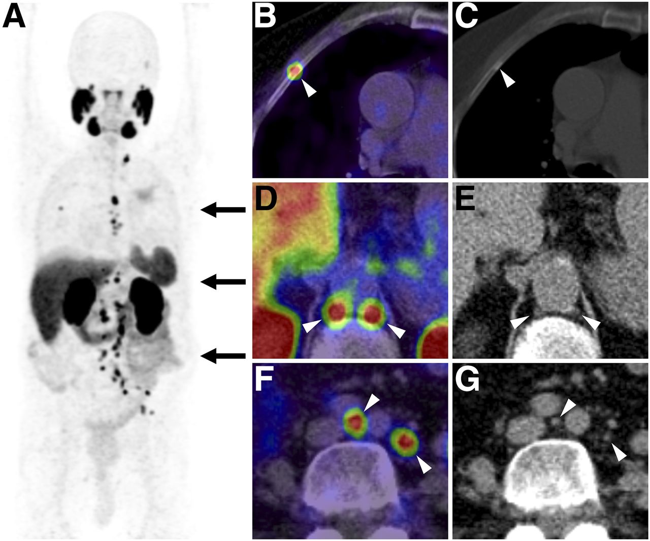
Images of patient 8, who presented with BCR after radical prostatectomy and salvage radiotherapy. 18F-PSMA-1007 PET/CT (maximum-intensity projection, A) detected recurrent disease with multiple bone metastases (B and C) and LN metastases (D–G) 8 y after initial diagnosis. All images were acquired 3 h after injection. Arrowheads indicate locations of tumors.

Clinical Impact

Of the 3 patients diagnosed with local recurrence, 2 had undergone prior radical prostatectomy and were eligible for salvage radiation with a local boost to PET-positive lesions. The patient diagnosed with local recurrence after primary radiotherapy underwent radical prostatectomy with concomitant lymphadenectomy, which confirmed organ-defined recurrence (pTXypN0). In patients 4, 7, 8, and 9, diagnosed with extrapelvic LN or bone metastases, systemic therapy was initiated. Notably, in routine management of BCR these patients would have been candidates for salvage radiation. Patient 5, presenting with a single PET-positive pelvic LN, received salvage surgery, which confirmed the PET/CT results histologically. On follow-up, PSA was undetectable and PSMA PET/CT did not reveal any tumor-suggestive findings.

Tumor Uptake Characteristics

18F-PSMA-1007 PET/CT detected 76 lesions (3 representing local recurrence, 50 representing LN metastases, and 23 representing bone metastases). The median SUVmax for all lesions at 1 h after injection was 7.00 (range, 2.04–40.01). SUVmax increased significantly by 3 h after injection (median, 11.35; range, 2.36–55.05; P < 0.001). Additionally, the ratio of median SUVmax to blood pool increased significantly between 1 and 3 h after injection (2.10 vs. 4.57, P < 0.001). Uptake dynamics and the ratio of SUVmax to blood pool for each lesion group are shown in Figure 3.

FIGURE 3.
  • Download figure
  • Open in new tab
  • Download powerpoint
FIGURE 3.

(A) Tumor SUVmax increased from 1 to 3 h after injection in all lesion groups. (B) Ratios of SUVmax to blood pool presented improved contrast at delayed time point. Data are mean and SE.

Volumetric Characteristics of PSMA-Positive LNs

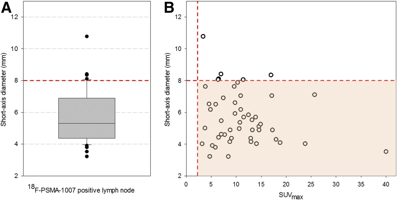
In total, 50 18F-PSMA-1007–positive LNs were evaluated. Their mean short-axis diameter was 5.7 mm (95% confidence interval, 5.2–6.1 mm), their mean long-axis diameter was 8.1 mm (95% confidence interval, 7.5–8.8 mm), and their mean volume was 0.3 mL (95% confidence interval, 0.2–0.4 mL).

Forty-four (88%) of 50 18F-PSMA-1007–positive LNs had a short-axis diameter of less than 8 mm. Figure 4 shows detailed volumetric characteristics and the relation between SUVmax and short-axis diameter.

FIGURE 4.
  • Download figure
  • Open in new tab
  • Download powerpoint
FIGURE 4.

(A) Analysis of short-axis diameter of 18F-PSMA-1007–positive LNs indicates that 18F-PSMA-1007 PET/CT detects micrometastases that could be missed on CT or MRI. (B) No dependency of SUVmax on lesion diameter was found. Orange dashed reference lines are shown for short-axis diameter of 8 mm and SUVmax of 2.0.

DISCUSSION

To our knowledge, this was the first study evaluating 18F-PSMA-1007 PET/CT in the setting of BCR of prostate cancer. The source of PSA elevation was successfully located in 9 of 12 patients (75%). All 3 patients with negative PET findings had PSA values of 0.5 ng/mL or less. However, 1 patient with positive PET findings had a PSA level of 0.08 ng/mL. These results suggest that 18F-PSMA-1007 has a limited sensitivity below a PSA level of 0.5 ng/mL—a limitation that may be general among PSMA tracers, as similar findings have been reported for 68Ga-PSMA-11 and 18F-DCFPyL (16). In patients undergoing 68Ga-PSMA-11 PET/CT, a sensitivity of 33%–46% has been reported for a PSA level of 0.5 ng/mL or less, improving to more than 80% for higher PSA levels (6,16).

Because the positron energy of 18F is lower than that of 68Ga (0.65 vs. 1.9 MeV), the theoretic achievable resolution of 18F is slightly better than that of 68Ga (17). Therefore, it is hypothesized that the sensitivity of PSMA PET could be further improved by the introduction of 18F-tracers. However, phantom measurements in human scanners found comparable results for both nuclides (18), and a study comparing 68Ga-PSMA-11 and 18F-DCFPyL found minimal differences between the two, with the sensitivity of both decreasing abruptly when PSA was below 0.5 ng/mL (16). Larger comparative trials are required to demonstrate those differences with regard to 18F-PSMA-1007. Nevertheless, the actual findings already imply that, rather than the applied radiolabel, it is the technical resolution limit of current PET/CT scanners (2–3 mm) that might be the limiting factor in the detection of micrometastases.

Assuming similar diagnostic accuracy, the actual added value of 18F-tracers might lie in the possibility of producing large-scale batches in a cyclotron and supplying them through a satellite center, because of the longer half-life. A specific advantage of 18F-PSMA-1007 for the detection of local recurrence may be its low urinary clearance. Additionally, although yet unproven, the similar chemical structure of 18F-PSMA-1007 might allow closer prediction of uptake of the therapeutic ligand PSMA-617, which can be used for PSMA-targeted radioligand therapy (19).

CONCLUSION

This study showed the potential of 18F-PSMA-1007 PET/CT for prostate cancer patients with BCR, confirming that nonvisualization of the urinary bladder facilitates the evaluation of lesions close to the urinary tract, such as local recurrence and pelvic LNs. Nevertheless, these initial results, although limited by a small number of patients, appear comparable to those of 68Ga-PSMA-11 PET/CT.

DISCLOSURE

There have been patent applications for PSMA-617 by Klaus Kopka and Uwe Haberkorn and for PSMA-1007 by Jens Cardinale, Uwe Haberkorn, Frederik Giesel, and Klaus Kopka. No other potential conflict of interest relevant to this article was reported.

Acknowledgments

We thank Martin Schäfer for synthesis of the tracer.

Footnotes

  • Published online Feb. 1, 2018.

  • © 2018 by the Society of Nuclear Medicine and Molecular Imaging.

REFERENCES

  1. 1.↵
    1. Rouvière O,
    2. Vitry T,
    3. Lyonnet D
    . Imaging of prostate cancer local recurrences: why and how? Eur Radiol. 2010;20:1254–1266.
    OpenUrlCrossRefPubMed
  2. 2.↵
    1. Afshar-Oromieh A,
    2. Avtzi E,
    3. Giesel FL,
    4. et al
    . The diagnostic value of PET/CT imaging with the 68Ga-labelled PSMA ligand HBED-CC in the diagnosis of recurrent prostate cancer. Eur J Nucl Med Mol Imaging. 2015;42:197–209.
    OpenUrlCrossRefPubMed
  3. 3.
    1. Eiber M,
    2. Maurer T,
    3. Souvatzoglou M,
    4. Beer AJ,
    5. Ruffani A,
    6. Haller B
    . Evaluation of hybrid 68Ga-PSMA ligand PET/CT in 248 patients with biochemical recurrence after radical prostatectomy. J Nucl Med. 2015;56:668–674.
    OpenUrlAbstract/FREE Full Text
  4. 4.
    1. Maurer T,
    2. Gschwend JE,
    3. Rauscher I,
    4. et al
    . Diagnostic efficacy of 68gallium-PSMA positron emission tomography compared to conventional imaging for lymph node staging of 130 consecutive patients with intermediate to high risk prostate cancer. J Urol. 2016;195:1436–1443.
    OpenUrlCrossRefPubMed
  5. 5.
    1. Perera M,
    2. Papa N,
    3. Christidis D,
    4. et al
    . Sensitivity, specificity, and predictors of positive 68Ga-prostate-specific membrane antigen positron emission tomography in advanced prostate cancer: a systematic review and meta-analysis. Eur Urol. 2016;70:926–937.
    OpenUrl
  6. 6.↵
    1. Afshar-Oromieh A,
    2. Holland-Letz T,
    3. Giesel FL,
    4. et al
    . Diagnostic performance of 68Ga-PSMA-11 (HBED-CC) PET/CT in patients with recurrent prostate cancer: evaluation in 1007 patients. Eur J Nucl Med Mol Imaging. 2017;44:1258–1268.
    OpenUrl
  7. 7.↵
    1. Morigi JJ,
    2. Stricker PD,
    3. van Leeuwen PJ,
    4. et al
    . Prospective comparison of 18F-fluoromethylcholine versus 68Ga-PSMA PET/CT in prostate cancer patients who have rising PSA after curative treatment and are being considered for targeted therapy. J Nucl Med. 2015;56:1185–1190.
    OpenUrlAbstract/FREE Full Text
  8. 8.↵
    1. Afshar-Oromieh A,
    2. Haberkorn U,
    3. Eder M,
    4. Eisenhut M,
    5. Zechmann CM
    . [68Ga]gallium-labelled PSMA ligand as superior PET tracer for the diagnosis of prostate cancer: comparison with 18F-FECH. Eur J Nucl Med Mol Imaging. 2012;39:1085–1086.
    OpenUrlCrossRefPubMed
  9. 9.↵
    1. Cho SY,
    2. Gage KL,
    3. Mease RC,
    4. et al
    . Biodistribution, tumor detection, and radiation dosimetry of 18F-DCFBC, a low-molecular-weight inhibitor of prostate-specific membrane antigen, in patients with metastatic prostate cancer. J Nucl Med. 2012;53:1883–1891.
    OpenUrlAbstract/FREE Full Text
  10. 10.↵
    1. Giesel FL,
    2. Hadaschik B,
    3. Cardinale J,
    4. et al
    . F-18 labelled PSMA-1007: biodistribution, radiation dosimetry and histopathological validation of tumor lesions in prostate cancer patients. Eur J Nucl Med Mol Imaging. 2017;44:678–688.
    OpenUrl
  11. 11.↵
    1. Eiber M,
    2. Weirich G,
    3. Holzapfel K,
    4. et al
    . Simultaneous 68Ga-PSMA HBED-CC PET/MRI improves the localization of primary prostate cancer. Eur Urol. 2016;70:829–836.
    OpenUrl
  12. 12.↵
    1. Afshar-Oromieh A,
    2. Haberkorn U,
    3. Schlemmer HP,
    4. et al
    . Comparison of PET/CT and PET/MRI hybrid systems using a 68Ga-labelled PSMA ligand for the diagnosis of recurrent prostate cancer: initial experience. Eur J Nucl Med Mol Imaging. 2014;41:887–897.
    OpenUrlCrossRefPubMed
  13. 13.↵
    1. Freitag MT,
    2. Radtke JP,
    3. Afshar-Oromieh A,
    4. et al
    . Local recurrence of prostate cancer after radical prostatectomy is at risk to be missed in 68Ga-PSMA-11-PET of PET/CT and PET/MRI: comparison with mpMRI integrated in simultaneous PET/MRI. Eur J Nucl Med Mol Imaging. 2017;44:776–787.
    OpenUrl
  14. 14.↵
    1. Giesel FL,
    2. Kesch C,
    3. Yun M,
    4. et al
    . 18F-PSMA-1007 PET/CT detects micrometastases in a patient with biochemically recurrent prostate cancer. Clin Genitourin Cancer. 2017;15:e497–e499.
    OpenUrl
  15. 15.↵
    1. Moltz JH,
    2. Bornemann L,
    3. Kuhnigk J-M,
    4. et al
    . Advanced segmentation techniques for lung nodules, liver metastases, and enlarged lymph nodes in CT scans. IEEE J Sel Top Signal Process. 2009;3:122–134.
    OpenUrlCrossRef
  16. 16.↵
    1. Dietlein F,
    2. Kobe C,
    3. Neubauer S,
    4. et al
    . PSA-stratified performance of 18F- and 68Ga-PSMA PET in patients with biochemical recurrence of prostate cancer. J Nucl Med. 2017;58:947–952.
    OpenUrlAbstract/FREE Full Text
  17. 17.↵
    1. Sanchez-Crespo A
    . Comparison of gallium-68 and fluorine-18 imaging characteristics in positron emission tomography. Appl Radiat Isot. 2013;76:55–62.
    OpenUrlCrossRef
  18. 18.↵
    1. Kemerink GJ,
    2. Visser MG,
    3. Franssen R,
    4. et al
    . Effect of the positron range of 18F, 68Ga and 124I on PET/CT in lung-equivalent materials. Eur J Nucl Med Mol Imaging. 2011;38:940–948.
    OpenUrlCrossRefPubMed
  19. 19.↵
    1. Giesel FL,
    2. Cardinale J,
    3. Schafer M,
    4. et al
    . 18F-labelled PSMA-1007 shows similarity in structure, biodistribution and tumour uptake to the theragnostic compound PSMA-617. Eur J Nucl Med Mol Imaging. 2016;43:1929–1930.
    OpenUrl
  • Received for publication May 22, 2017.
  • Accepted for publication December 19, 2017.
PreviousNext
Back to top

In this issue

Journal of Nuclear Medicine: 59 (4)
Journal of Nuclear Medicine
Vol. 59, Issue 4
April 1, 2018
  • Table of Contents
  • Table of Contents (PDF)
  • About the Cover
  • Index by author
Print
Download PDF
Article Alerts
Sign In to Email Alerts with your Email Address
Email Article

Thank you for your interest in spreading the word on Journal of Nuclear Medicine.

NOTE: We only request your email address so that the person you are recommending the page to knows that you wanted them to see it, and that it is not junk mail. We do not capture any email address.

Enter multiple addresses on separate lines or separate them with commas.
Biochemical Recurrence of Prostate Cancer: Initial Results with [18F]PSMA-1007 PET/CT
(Your Name) has sent you a message from Journal of Nuclear Medicine
(Your Name) thought you would like to see the Journal of Nuclear Medicine web site.
Citation Tools
Biochemical Recurrence of Prostate Cancer: Initial Results with [18F]PSMA-1007 PET/CT
Frederik L. Giesel, Leon Will, Claudia Kesch, Martin Freitag, Christophe Kremer, Jonas Merkle, Oliver C. Neels, Jens Cardinale, Boris Hadaschik, Markus Hohenfellner, Klaus Kopka, Uwe Haberkorn, Clemens Kratochwil
Journal of Nuclear Medicine Apr 2018, 59 (4) 632-635; DOI: 10.2967/jnumed.117.196329

Citation Manager Formats

  • BibTeX
  • Bookends
  • EasyBib
  • EndNote (tagged)
  • EndNote 8 (xml)
  • Medlars
  • Mendeley
  • Papers
  • RefWorks Tagged
  • Ref Manager
  • RIS
  • Zotero
Share
Biochemical Recurrence of Prostate Cancer: Initial Results with [18F]PSMA-1007 PET/CT
Frederik L. Giesel, Leon Will, Claudia Kesch, Martin Freitag, Christophe Kremer, Jonas Merkle, Oliver C. Neels, Jens Cardinale, Boris Hadaschik, Markus Hohenfellner, Klaus Kopka, Uwe Haberkorn, Clemens Kratochwil
Journal of Nuclear Medicine Apr 2018, 59 (4) 632-635; DOI: 10.2967/jnumed.117.196329
Twitter logo Facebook logo LinkedIn logo Mendeley logo
  • Tweet Widget
  • Facebook Like
  • Google Plus One
Bookmark this article

Jump to section

  • Article
    • Abstract
    • MATERIALS AND METHODS
    • RESULTS
    • DISCUSSION
    • CONCLUSION
    • DISCLOSURE
    • Acknowledgments
    • Footnotes
    • REFERENCES
  • Figures & Data
  • Info & Metrics
  • PDF

Related Articles

  • This Month in JNM
  • PubMed
  • Google Scholar

Cited By...

  • Intraoperative 68Ga-PSMA Cerenkov Luminescence Imaging for Surgical Margins in Radical Prostatectomy: A Feasibility Study
  • Detection Efficacy of 18F-PSMA-1007 PET/CT in 251 Patients with Biochemical Recurrence of Prostate Cancer After Radical Prostatectomy
  • Interobserver Agreement for the Standardized Reporting System PSMA-RADS 1.0 on 18F-DCFPyL PET/CT Imaging
  • Google Scholar

More in this TOC Section

Theranostics

  • Determination of the Intralesional Distribution of Theranostic 124I-Omburtamab Convection-Enhanced Delivery in Treatment of Diffuse Intrinsic Pontine Glioma
  • Evidence-Based Clinical Protocols to Monitor Efficacy of [177Lu]Lu-PSMA Radiopharmaceutical Therapy in Metastatic Castration-Resistant Prostate Cancer Using Real-World Data
  • 177Lu-Labeled Anticlaudin 6 Monoclonal Antibody for Targeted Therapy in Esophageal Cancer
Show more Theranostics

Clinical

  • Detection of Early Progression with 18F-DCFPyL PET/CT in Men with Metastatic Castration-Resistant Prostate Cancer Receiving Bipolar Androgen Therapy
  • TauIQ: A Canonical Image Based Algorithm to Quantify Tau PET Scans
  • Dual PET Imaging in Bronchial Neuroendocrine Neoplasms: The NETPET Score as a Prognostic Biomarker
Show more Clinical

Similar Articles

Keywords

  • Peptides
  • PET/CT
  • biochemical recurrence
  • 18F-PSMA-1007
  • PSMA-PET
  • prostate cancer
SNMMI

© 2025 SNMMI

Powered by HighWire