
Main menu

  • Home
  • Content
    • Current
    • Ahead of print
    • Past Issues
    • JNM Supplement
    • SNMMI Annual Meeting Abstracts
    • Continuing Education
    • JNM Podcasts
  • Subscriptions
    • Subscribers
    • Institutional and Non-member
    • Rates
    • Journal Claims
    • Corporate & Special Sales
  • Authors
    • Submit to JNM
    • Information for Authors
    • Assignment of Copyright
    • AQARA requirements
  • Info
    • Reviewers
    • Permissions
    • Advertisers
  • About
    • About Us
    • Editorial Board
    • Contact Information
  • More
    • Alerts
    • Feedback
    • Help
    • SNMMI Journals
  • SNMMI
    • JNM
    • JNMT
    • SNMMI Journals
    • SNMMI

User menu

  • Subscribe
  • My alerts
  • Log in
  • My Cart

Search

  • Advanced search
Journal of Nuclear Medicine
  • SNMMI
    • JNM
    • JNMT
    • SNMMI Journals
    • SNMMI
  • Subscribe
  • My alerts
  • Log in
  • My Cart
Journal of Nuclear Medicine

Advanced Search

  • Home
  • Content
    • Current
    • Ahead of print
    • Past Issues
    • JNM Supplement
    • SNMMI Annual Meeting Abstracts
    • Continuing Education
    • JNM Podcasts
  • Subscriptions
    • Subscribers
    • Institutional and Non-member
    • Rates
    • Journal Claims
    • Corporate & Special Sales
  • Authors
    • Submit to JNM
    • Information for Authors
    • Assignment of Copyright
    • AQARA requirements
  • Info
    • Reviewers
    • Permissions
    • Advertisers
  • About
    • About Us
    • Editorial Board
    • Contact Information
  • More
    • Alerts
    • Feedback
    • Help
    • SNMMI Journals
  • View or Listen to JNM Podcast
  • Visit JNM on Facebook
  • Join JNM on LinkedIn
  • Follow JNM on Twitter
  • Subscribe to our RSS feeds
Research ArticleOncology

Efficacy of Radioembolization with 166Ho-Microspheres in Salvage Patients with Liver Metastases: A Phase 2 Study

Jip F. Prince, Maurice A.A.J. van den Bosch, Johannes F.W. Nijsen, Maarten L.J. Smits, Andor F. van den Hoven, Stavros Nikolakopoulos, Frank J. Wessels, Rutger C.G. Bruijnen, Manon N.G.J.A. Braat, Bernard A. Zonnenberg and Marnix G.E.H. Lam
Journal of Nuclear Medicine April 2018, 59 (4) 582-588; DOI: https://doi.org/10.2967/jnumed.117.197194
Jip F. Prince
1Department of Radiology and Nuclear Medicine, University Medical Center Utrecht, Utrecht, The Netherlands; and
  • Find this author on Google Scholar
  • Find this author on PubMed
  • Search for this author on this site
Maurice A.A.J. van den Bosch
1Department of Radiology and Nuclear Medicine, University Medical Center Utrecht, Utrecht, The Netherlands; and
  • Find this author on Google Scholar
  • Find this author on PubMed
  • Search for this author on this site
Johannes F.W. Nijsen
1Department of Radiology and Nuclear Medicine, University Medical Center Utrecht, Utrecht, The Netherlands; and
  • Find this author on Google Scholar
  • Find this author on PubMed
  • Search for this author on this site
Maarten L.J. Smits
1Department of Radiology and Nuclear Medicine, University Medical Center Utrecht, Utrecht, The Netherlands; and
  • Find this author on Google Scholar
  • Find this author on PubMed
  • Search for this author on this site
Andor F. van den Hoven
1Department of Radiology and Nuclear Medicine, University Medical Center Utrecht, Utrecht, The Netherlands; and
  • Find this author on Google Scholar
  • Find this author on PubMed
  • Search for this author on this site
Stavros Nikolakopoulos
2Department of Biostatistics, Julius Center for Health Sciences and Primary Care, University Medical Center Utrecht, Utrecht, The Netherlands
  • Find this author on Google Scholar
  • Find this author on PubMed
  • Search for this author on this site
Frank J. Wessels
1Department of Radiology and Nuclear Medicine, University Medical Center Utrecht, Utrecht, The Netherlands; and
  • Find this author on Google Scholar
  • Find this author on PubMed
  • Search for this author on this site
Rutger C.G. Bruijnen
1Department of Radiology and Nuclear Medicine, University Medical Center Utrecht, Utrecht, The Netherlands; and
  • Find this author on Google Scholar
  • Find this author on PubMed
  • Search for this author on this site
Manon N.G.J.A. Braat
1Department of Radiology and Nuclear Medicine, University Medical Center Utrecht, Utrecht, The Netherlands; and
  • Find this author on Google Scholar
  • Find this author on PubMed
  • Search for this author on this site
Bernard A. Zonnenberg
1Department of Radiology and Nuclear Medicine, University Medical Center Utrecht, Utrecht, The Netherlands; and
  • Find this author on Google Scholar
  • Find this author on PubMed
  • Search for this author on this site
Marnix G.E.H. Lam
1Department of Radiology and Nuclear Medicine, University Medical Center Utrecht, Utrecht, The Netherlands; and
  • Find this author on Google Scholar
  • Find this author on PubMed
  • Search for this author on this site
  • Article
  • Figures & Data
  • Supplemental
  • Info & Metrics
  • PDF
Loading

Article Figures & Data

Figures

  • Tables
  • Additional Files
  • FIGURE 1.
    • Download figure
    • Open in new tab
    • Download powerpoint
    FIGURE 1.

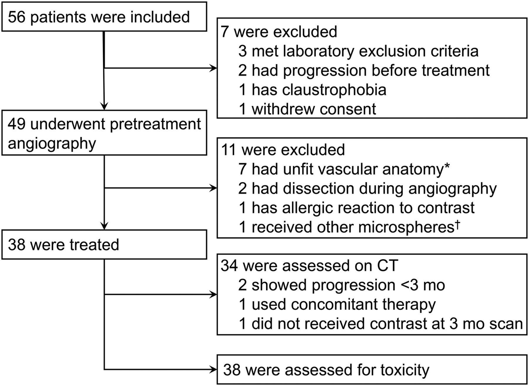
    Flowchart of study. *Metastases could not be adequately targeted, extrahepatic tissue was also targeted, or there were more than 3 injection positions. †Laboratory experiments indicated need to optimize microsphere production process; treatments were halted until optimization was complete.
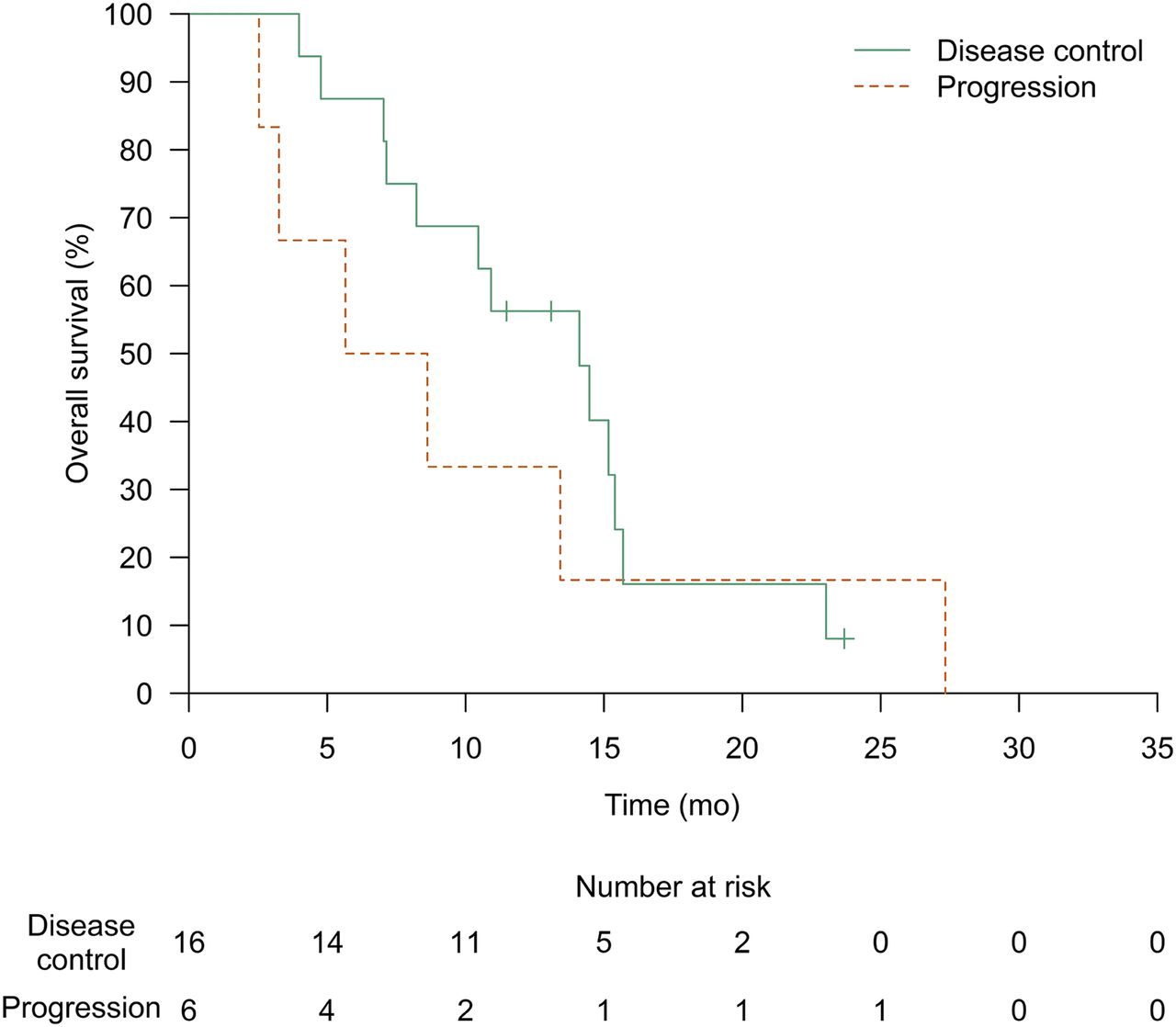
  • FIGURE 2.
    • Download figure
    • Open in new tab
    • Download powerpoint
    FIGURE 2.

    Kaplan–Meier estimate of median overall survival was 14.1 mo (95% CI, 8.2 mo–∞) for patients with colorectal disease and disease control of target lesions at 3 mo, and 7.1 mo (95% CI, 3.3 mo–∞) for patients with progressive target lesions at 3 mo (P = 0.44). Data of 1 patient are missing (Fig. 1).

  • FIGURE 3.
    • Download figure
    • Open in new tab
    • Download powerpoint
    FIGURE 3.

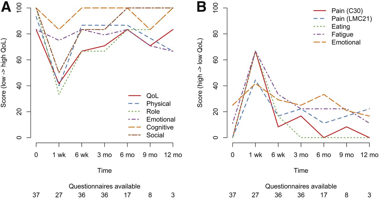
    (A) Median quality-of-life (QoL) scores per time point (higher score = better QoL). (B) Median symptom scores per time point (higher score = worse symptoms).

Tables

  • Figures
  • Additional Files
    • View popup
    TABLE 1

    Baseline Characteristics of Treated Patients (n = 38)

    CharacteristicValue
    Age (y)66 (41–84)
    Sex
     Male22 (58%)
     Female16 (42%)
    World Health Organization performance  status
     032 (84%)
     15 (13%)
     21 (3%)
    Primary malignancy
     Colorectal23 (61%)
     Breast4 (11%)
     Cholangiocarcinoma4 (11%)
     Neuroendocrine tumor2 (5%)
     Uveal melanoma2 (5%)
     Pancreatic cancer, gastric cancer,  or thymoma3 (8%)
    Time since diagnosis (mo)28 (4–95)
    Occurrence of liver metastases
     Synchronous19 (50%)
     Metachronous19 (50%)
    Time since liver metastases (mo)18 (3–92)
    Extrahepatic disease on 18F-FDG  PET/CT
     Lung8/10 (80%)
     Lymph node4/10 (40%)
     Skeletal2/10 (20%)
     Liver mass (kg)2.0 (1.2–4.0)
    Tumor load on contrast-enhanced CT
     0%–25%30 (79%)
     25%–50%6 (16%)
     >50%2 (5%)
    • Qualitative data are expressed as numbers followed by percentages in parentheses; continuous data are expressed as median followed by range in parentheses.

    • View popup
    TABLE 2

    Treatment Characteristics of Colorectal Cancer Patients (n = 23)

    CharacteristicValue
    Chemotherapy (prior to study inclusion)
     Total23 (100%)
     Capecitabine22 (96%)
     Oxaliplatin21 (91%)
     Bevacizumab14 (61%)
     Irinotecan11 (48%)
     5-fluorouracil6 (26%)
     Leucovorin5 (22%)
     Panitumumab4 (17%)
     Cetuximab1 (4%)
     Dabrafenib1 (4%)
     Tegafur-uracil1 (4%)
    Targeted part of liver
     Whole liver36 (95%)
     Right lobe2 (5%)
     Left lobe0 (0%)
    Treatment sessions
     136 (95%)
     22 (5%)
    Specific activity (MBq/mg)*11.26 (4.95–21.34)
    Infused activity (MBq)*6,412 (2,213–13,189)
    Administration of treatment  dose†96% (41%–99%)
    Adequate administration of  activity (>90%)‡26/37 (70%)
    Absorbed liver dose (Gy)51 (26–69)
    Predicted lung shunt  (99mTc-MAA)3.2% (0.01%–19.3%)
    Actual lung shunt (166Ho)0.02% (0%–0.7%)
    • ↵* All injections combined (including scout dose), at respective time of injection.

    • ↵† Percentage of prepared.

    • ↵‡ In other patients, stasis occurred or infusion was stopped because of pain.

    • Qualitative data are expressed as numbers followed by percentages in parentheses; continuous data are expressed as median followed by range in parentheses.

    • View popup
    TABLE 3

    Response on Contrast-Enhanced CT

    Response categoryTarget lesionsLiver-specificAbdomen
    3 mo
     Complete response———
     Partial response5 (14)5 (14)5 (14)
     Stable disease22 (59)13 (35)9 (24)
     Progressive disease*10 (27)19 (51)23 (62)
     Total37 (100)37 (100)37 (100)
    6 mo
     Complete response1 (3)1 (3)1 (3)
     Partial response2 (5)2 (5)2 (5)
     Stable disease10 (26)4 (11)3 (8)
     Progressive disease*25 (66)31 (82)32 (84)
     Total38 (100)38 (100)38 (100)
    9 mo
     Complete response1 (3)1 (3)1 (3)
     Partial response1 (3)——
     Stable disease2 (5)1 (3)1 (3)
     Progressive disease*34 (89)36 (95)36 (95)
     Total38 (100)38 (100)38 (100)
    12 mo
     Complete response———
     Partial response———
     Stable disease2 (5)——
     Progressive disease*36 (95)38 (100)38 (100)
     Total38 (100)38 (100)38 (100)
    • ↵* Not evaluable, or disease progression was inferred for patients with missing data.

    • Data are n followed by percentage in parentheses. Timing was at median of 90 d (range, 76–104 d) after treatment at 3 mo, 181 d (152–195) at 6 mo, 278 d (252–312) at 9 mo, and 369 d (368–369) at 12 mo.

    • View popup
    TABLE 4

    Adverse Events—Clinical

    Adverse eventAny time≤1 wk>1 wkGrade 3 or 4
    Nausea74% (28)71% (27)34% (13)8% (3)
    Abdominal pain71% (27)68% (26)47% (18)18% (7)
    Fatigue66% (25)26% (10)61% (23)3% (1)
    Vomiting66% (25)58% (22)18% (7)3% (1)
    Back pain34% (13)24% (9)11% (4)0
    Anorexia26% (10)13% (5)21% (8)0
    Edema limbs18% (7)018% (7)0
    Fever18% (7)11% (4)11% (4)0
    Constipation16% (6)5% (2)11% (4)0
    Dizziness16% (6)11% (4)11% (4)0
    Allergic reaction13% (5)13% (5)5% (2)0
    Arthralgia13% (5)3% (1)11% (4)0
    Dyspnea13% (5)013% (5)0
    Shoulder pain13% (5)5% (2)11% (4)0
    Ascites11% (4)011% (4)3% (1)
    Chills11% (4)011% (4)0
    Dysgeusia11% (4)3% (1)11% (4)0
    Gastric stenosis3% (1)03% (1)3% (1)
    Hepatic failure*3% (1)03% (1)3% (1)
    Liver abscesses3% (1)03% (1)3% (1)
    Paroxysmal atrial tachycardia3% (1)03% (1)3% (1)
    Thoracic pain3% (1)3% (1)3% (1)3% (1)
    Upper gastrointestinal hemorrhage3% (1)03% (1)3% (1)
    • ↵* We suspected mix of disease progression and radioembolization induced liver disease. This patient is also included in parameter “ascites.”

    • Numbers of events are in parentheses. Adverse events are those with incidence of >10% or CTCAE grade 3 or 4, after treatment, regardless of relation with 166Ho radioembolization.

    • View popup
    TABLE 5

    Adverse Events—Laboratory Examinations

    Adverse eventGrade 1–2Grade 3Grade 4NA
    γ-glutamyl transferase62840
    Lymphocytopenia15610
    Bilirubin12010
    Alkaline phosphatase32600
    Aspartate aminotransferase35300
    Alanine aminotransferase28200
    Lactate dehydrogenase27000
    Hemoglobin26000
    Ammonia22000
    Albumin20000
    Platelet count19000
    Erythrocytes15000
    Sodium15002
    Leukocytes14000
    Creatinine12000
    Potassium9002
    Total protein9004
    Calcium (ionized)8003
    Magnesium6004
    Phosphorus6003
    Urea6003
    Chloride5004
    Bicarbonate4004
    • NA = not available.

    • Maximum CTCAE grade of adverse event during follow-up after treatment is shown; some of these adverse events were preexistent.

Additional Files

  • Figures
  • Tables
  • Supplemental Data

    Files in this Data Supplement:

    • Supplemental Data
PreviousNext
Back to top

In this issue

Journal of Nuclear Medicine: 59 (4)
Journal of Nuclear Medicine
Vol. 59, Issue 4
April 1, 2018
  • Table of Contents
  • Table of Contents (PDF)
  • About the Cover
  • Index by author
Print
Download PDF
Article Alerts
Sign In to Email Alerts with your Email Address
Email Article

Thank you for your interest in spreading the word on Journal of Nuclear Medicine.

NOTE: We only request your email address so that the person you are recommending the page to knows that you wanted them to see it, and that it is not junk mail. We do not capture any email address.

Enter multiple addresses on separate lines or separate them with commas.
Efficacy of Radioembolization with 166Ho-Microspheres in Salvage Patients with Liver Metastases: A Phase 2 Study
(Your Name) has sent you a message from Journal of Nuclear Medicine
(Your Name) thought you would like to see the Journal of Nuclear Medicine web site.
Citation Tools
Efficacy of Radioembolization with 166Ho-Microspheres in Salvage Patients with Liver Metastases: A Phase 2 Study
Jip F. Prince, Maurice A.A.J. van den Bosch, Johannes F.W. Nijsen, Maarten L.J. Smits, Andor F. van den Hoven, Stavros Nikolakopoulos, Frank J. Wessels, Rutger C.G. Bruijnen, Manon N.G.J.A. Braat, Bernard A. Zonnenberg, Marnix G.E.H. Lam
Journal of Nuclear Medicine Apr 2018, 59 (4) 582-588; DOI: 10.2967/jnumed.117.197194

Citation Manager Formats

  • BibTeX
  • Bookends
  • EasyBib
  • EndNote (tagged)
  • EndNote 8 (xml)
  • Medlars
  • Mendeley
  • Papers
  • RefWorks Tagged
  • Ref Manager
  • RIS
  • Zotero
Share
Efficacy of Radioembolization with 166Ho-Microspheres in Salvage Patients with Liver Metastases: A Phase 2 Study
Jip F. Prince, Maurice A.A.J. van den Bosch, Johannes F.W. Nijsen, Maarten L.J. Smits, Andor F. van den Hoven, Stavros Nikolakopoulos, Frank J. Wessels, Rutger C.G. Bruijnen, Manon N.G.J.A. Braat, Bernard A. Zonnenberg, Marnix G.E.H. Lam
Journal of Nuclear Medicine Apr 2018, 59 (4) 582-588; DOI: 10.2967/jnumed.117.197194
Twitter logo Facebook logo LinkedIn logo Mendeley logo
  • Tweet Widget
  • Facebook Like
  • Google Plus One
Bookmark this article

Jump to section

  • Article
    • Abstract
    • MATERIALS AND METHODS
    • RESULTS
    • DISCUSSION
    • CONCLUSION
    • DISCLOSURE
    • Acknowledgments
    • Footnotes
    • REFERENCES
  • Figures & Data
  • Supplemental
  • Info & Metrics
  • PDF

Related Articles

  • This Month in JNM
  • PubMed
  • Google Scholar

Cited By...

  • Safety and Efficacy of 166Ho Radioembolization in Hepatocellular Carcinoma: The HEPAR Primary Study
  • Dose-Effect Relationships of 166Ho Radioembolization in Colorectal Cancer
  • First Evidence for a Dose-Response Relationship in Patients Treated with 166Ho Radioembolization: A Prospective Study
  • Prediction of Tumor Control in 90Y Radioembolization by Logit Models with PET/CT-Based Dose Metrics
  • Developing a Roadmap for Interventional Oncology
  • Google Scholar

More in this TOC Section

Oncology

  • Comparative PSMA expression at early (1hour) vs late (2hour) in primary and secondary sites of involvement in prostate cancer.
  • Imaging spectrum of peritoneal carcinomatosis associated with various etiologies on 18F-FDG PET/CT
  • Hybrid Imaging Features of Musculoskeletal Tumors
Show more Oncology

Clinical

  • Comparative PSMA expression at early (1hour) vs late (2hour) in primary and secondary sites of involvement in prostate cancer.
  • Imaging spectrum of peritoneal carcinomatosis associated with various etiologies on 18F-FDG PET/CT
  • Hybrid Imaging Features of Musculoskeletal Tumors
Show more Clinical

Similar Articles

Keywords

  • gastrointestinal
  • radionuclide therapy
  • radioembolization
  • SIRT
  • holmium
  • liver
SNMMI

© 2025 SNMMI

Powered by HighWire