
Main menu

  • Home
  • Content
    • Current
    • Ahead of print
    • Past Issues
    • JNM Supplement
    • SNMMI Annual Meeting Abstracts
    • Continuing Education
    • JNM Podcasts
  • Subscriptions
    • Subscribers
    • Institutional and Non-member
    • Rates
    • Journal Claims
    • Corporate & Special Sales
  • Authors
    • Submit to JNM
    • Information for Authors
    • Assignment of Copyright
    • AQARA requirements
  • Info
    • Reviewers
    • Permissions
    • Advertisers
  • About
    • About Us
    • Editorial Board
    • Contact Information
  • More
    • Alerts
    • Feedback
    • Help
    • SNMMI Journals
  • SNMMI
    • JNM
    • JNMT
    • SNMMI Journals
    • SNMMI

User menu

  • Subscribe
  • My alerts
  • Log in
  • My Cart

Search

  • Advanced search
Journal of Nuclear Medicine
  • SNMMI
    • JNM
    • JNMT
    • SNMMI Journals
    • SNMMI
  • Subscribe
  • My alerts
  • Log in
  • My Cart
Journal of Nuclear Medicine

Advanced Search

  • Home
  • Content
    • Current
    • Ahead of print
    • Past Issues
    • JNM Supplement
    • SNMMI Annual Meeting Abstracts
    • Continuing Education
    • JNM Podcasts
  • Subscriptions
    • Subscribers
    • Institutional and Non-member
    • Rates
    • Journal Claims
    • Corporate & Special Sales
  • Authors
    • Submit to JNM
    • Information for Authors
    • Assignment of Copyright
    • AQARA requirements
  • Info
    • Reviewers
    • Permissions
    • Advertisers
  • About
    • About Us
    • Editorial Board
    • Contact Information
  • More
    • Alerts
    • Feedback
    • Help
    • SNMMI Journals
  • View or Listen to JNM Podcast
  • Visit JNM on Facebook
  • Join JNM on LinkedIn
  • Follow JNM on Twitter
  • Subscribe to our RSS feeds
Research ArticleNeurology

Impact of Endothelial 18-kDa Translocator Protein on the Quantification of 18F-DPA-714

Catriona Wimberley, Sonia Lavisse, Vincent Brulon, Marie-Anne Peyronneau, Claire Leroy, Benedetta Bodini, Philippe Remy, Bruno Stankoff, Irène Buvat and Michel Bottlaender
Journal of Nuclear Medicine February 2018, 59 (2) 307-314; DOI: https://doi.org/10.2967/jnumed.117.195396
Catriona Wimberley
1IMIV, Inserm, CEA, Université Paris-Sud, Université Paris Saclay, Orsay, France
  • Find this author on Google Scholar
  • Find this author on PubMed
  • Search for this author on this site
Sonia Lavisse
2MIRCen, CEA and CNRS-UMR9199, Université Paris-Sud, Fontenay-aux-Roses, France
  • Find this author on Google Scholar
  • Find this author on PubMed
  • Search for this author on this site
Vincent Brulon
1IMIV, Inserm, CEA, Université Paris-Sud, Université Paris Saclay, Orsay, France
  • Find this author on Google Scholar
  • Find this author on PubMed
  • Search for this author on this site
Marie-Anne Peyronneau
1IMIV, Inserm, CEA, Université Paris-Sud, Université Paris Saclay, Orsay, France
  • Find this author on Google Scholar
  • Find this author on PubMed
  • Search for this author on this site
Claire Leroy
1IMIV, Inserm, CEA, Université Paris-Sud, Université Paris Saclay, Orsay, France
  • Find this author on Google Scholar
  • Find this author on PubMed
  • Search for this author on this site
Benedetta Bodini
3Institut du Cerveau et de la Moelle épinière, Inserm, U 1127, F-75013, Paris, France, and Sorbonne University, UPMC Université Paris 06, UMR S 1127, Paris, France
  • Find this author on Google Scholar
  • Find this author on PubMed
  • Search for this author on this site
Philippe Remy
2MIRCen, CEA and CNRS-UMR9199, Université Paris-Sud, Fontenay-aux-Roses, France
4Neurology Department, Centre Expert Parkinson, and NeurATRIS, CHU Henri Mondor, AP-HP and Université Paris-Est Créteil, France; and
  • Find this author on Google Scholar
  • Find this author on PubMed
  • Search for this author on this site
Bruno Stankoff
3Institut du Cerveau et de la Moelle épinière, Inserm, U 1127, F-75013, Paris, France, and Sorbonne University, UPMC Université Paris 06, UMR S 1127, Paris, France
  • Find this author on Google Scholar
  • Find this author on PubMed
  • Search for this author on this site
Irène Buvat
1IMIV, Inserm, CEA, Université Paris-Sud, Université Paris Saclay, Orsay, France
  • Find this author on Google Scholar
  • Find this author on PubMed
  • Search for this author on this site
Michel Bottlaender
1IMIV, Inserm, CEA, Université Paris-Sud, Université Paris Saclay, Orsay, France
5Neurospin, CEA, Gif-sur-Yvette, France
  • Find this author on Google Scholar
  • Find this author on PubMed
  • Search for this author on this site
  • Article
  • Figures & Data
  • Supplemental
  • Info & Metrics
  • PDF
Loading

Article Figures & Data

Figures

  • Additional Files
  • FIGURE 1.
    • Download figure
    • Open in new tab
    • Download powerpoint
    FIGURE 1.

    2TC (A) and 2TC-1K (B) models (proposed by Rizzo et al. (19)). Each compartment represents pool of radioligand concentration: CPL (concentration within plasma), CND (nondisplaceable concentration), CS (concentration specifically bound within brain tissue), and CVASC (concentration within vascular compartment). Dotted line indicates concentrations captured by PET scan, and hatched area indicates vascular fraction.

  • FIGURE 2.
    • Download figure
    • Open in new tab
    • Download powerpoint
    FIGURE 2.

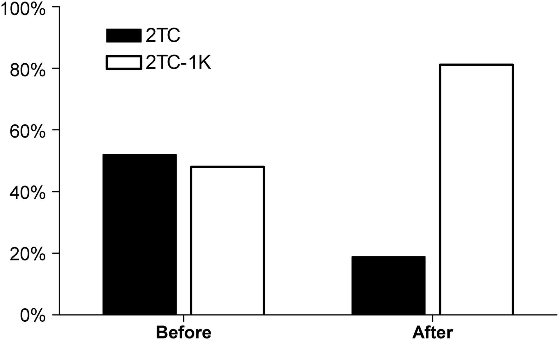
    Percentage of 2TC and 2TC-1K time–activity curves preferred by AIC for each of the 2 models before and after noise reduction.

  • FIGURE 3.
    • Download figure
    • Open in new tab
    • Download powerpoint
    FIGURE 3.

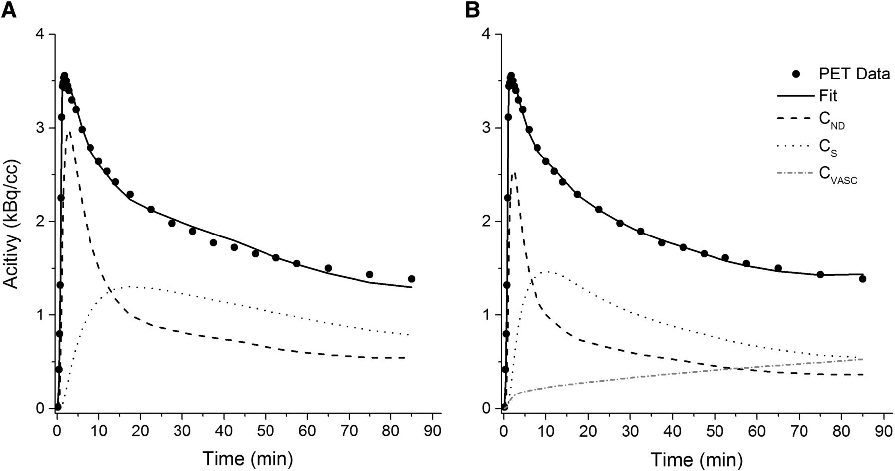
    Model fits for 1 representative time–activity curve with 2TC (A) and 2TC-1K (B) models. CND = nondisplaceable concentration, CS = concentration specifically bound within brain tissue, CVASC = concentration within vascular compartment.

  • FIGURE 4.
    • Download figure
    • Open in new tab
    • Download powerpoint
    FIGURE 4.

    Mean parameter estimates for 2TC and 2TC-1K models in some brain regions of high-affinity-binders. Parameters shown are VT (A), VND (B), and BPND (C).

  • FIGURE 5.
    • Download figure
    • Open in new tab
    • Download powerpoint
    FIGURE 5.

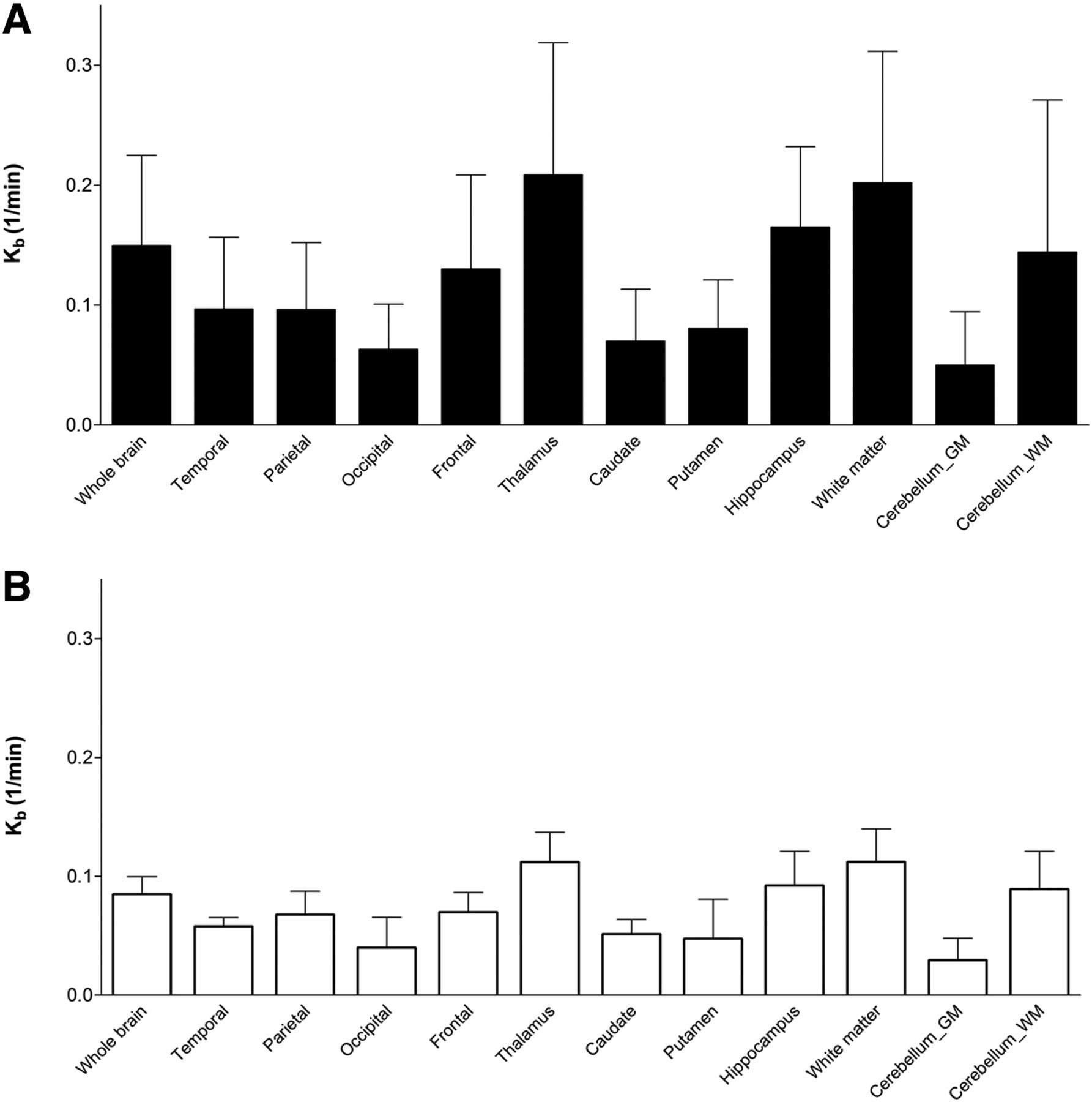
    Mean Kb estimates for 2TC-1K model for each brain region for high-affinity (A) and mixed-affinity (B) binders. GM = gray matter; WM = white matter.

  • FIGURE 6.
    • Download figure
    • Open in new tab
    • Download powerpoint
    FIGURE 6.

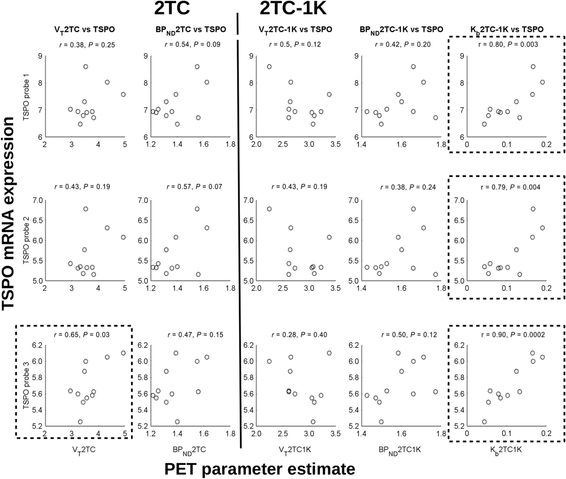
    Correlations of kinetic parameter estimates (all subjects) from 18F-DPA-714 PET scans with TSPO mRNA expression from AHB atlas. (Left) VT and BPND from 2TC. (Right) VT, BPND, and Kb from 2TC-1K. Plots surrounded by box show significant correlations.
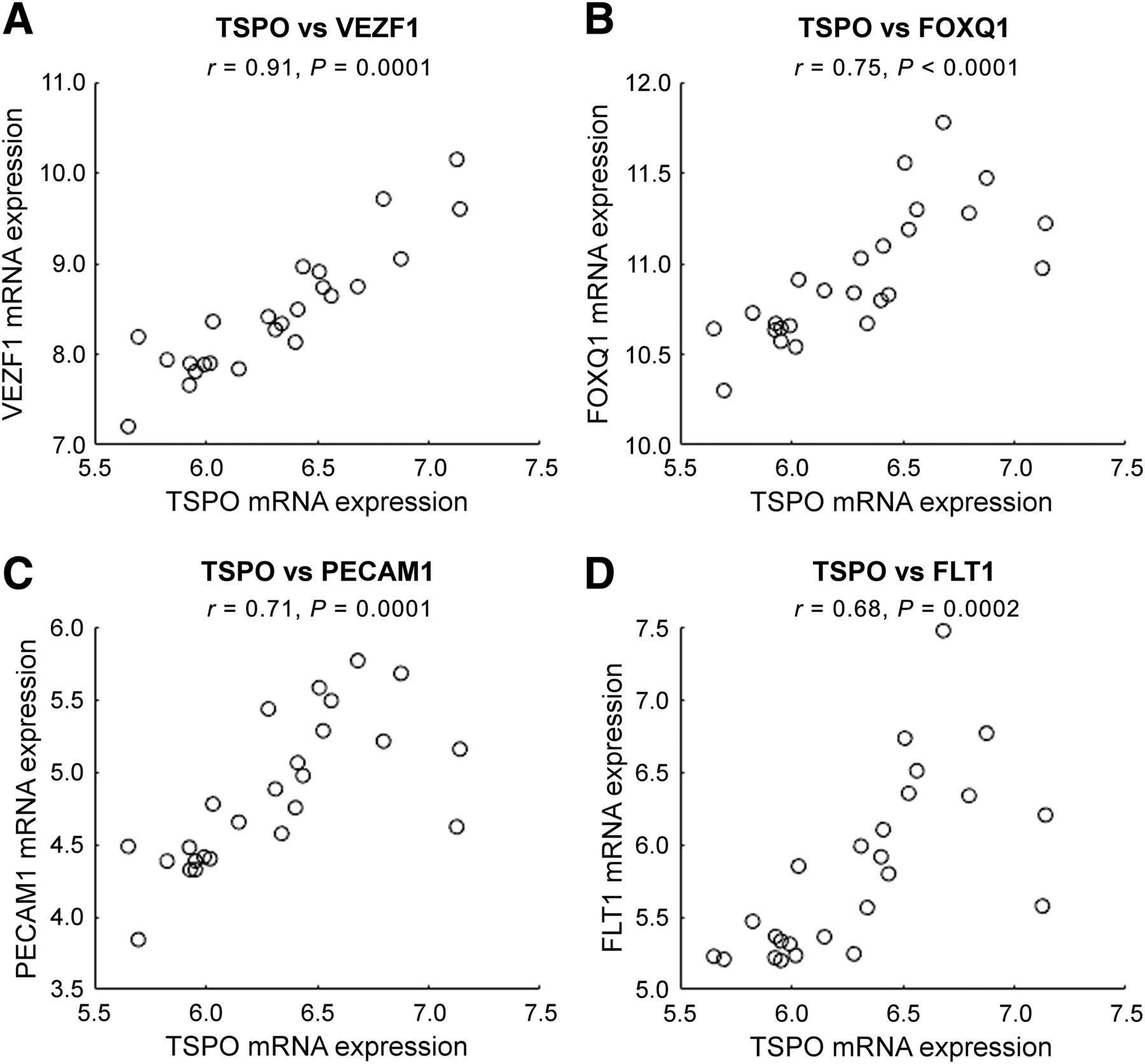
  • FIGURE 7.
    • Download figure
    • Open in new tab
    • Download powerpoint
    FIGURE 7.

    Regional correlations over 25 different regions from AHB atlas between average TSPO mRNA expression (x-axis) and mRNA expression for 4 different genes specific to endothelial cells (y-axis): vascular endothelial zinc finger 1 (VEZF1) (A), forkhead box Q1 (FOXQ1) (B), platelet/endothelial cell adhesion molecule 1 (PECAM1) (C), and FMS-related tyrosine kinase 1 (FLT1) (D).

Additional Files

  • Figures
  • Supplemental Data

    Files in this Data Supplement:

    • Supplemental Data
PreviousNext
Back to top

In this issue

Journal of Nuclear Medicine: 59 (2)
Journal of Nuclear Medicine
Vol. 59, Issue 2
February 1, 2018
  • Table of Contents
  • Table of Contents (PDF)
  • About the Cover
  • Index by author
Print
Download PDF
Article Alerts
Sign In to Email Alerts with your Email Address
Email Article

Thank you for your interest in spreading the word on Journal of Nuclear Medicine.

NOTE: We only request your email address so that the person you are recommending the page to knows that you wanted them to see it, and that it is not junk mail. We do not capture any email address.

Enter multiple addresses on separate lines or separate them with commas.
Impact of Endothelial 18-kDa Translocator Protein on the Quantification of 18F-DPA-714
(Your Name) has sent you a message from Journal of Nuclear Medicine
(Your Name) thought you would like to see the Journal of Nuclear Medicine web site.
Citation Tools
Impact of Endothelial 18-kDa Translocator Protein on the Quantification of 18F-DPA-714
Catriona Wimberley, Sonia Lavisse, Vincent Brulon, Marie-Anne Peyronneau, Claire Leroy, Benedetta Bodini, Philippe Remy, Bruno Stankoff, Irène Buvat, Michel Bottlaender
Journal of Nuclear Medicine Feb 2018, 59 (2) 307-314; DOI: 10.2967/jnumed.117.195396

Citation Manager Formats

  • BibTeX
  • Bookends
  • EasyBib
  • EndNote (tagged)
  • EndNote 8 (xml)
  • Medlars
  • Mendeley
  • Papers
  • RefWorks Tagged
  • Ref Manager
  • RIS
  • Zotero
Share
Impact of Endothelial 18-kDa Translocator Protein on the Quantification of 18F-DPA-714
Catriona Wimberley, Sonia Lavisse, Vincent Brulon, Marie-Anne Peyronneau, Claire Leroy, Benedetta Bodini, Philippe Remy, Bruno Stankoff, Irène Buvat, Michel Bottlaender
Journal of Nuclear Medicine Feb 2018, 59 (2) 307-314; DOI: 10.2967/jnumed.117.195396
Twitter logo Facebook logo LinkedIn logo Mendeley logo
  • Tweet Widget
  • Facebook Like
  • Google Plus One
Bookmark this article

Jump to section

  • Article
    • Abstract
    • MATERIALS AND METHODS
    • RESULTS
    • DISCUSSION
    • CONCLUSION
    • DISCLOSURE
    • Acknowledgments
    • Footnotes
    • REFERENCES
  • Figures & Data
  • Supplemental
  • Info & Metrics
  • PDF

Related Articles

  • This Month in JNM
  • PubMed
  • Google Scholar

Cited By...

  • Deciphering sources of PET signals in the tumor microenvironment of glioblastoma at cellular resolution
  • Brain and systemic inflammation in de novo Parkinsons disease
  • Structural and Clinical Correlates of a Periventricular Gradient of Neuroinflammation in Multiple Sclerosis
  • 18F-PBR111 PET Imaging in Healthy Controls and Schizophrenia: Test-Retest Reproducibility and Quantification of Neuroinflammation
  • Head-to-Head Comparison of 11C-PBR28 and 18F-GE180 for Quantification of the Translocator Protein in the Human Brain
  • Google Scholar

More in this TOC Section

Neurology

  • Dopamine D1 Receptor Agonist PET Tracer Development: Assessment in Nonhuman Primates
  • Hypermetabolism on Pediatric PET Scans of Brain Glucose Metabolism: What Does It Signify?
  • TauIQ: A Canonical Image Based Algorithm to Quantify Tau PET Scans
Show more Neurology

Clinical

  • Dopamine D1 Receptor Agonist PET Tracer Development: Assessment in Nonhuman Primates
  • Hypermetabolism on Pediatric PET Scans of Brain Glucose Metabolism: What Does It Signify?
  • TauIQ: A Canonical Image Based Algorithm to Quantify Tau PET Scans
Show more Clinical

Similar Articles

Keywords

  • PET
  • neuroinflammation
  • TSPO
  • 18F-DPA-714
  • quantification
SNMMI

© 2025 SNMMI

Powered by HighWire