


Article Figures & Data
Figures
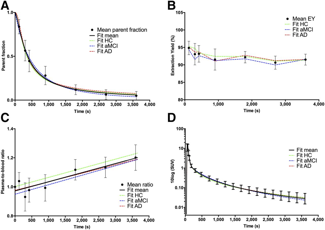
- FIGURE 1.
(A) Amount of unchanged 18F-AV45 (i.e., mean parent fraction ± SD, ●) at different time points after injection for all subjects investigated; black solid line represents Watabe curve fit. Colored, dotted lines represent Watabe curve fits to group-averaged parent fractions. (B) Extraction yield (EY). (C) Plasma–to–whole-blood ratios fitted with linear function. (D) Metabolite-corrected plasma input functions.
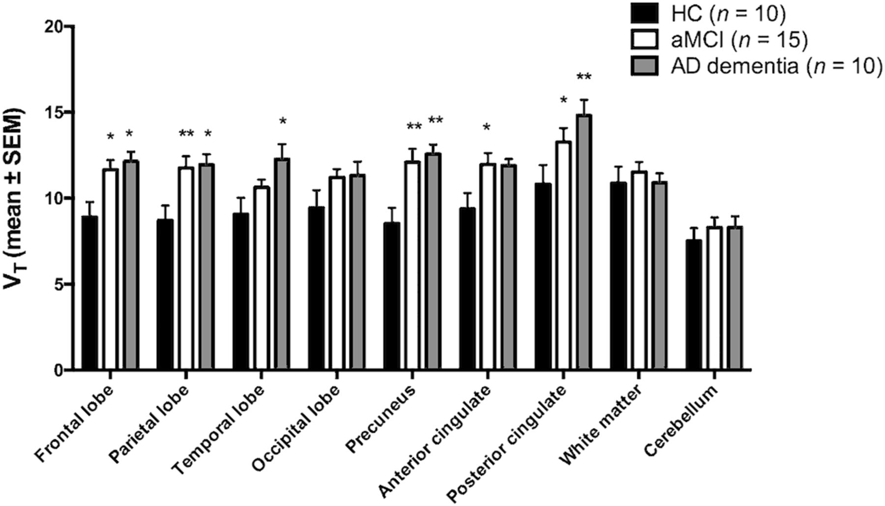
- FIGURE 2.
Regional 18F-AV45 VT (mean ± SEM) values for the 3 groups. Asterisks denote significant differences with HC group (*P < 0.05; **P < 0.01).
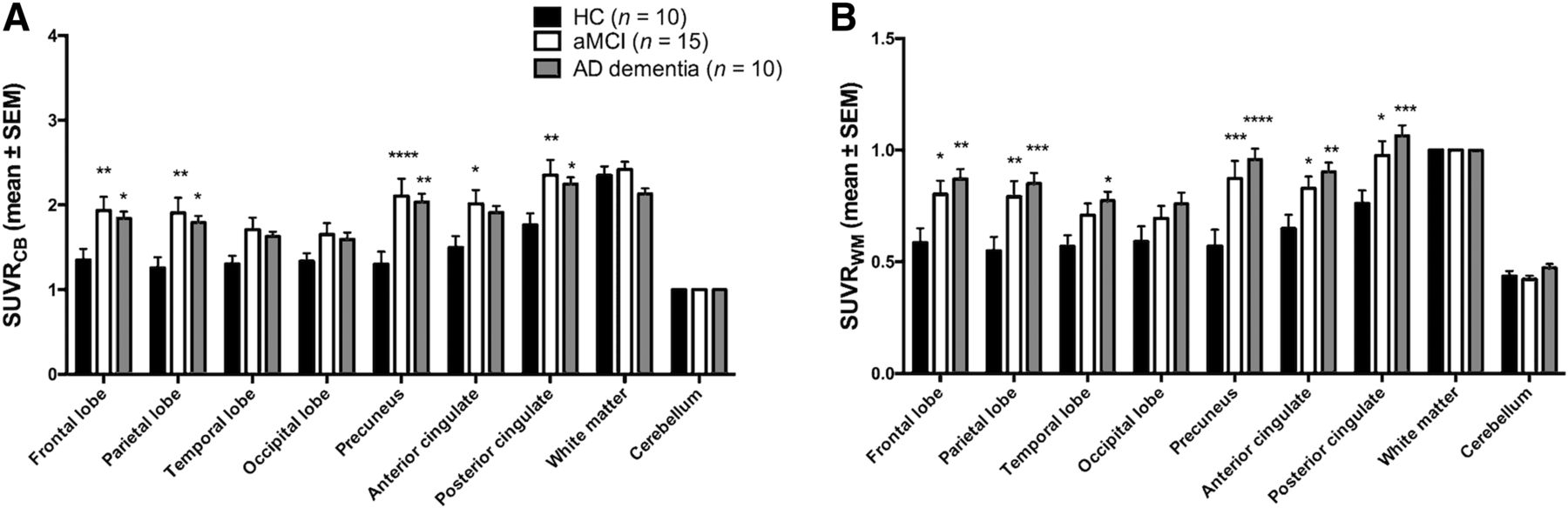
- FIGURE 3.
Regional 18F-AV45 SUVRCB (A) and SUVRWM (B) (mean ± SEM) values for the 3 groups. Asterisks denote significant differences with HC group (*P < 0.05; **P < 0.01; ***P < 0.001; ****P < 0.0001).
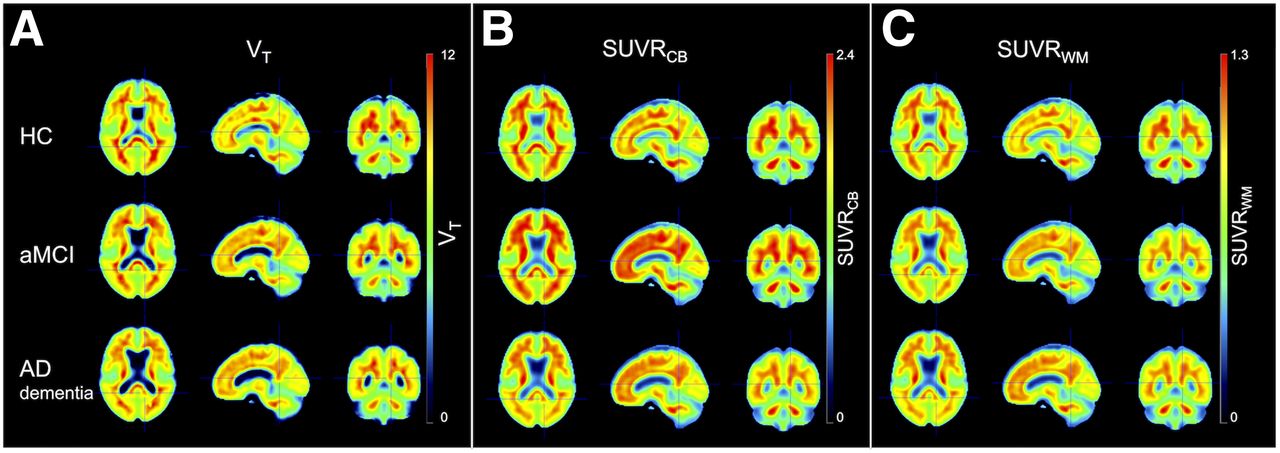
- FIGURE 4.
Average spatially normalized 18F-AV45 VT (A), SUVRCB (B), and SUVRWM (C) images for the 3 groups.
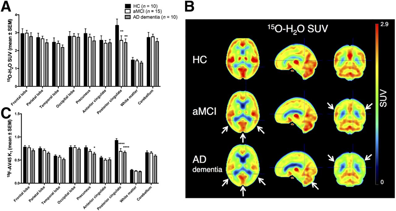
- FIGURE 5.
(A) Regional 15O-water SUV (mean ± SEM) values for the 3 groups. (B) Average spatially normalized 15O-water SUV images. White arrows indicate regions with reduced flow compared with controls (parietotemporal cortex, posterior cingulate cortex, cerebellum). (C) 18F-AV45 delivery rate K1 (mean ± SEM). Asterisks denote significant differences with HC group (*P < 0.05; **P < 0.01; ****P < 0.0001).


Tables
Parameter HC (n = 10) aMCI (n = 18) AD (n = 10) Age (y) 69 ± 6 73 ± 8 73 ± 5 Sex Male 4 9 7 Female 6 9 3 Education (y) 14 ± 2 11 ± 4 12 ± 4 MMSE 29 ± 1 25 ± 3* 22 ± 4* *Significantly different from controls (1-way ANOVA, corrected for multiple comparisons via Dunnett, P < 0.05).
MMSE = Mini-Mental State Examination.
Data are mean ± SD, unless otherwise stated.
Brain region Vb K1 k2 k3 k4 VT Precuneus HC 0.05 ± 0.01 0.77 ± 0.12 0.17 ± 0.02 0.02 ± 0.01 0.02 ± 0.01 8.5 ± 2.9 aMCI 0.05 ± 0.01 0.69 ± 0.15 0.14 ± 0.04 0.03 ± 0.01 0.02 ± 0.005 12.1* ± 3.0 AD 0.05 ± 0.01 0.63* ± 0.10 0.13* ± 0.03 0.03 ± 0.01 0.02 ± 0.007 12.6* ± 1.6 CB HC 0.05 ± 0.01 0.66 ± 0.12 0.17 ± 0.03 0.01 ± 0.01 0.02 ± 0.01 7.5 ± 2.3 aMCI 0.06 ± 0.01 0.65 ± 0.11 0.17 ± 0.03 0.01 ± 0.01 0.01 ± 0.01 8.3 ± 2.3 AD 0.05 ± 0.01 0.59 ± 0.09 0.16 ± 0.03 0.01 ± 0.003 0.01 ± 0.01 8.3 ± 2.0 WM HC 0.02 ± 0.01 0.28 ± 0.06 0.14 ± 0.04 0.19 ± 0.10 0.04 ± 0.01 10.9 ± 3.0 aMCI 0.02 ± 0.01 0.26 ± 0.05 0.13 ± 0.04 0.16 ± 0.11 0.03 ± 0.02 11.5 ± 2.3 AD 0.02 ± 0.01 0.25 ± 0.04 0.11 ± 0.04 0.12* ± 0.04 0.03 ± 0.01 10.9 ± 1.7 ↵* Significantly different from controls (1-way ANOVA, corrected for multiple comparisons via Dunnett, P < 0.05).
- TABLE 3
Pearson Correlation Coefficient of SUVRCB and SUVRWM with VT Across All Diagnostic Groups
Brain region VT – SUVRCB VT – SUVRWM Frontal lobe 0.72* 0.82* Parietal lobe 0.76* 0.85* Temporal lobe 0.51† 0.70* Occipital lobe 0.69* 0.79* Precuneus 0.81* 0.89* Anterior cingulate 0.79* 0.74* Posterior cingulate 0.54‡ 0.63* Symbols denote significant correlations (*P < 0.0001, †P = 0.002, ‡P = 0.0009).
Supplemental Data
Files in this Data Supplement:


Jump to section
Related Articles
Cited By...
- Amyloid-PET of the white matter: relationship to free water, fiber integrity, and cognition in patients with dementia and small vessel disease
- White Matter Reference Region in PET Studies of 11C-Pittsburgh Compound B Uptake: Effects of Age and Amyloid-{beta} Deposition
- Validation of Noninvasive Tracer Kinetic Analysis of 18F-Florbetaben PET Using a Dual-Time-Window Acquisition Protocol