


Article Figures & Data
Figures
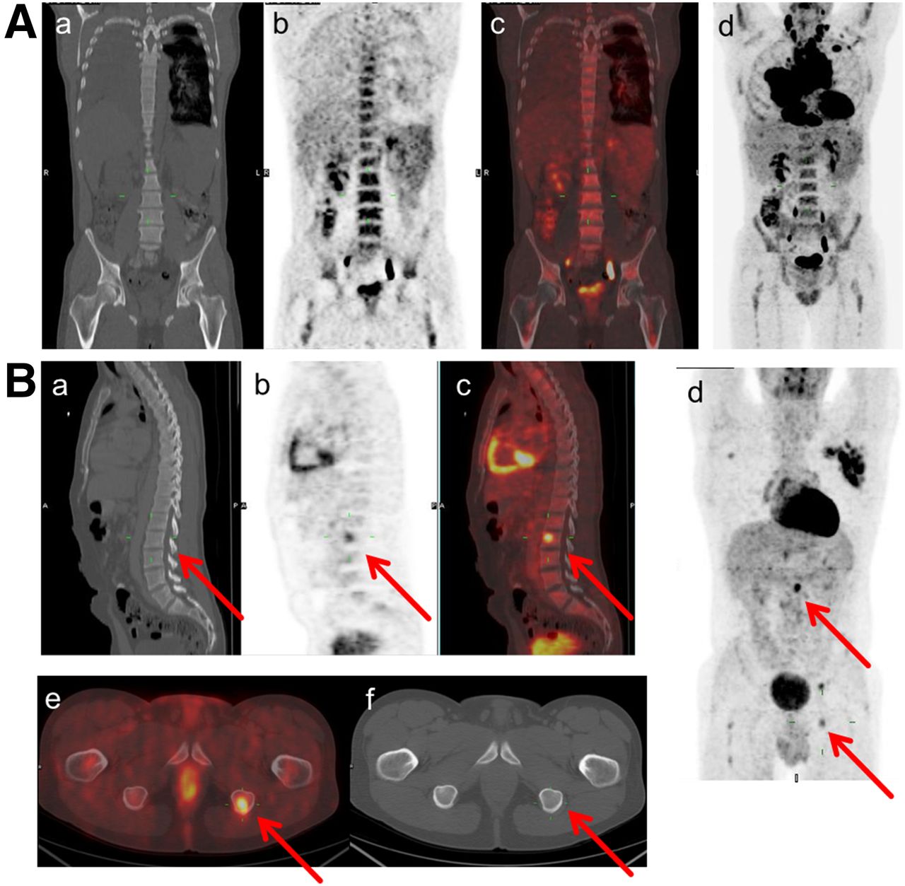
- FIGURE 1.
(A) Example of pure diffuse BM uptake > liver uptake in baseline 18F-FDG PET (coronal CT slices [a], PET [b] and fused PET/CT [c], and maximum-intensity projection [d]). BMB was negative, and patient was in complete remission (follow-up, +81 mo). (B) Example of multifocal BMI, with 3 focal BM lesions (L2, left ischium, and right scapula) in baseline 18F-FDG PET, without corresponding CT abnormalities (sagittal slices of CT [a], PET [b] and fused PET/CT [c], maximum-intensity projection [d], axial fused PET/CT [e], and CT slices [f]). BMB was negative, and patient relapsed 6 mo after end of ABVD treatment.
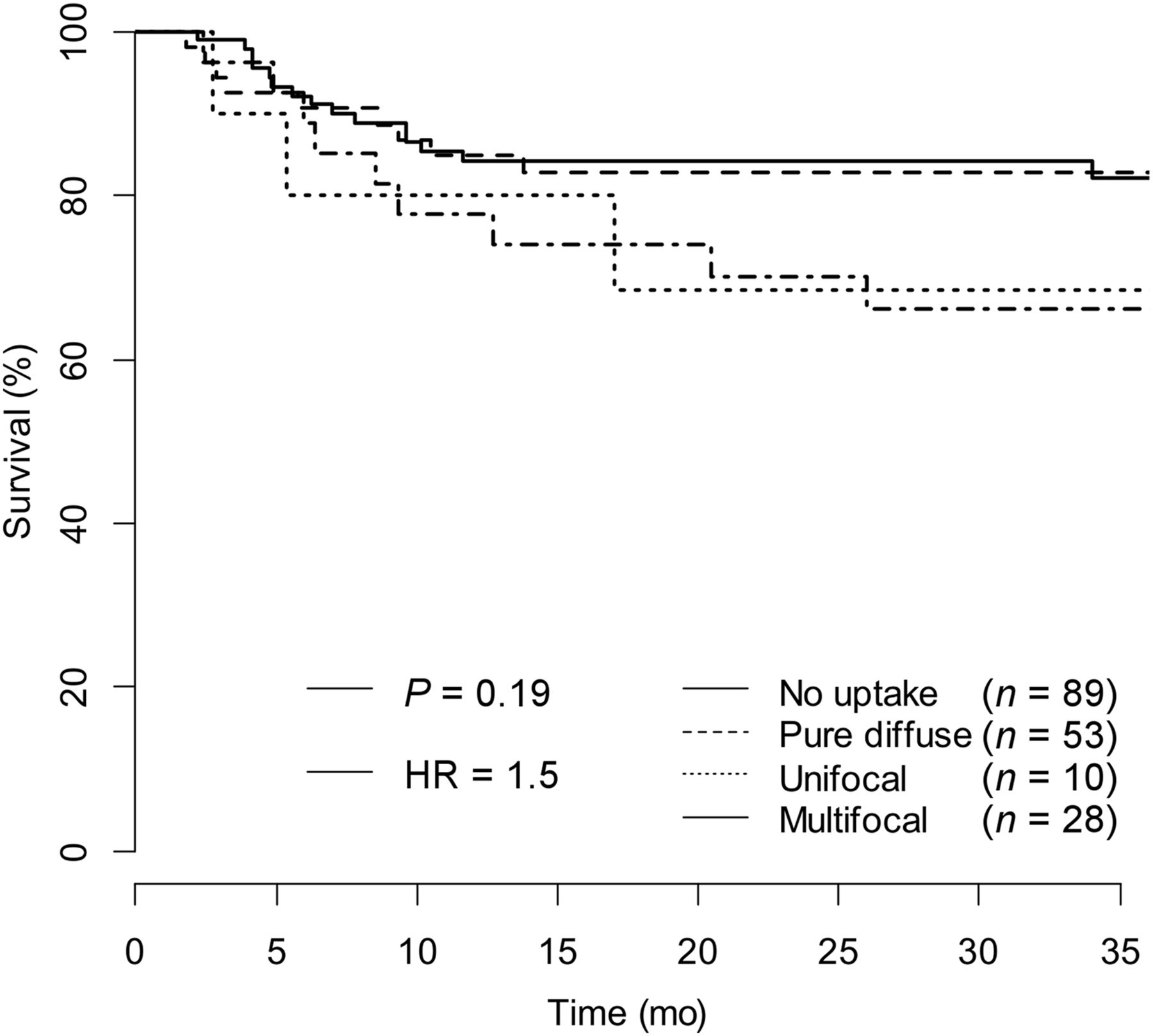
- FIGURE 2.
Kaplan–Meier PFS curves of different BM 18F-FDG uptake patterns: no 18F-FDG uptake, pure diffuse 18F-FDG uptake > liver, unifocal 18F-FDG uptake, and multifocal 18F-FDG uptake. Log-rank P = 0.19.
- FIGURE 3.
Kaplan–Meier PFS curves of focal PET lesions and nonfocal BM uptake groups (pure dPET+ and nPET+). Log-rank P = 0.03.

Tables
Characteristic Total (n = 180) IVS (n = 133) Polish study (n = 47) P Median age (y) 38.6; range, 19–82.5 39; range, 32.6–48.3 37.7; range, 26–49.8 0.101 Male-to-female ratio 0.8% 0.9% 0.6% 0.515 B symptoms (n) 123 (68.3) 88 (66.2) 35 (74.5) 0.385 Histology 0.275 Scleronodular 142 (78.8) 101 (75.9) 41 (87.2) Lymphocyte rich 16 (8.8) 15 (11.3) 1 (2.1) Mixed cellularity 3 (1.6) 2 (1.5) 1 (2.1) Lymph depletion 13 (7.2) 9 (6.8) 4 (8.5) Lymph predominance 2 (1.1) 2 (1.5) 0 (0) Undetermined 4 (2.2) 4 (3) 0 (0) Ann Arbor stage (n) 0.003 II 62 (34.4) 52 (39.1) 10 (21.3) III 58 (32.2) 46 (34.6) 12 (25.5) IV 60 (33.3) 35 (26.3) 25 (53.2) Mediastinal bulk tumor (n) 78 (43.3) 54 (40.6) 23 (48.9) 0.412 International prognostic score > 2 (n) 55 (30.5) 33 (24.8) 22 (48.9) 0.005 Extranodal involvement (n) 61 (33.9) 40 (30.1) 21 (44.7) 0.101 Radiotherapy (n)* 68 (37.7) 53 (43.8) 15 (33.3) 0.432 ↵* Missing data in 15 patients.
Data in parentheses are percentages.
BM 18F-FDG uptake characteristic n No. of positive BMB BM 18F-FDG uptake < liver (nPET)* 89 (49.4) 5 (2.8) Focal BM uptake: unifocal (n = 10) and multifocal (n = 28) Without diffuse BM uptake (pure fPET+) 17 (9.4) 4 (2.2) With diffuse BM uptake (f/dPET+) 21 (11.7) 2 (1.1) Pure diffuse BM uptake (pure dPET+) 53 (29.4) 1 (0.6) Total 180 (100) 12 (6.6) ↵* Missing data of BMB for 2 patients in this group.
Data in parentheses are percentages.
Supplemental Data
Files in this Data Supplement:


