


Article Figures & Data
Figures
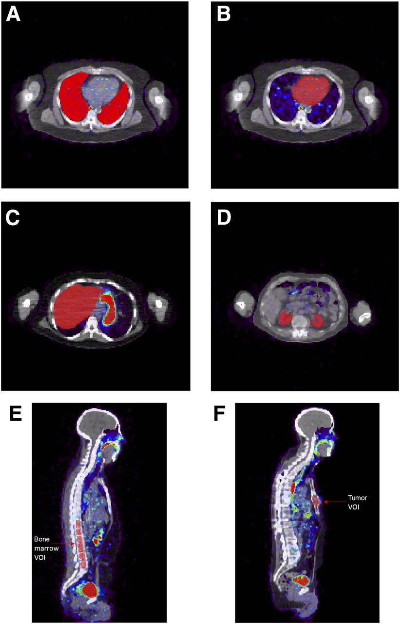
- FIGURE 1.
Transverse and sagittal views of patients’ CT and PET images (superimposed) and drawn VOIs covering lungs (A), heart (B), liver (C), kidneys (D), bone marrow (E), and tumor (F).
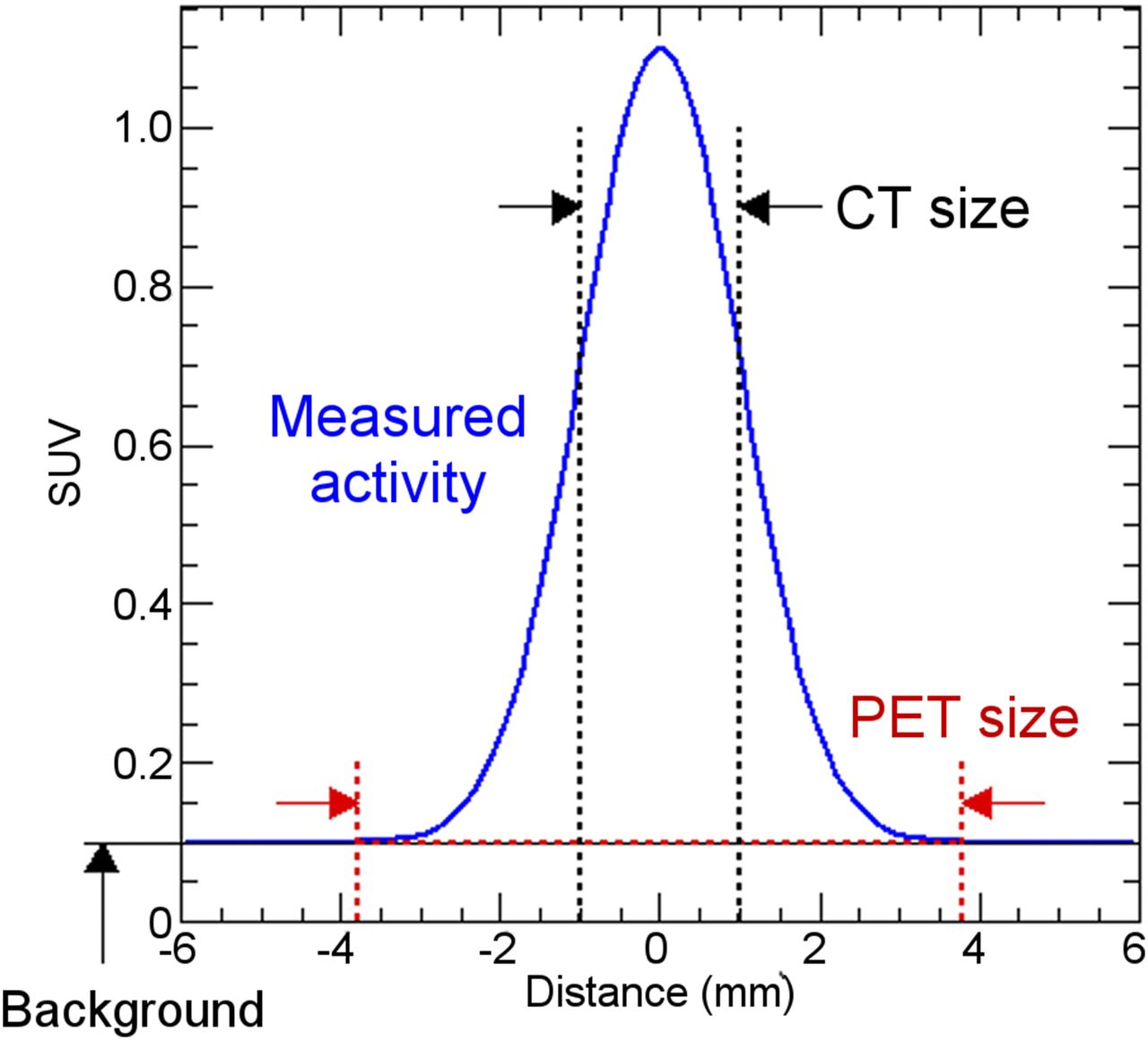
- FIGURE 2.
Activity measurement method in tumors.
- FIGURE 3.
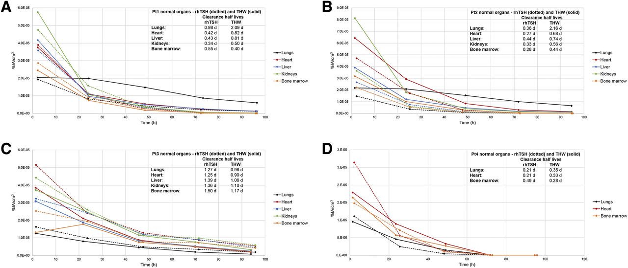
Normal organ %IA/cm3 vs. time (hr) after injection of 124I for rhTSH and THW for patient 1 (A), patient 2 (B), patient 3 (C), and patient 4 (D). Pt = patient.
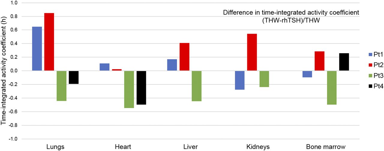
- FIGURE 4.
Difference in time-integrated activity coefficient (THW − rhTSH)/THW.
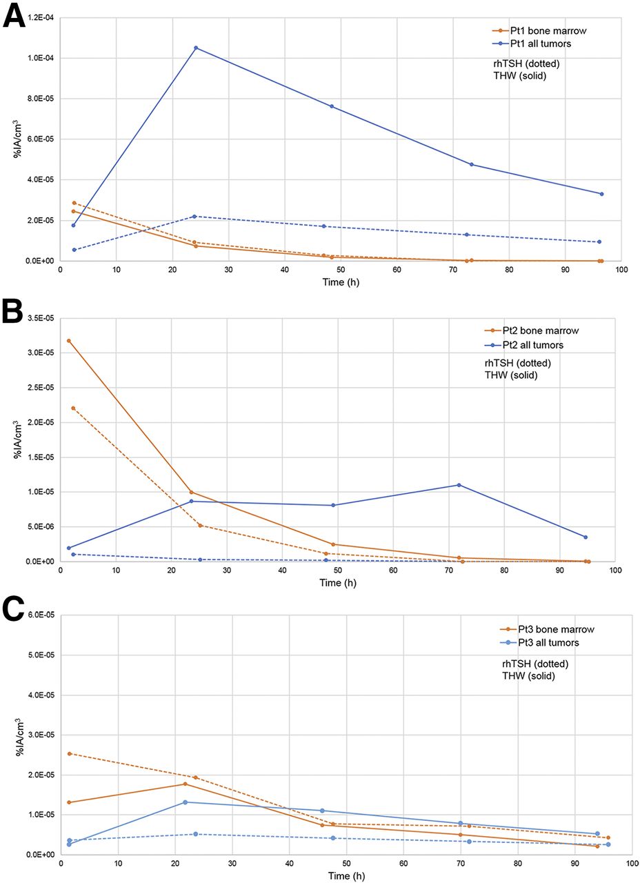
- FIGURE 5.
Aggregate tumor and bone marrow 124I activity concentration (%IA/cm3), rhTSH (dotted) and THW (solid) study for patient 1 (A), patient 2 (B), and patient 3 (C).


Tables
Characteristic Patient 1 Patient 2 Patient 3 Patient 4 Age (y) 47 30 63 51 Sex M M M M Thyroglobulin level (ng/mL) 170*; 324† NA*; 1,527† 3,735*; 2,480† 86*; 58† TSH level (μIU/mL) 98*; 82.9† 139*; 142† 89*; 124† 64*; 72† Urine iodine level (μg/L) 182‡; 85† 75‡; 630† 99‡; NA† 28‡; 21† Thyroxine dose before and during rhTSH stimulation (μg) 125 125 139 200 124I activity (MBq) 131I activity Patient no. rhTSH THW mCi MBq 1 30 29 319 11,803 2 61 65 398 14,726 3 36 33 198 7,326 4 37 33 326 12,062 - TABLE 3
Absorbed Dose per Unit Administered Activity of 131I for Each Study (rhTSH and THW) and Ratio in Absorbed Dose per Unit Administered Activity (THW/rhTSH)
Organ VOI Patient Lungs Heart Liver Kidneys Bone marrow 1 Absorbed dose per unit administered activity for rhTSH (mGy/MBq) 0.21 0.15 0.15 0.19 0.13 Absorbed dose per unit administered activity for THW (mGy/MBq) 0.58 0.21 0.19 0.17 0.11 THW/rhTSH ratio 2.8 1.4 1.3 0.9 0.85 2 Absorbed dose per unit administered activity for rhTSH (mGy/MBq) 0.11 0.27 0.11 0.11 0.086 Absorbed dose per unit administered activity for THW (mGy/MBq) 0.66 0.33 0.20 0.23 0.14 THW/rhTSH ratio 6.0 1.2 1.8 2.1 1.6 3 Absorbed dose per unit administered activity for rhTSH (mGy/MBq) 0.44 0.46 0.40 0.44 0.33 Absorbed dose per unit administered activity for THW (mGy/MBq) 0.25 0.28 0.24 0.32 0.22 THW/rhTSH ratio 0.57 0.61 0.60 0.73 0.67 4 Absorbed dose per unit administered activity for rhTSH (mGy/MBq) 0.10 0.11 NA NA 0.068 Absorbed dose per unit administered activity for THW (mGy/MBq) 0.11 0.10 NA NA 0.080 THW/rhTSH ratio 1.1 0.91 NA NA 1.2 NA = not applicable.
- TABLE 4
Calculated Tumor Absorbed Dose per Unit Administered Activity of 131I and Ratio in Tumor-Absorbed Dose (THW/rhTSH)
Tumor no. Tumor volume (cm3) Absorbed dose per unit administered activity (mGy/MBq) for rhTSH Absorbed dose per unit administered activity (mGy/MBq) for THW THW/rhTSH ratio Patient 1 1 1.5 2.8 8.6 3.1 2 0.98 11 18 1.6 3 0.46 3.8 20 5.3 4 0.87 2.6 18 7.0 5 0.50 17 82 4.9 6 1.2 9.7 27 2.7 7 1.1 21 40 1.9 8 1.7 0.84 4.6 5.5 9 0.89 3.8 12 3.3 Patient 2 1 0.67 0.045 1.3 28 2 0.38 0.089 2.2 25 Patient 3 1 2.2 5.2 20 3.9 2 11 0.66 1.1 1.7 3 2.9 0.36 0.56 1.5 4 3.8 1.9 1.6 0.83 5 34 4.8 4.8 1.0 6 12 5.4 0.92 0.17 7 1.5 19 21 1.1 8 1.7 0.34 0.42 1.2 9 4.3 0.20 0.12 0.63 10 47 1.9 2.1 1.1 11 9.0 0.36 0.65 1.8
Supplemental Data
Files in this Data Supplement:


Jump to section
Related Articles
Cited By...
- Analysis of Residence Time, Effective Half-Life, and Internal Dosimetry Before Radioiodine Therapy
- Diagnostic Performance of 124I-Metaiodobenzylguanidine PET/CT in Patients with Pheochromocytoma
- SNMMI Procedure Standard/EANM Practice Guideline for Nuclear Medicine Evaluation and Therapy of Differentiated Thyroid Cancer: Abbreviated Version
- Management of Differentiated Thyroid Cancer: The Standard of Care
- Dosimetry in Clinical Radiopharmaceutical Therapy of Cancer: Practicality Versus Perfection in Current Practice
- Comparative Dosimetry for 68Ga-DOTATATE: Impact of Using Updated ICRP Phantoms, S Values, and Tissue-Weighting Factors
- Treatment of refractory thyroid cancer