


Article Figures & Data
Figures
- FIGURE 1.
Topographic representation of clusters in which 18F-FDG uptake was significantly lower in MCI converters (n = 95) than in MCI nonconverters (n = 27) (threshold P < 0.05, corrected for multiple comparisons with familywise error option). Clusters are superimposed on Montreal Neurologic Institute template in coronal (left), sagittal (middle), and transversal (right) views.
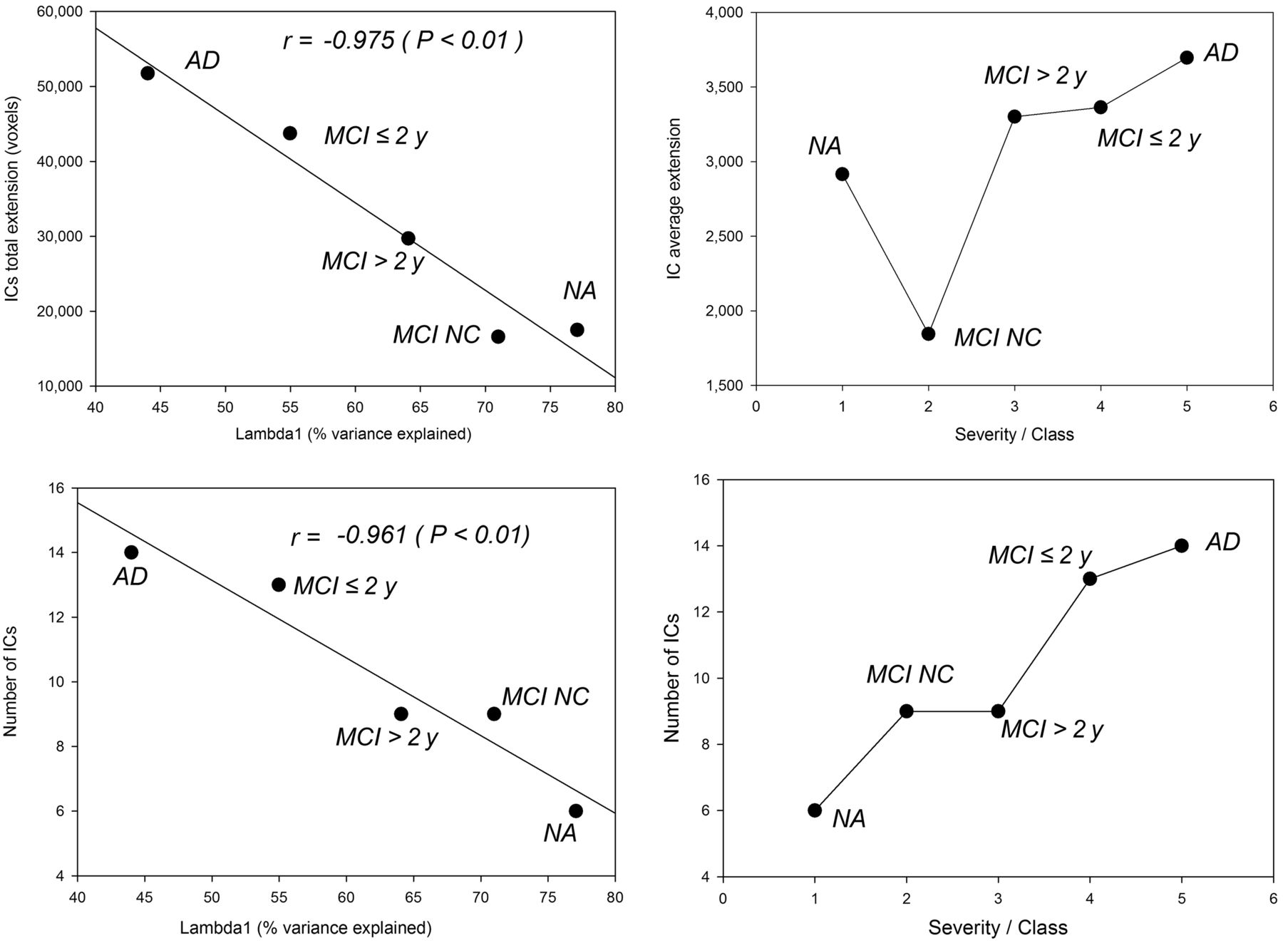
- FIGURE 2.
(Left) For each severity class, negative correlations between percentage of variance explained by first principal component (λ1) and generation of local circuits expressed as total independent-component extent in voxels (top) and number of independent components (bottom). (Right) Relations between disease severity class and average independent-component extent (top) and number of independent components (bottom). MCI > 2 y = MCI converters after more than 2 y; IC = independent component; MCI ≤ 2 y = MCI converters within 2 y; NA = normally aging individuals; MCI NC = MCI nonconverters.
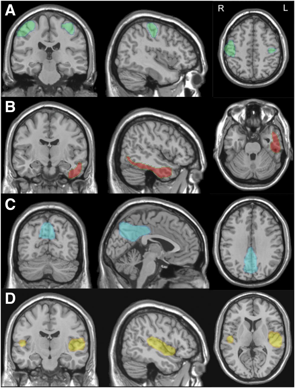
- FIGURE 3.
Topographic representations of independent components identifying sensorimotor cortex (A), left temporal cortex (B), posterior cingulate cortex/precuneus (C), and sylvian temporal cortex (D) on brain surfaces. Regions obtained from ICA have been superimposed on Montreal Neurologic Institute template in coronal (left), sagittal (middle), and transversal (right) views.
- FIGURE 4.
Receiver-operating-characteristic curves obtained by SVM classifier as applied to 3 different datasets.


Tables
Group Education (y) Age at PET (y) MMSE* Sex Normal aging 10.0 ± 4.1 68.8 ± 9.7 29.1 ± 0.9 12 M, 32 F Nonconverting MCI 8.9 ± 3.7 71.9 ± 6.4 26.8 ± 1.5 16 M, 12 F MCI > 2 y 10.4 ± 5.0 74.7 ± 7.0 26.3 ± 1.6 8 M, 28 F MCI ≤ 2 y 9.9 ± 4.5 75.5 ± 6.5 25.8 ± 1.9 22 M, 36 F AD 7.4 ± 4.2 73.4 ± 7.4 19.2 ± 4.0 18 M, 36 F ↵* Normalized for education
MCI > 2 y = MCI converting after more than 2 y; MCI ≤ 2 y = MCI converting within 2 y.
Qualitative data are expressed as numbers; continuous data are expressed as mean ± SD.
- TABLE 2
Independent Components Identified as Pathophysiologically Significant in Each Group
NA NC MCI MCI > 2 y MCI ≤ 2 y AD IC Size Regions IC Size Regions IC Size Regions IC Size Regions IC Size Regions 3 2,309 DLFC+MFG 6 907 SMA, premotor, and BA9 4 2,408 PCC+iPL (postDMN) 6 3,210 PCC+iPL (postDMN) 14 813 Basal ganglia 7 1,277 Basal ganglia 19 2,960 Basal ganglia+thalamic 15 5,083 Primary visual 16 3,117 Primary visual 7 5,346 Primary visual 10 5,721 Primary visual 1&9 8,783 Primary visual 1 5,971 Cerebellum 14 2,679 Cerebellum 4 5,358 Cerebellum 4 5,183 Cerebellum 3 5,178 Cerebellum 7 1,470 L sPL 1 1,756 L sPL+L DLFC+L T 8 891 R sensorimotor 16 1,493 R sensorimotor 19 2,370 Sensorimotor 10 3,588 Sensorimotor 15 1,702 Sylvian temporal 3 4,161 Sylvian temporal 3 4,464 Sylvian temporal 8 3,121 Sylvian temporal 1 1,038 L DLFC 2 1,700 iPL+O 12 787 VLFC 9 4,691 R iPL+R T 5 5,190 R iPL+R T 5 4,466 iPL+R T 17 2,922 R iPL+R O+R T 15 4,000 R iPL+R O 12 1,748 Thalami 13 2,231 L O 1 1,129 R MTL 6 1,357 MTL 2 2,565 VLFC 18 3,888 VLFC 6 4,427 sPL 13 5,119 sPL 12 3,700 PCC 7 3,343 PCC+PC 8 1,894 O 9 4,281 L sPL+PC+L T 13 1,526 DLFC 14 2,978 L T 16 2,959 L T Voxel extent 17,491 16,594 29,706 43,727 51,740 Number 9 9 9 13 14 NA = normal aging; NC = nonconverting; MCI > 2 y = MCI converting after more than 2 y; MCI ≤ 2 y = MCI converting within 2 y; IC = independent component; DLFC = dorsolateral frontal cortex; MFG = medial frontal gyrus; SMA = supplementary motor area; BA = Brodmann area; PCC = posterior cingulate cortex; iPL = inferior parietal lobule; postDNM = posterior default-mode network; L = left; sPL superior parietal lobule; T = temporal; R = right; O = occipital; VLFC = ventrolateral frontal cortex; MTL = mesial temporal lobe; PC = precuneus.
NA vs. (converting MCI+AD) Nonconverting MCI vs. (converting MCI+AD) 1 component 4 components 1 component 4 components Parameter Exp. CI Exp. CI Exp. CI Exp. CI Model performance Sensitivity 75.8 70.1–82.7 90.1 85.9–95.3 81.9 75.7–88.6 83.2 77.2–89.2 Specificity 83.3 72.1–94.6 88.1 78.3–97.9 77.8 62.1–93.5 85.2 71.8–98.6 Accuracy 77.5 71.6–83.4 90.0 85.8–94.3 81.3 75.5–87.0 83.5 78.0–89.0 ROC AUC 85.5 79.3–90.6 93.1 88.0–95.7 87.2 80.4–92.7 89.4 83.3–93.3 Within-group classification NA AD NA AD NC AD NC AD NA 81.0 19.0 92.9 7.1 81.0* 19.0* 88.1* 11.9* Nonconverters 66.7* 33.3* 88.2* 14.8* 85.2 14.8 96.3 3.7 Early MCI 29.7 70.3 18.9 81.1 35.1 64.9 29.7 70.3 Late MCI 27.6 72.4 19.0 81.0 29.3 70.7 20.7 79.3 AD 13.0 87.0 14.8 85.2 13.0 87.0 14.8 85.2 * Not involved in training step.
Exp. = expected value; CI = confidence interval; ROC AUC = area under receiver-operating-characteristic curve; NA = normal aging.
Discriminant models are as evaluated by leave-one-out cross-validation considering partitions into two contrasting groups: NA vs. all AD and nonconverting MCI vs. all AD. Linear discrimination was applied to best discriminant region (1 component), which in both cases was left temporal cortex. Four-component models were based on SVM method and involved sensorimotor cortex, left temporal cortex, posterior cingulate cortex/precuneus, and sylvian temporal cortex. Two-level discrimination as obtained by each model for each group is reported (within-group classification). Data are percentages.
Supplemental Data
Files in this Data Supplement:


Jump to section
Related Articles
Cited By...
- No citing articles found.