


Article Figures & Data
Figures
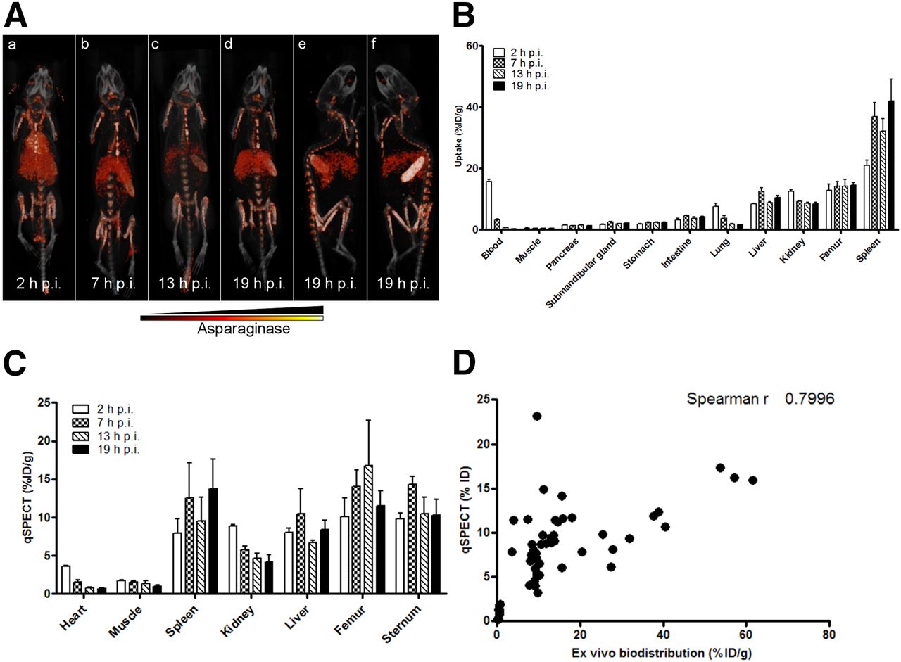
- FIGURE 1.
Asparaginase rapidly accumulates in spleen, liver, and bone marrow. (A) Ventral (a–d) and lateral (e and f) 3-dimensional volume projections of fused SPECT/CT scans of mice injected with 111In-labeled asparaginase at indicated hours after injection (h p.i.). (B) Ex vivo quantification of 111In-labeled asparaginase uptake in various organs at indicated hours after injection. Data represent mean ± SEM of n = 5 mice per time point. (C) Biodistribution of asparaginase as quantified from SPECT images. n = 3 per time point. (D) Uptake of 111In-labeled asparaginase as quantified from small-animal SPECT images plotted against values derived from ex vivo biodistribution studies.
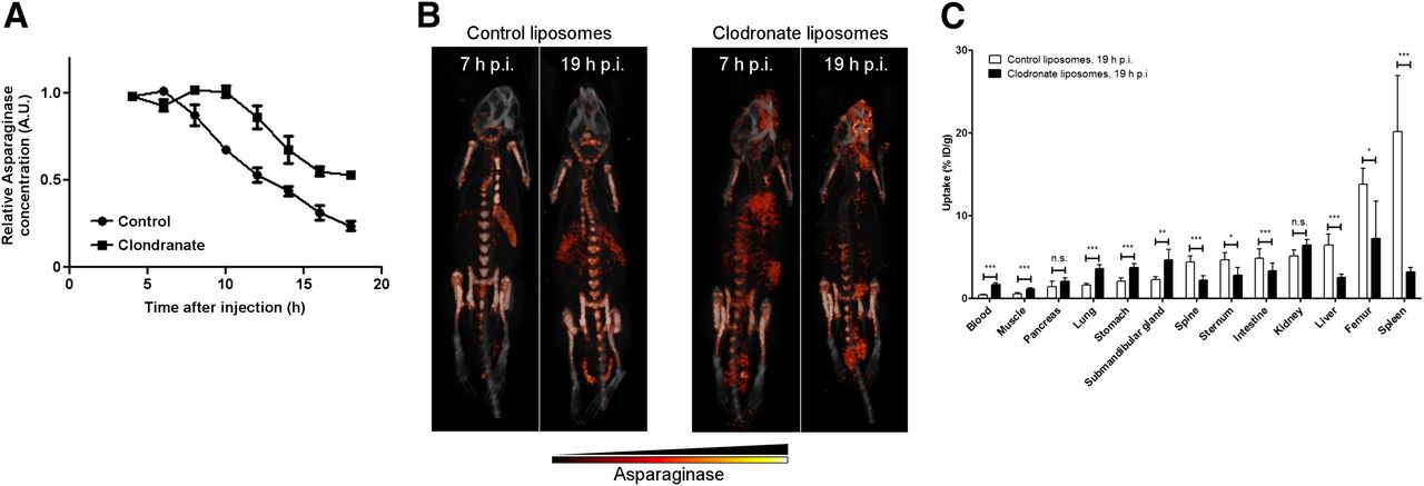
- FIGURE 2.
Depletion of macrophages affects asparaginase pharmacokinetics and biodistribution. (A) Residual serum asparaginase activity at indicated times after injection in mice. Mice were treated with single dose of control or clodronate liposomes 24 h before asparaginase injection. At indicated times after injection of unlabeled asparaginase, serum samples were taken and asparaginase activity was determined as described in “Materials and Methods” section. Data represent mean ± SEM of n = 3 mice per time point per treatment group. ANOVA statistical analysis was applied to test for significance (P < 0.05). (B) Ventral 3-dimensional volume projections of fused SPECT/CT scans of mice treated with single dose of control or clodronate liposomes 24 h before 111In-labeled asparaginase injection. Scans were obtained at 7 or 19 h after injection (h p.i.) with 111In-labeled asparaginase. (C) Ex vivo quantification of 111In-labeled asparaginase uptake in various organs of mice pretreated with control or clodronate liposomes, 19 h after injection of asparaginase. Data represent mean ± SEM of n = 5 mice per time point per treatment group. Statistical significance was determined by unpaired 2-sided t test. *P < 0.05. **P < 0.01. ***P < 0.001. n.s. = not significant.
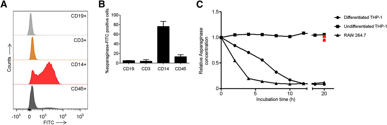
- FIGURE 3.
Human macrophages bind and degrade asparaginase. (A) Histograms showing binding of asparaginase to various cellular subsets in PBMCs. Freshly isolated PBMCs were incubated with FITC-labeled asparaginase and subsequently identified on basis of indicated cell surface markers using flow cytometry. Lineages were defined as follows: CD45, all nucleated cells; CD14, monocytes/macrophages; CD3, T cells; CD19, B cells. (B) Average of percentage of peripheral blood mononuclear cell subsets binding FITC-labeled asparaginase (n = 2). (C) Asparaginase degradation in lysates of cell lines. Asparaginase was incubated in lysate of human undifferentiated, monocytelike THP-1 cells (▪); differentiated, macrophage like THP-1 cells (●); and murine macrophage cell line RAW264.7 (▲). After incubation, residual ASNase activity was assayed as described in “Materials and Methods” section. Cathepsin inhibitor CA-074 was included to selected samples to confirm contribution of cathepsin B in this degradation (red data points).
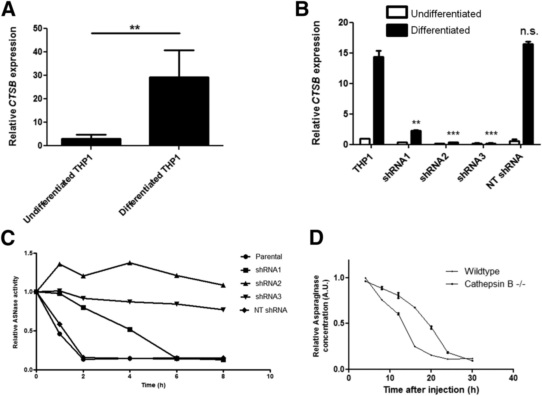
- FIGURE 4.
Cathepsin B is required for asparaginase degradation and controls asparaginase pharmacokinetics. (A) Real-time quantitative polymerase chain reaction analysis of cathepsin B messenger RNA expression in undifferentiated and PMA-induced differentiated THP-1 cells, normalized for expression of TATA binding protein. Data represent mean ± SEM of n = 4. Statistical significance was determined by unpaired 2-sided t test. **P < 0.01. (B) Real-time quantitative polymerase chain reaction analysis of cathepsin B messenger RNA expression in undifferentiated and PMA-induced differentiated THP-1 cells transduced with shRNAs targeting cathepsin B or control, nontargeting (NT) shRNA. Data are normalized for TBP expression and shown as mean ± SD of n = 2. Statistical significance was determined by unpaired 2-sided t test. **P < 0.01. (C) Asparaginase degradation in lysates of cell lines. Asparaginase was incubated in lysate of differentiated THP-1 cells, transduced with control of cathepsin B–targeting shRNAs. After incubation, residual ASNase activity was assayed as described in “Materials and Methods” section. Representative example of multiple experiments (n = 3) is shown. (D) Residual serum asparaginase activity at indicated times after injection in mice. Cathepsin B knockout mice and age- and sex-matched control were injected with unlabeled asparaginase. At indicated times after injection of unlabeled asparaginase, serum samples were taken and asparaginase activity was determined as described in “Materials and Methods” section. Data represent mean ± SD of n = 2 mice per time point per treatment group. ANOVA statistical analysis was applied to test for significance (P < 0.05). n.s. = not significant.


Additional Files
Supplemental Data
Files in this Data Supplement:


