
Main menu

  • Home
  • Content
    • Current
    • Ahead of print
    • Past Issues
    • JNM Supplement
    • SNMMI Annual Meeting Abstracts
    • Continuing Education
    • JNM Podcasts
  • Subscriptions
    • Subscribers
    • Institutional and Non-member
    • Rates
    • Journal Claims
    • Corporate & Special Sales
  • Authors
    • Submit to JNM
    • Information for Authors
    • Assignment of Copyright
    • AQARA requirements
  • Info
    • Reviewers
    • Permissions
    • Advertisers
  • About
    • About Us
    • Editorial Board
    • Contact Information
  • More
    • Alerts
    • Feedback
    • Help
    • SNMMI Journals
  • SNMMI
    • JNM
    • JNMT
    • SNMMI Journals
    • SNMMI

User menu

  • Subscribe
  • My alerts
  • Log in
  • My Cart

Search

  • Advanced search
Journal of Nuclear Medicine
  • SNMMI
    • JNM
    • JNMT
    • SNMMI Journals
    • SNMMI
  • Subscribe
  • My alerts
  • Log in
  • My Cart
Journal of Nuclear Medicine

Advanced Search

  • Home
  • Content
    • Current
    • Ahead of print
    • Past Issues
    • JNM Supplement
    • SNMMI Annual Meeting Abstracts
    • Continuing Education
    • JNM Podcasts
  • Subscriptions
    • Subscribers
    • Institutional and Non-member
    • Rates
    • Journal Claims
    • Corporate & Special Sales
  • Authors
    • Submit to JNM
    • Information for Authors
    • Assignment of Copyright
    • AQARA requirements
  • Info
    • Reviewers
    • Permissions
    • Advertisers
  • About
    • About Us
    • Editorial Board
    • Contact Information
  • More
    • Alerts
    • Feedback
    • Help
    • SNMMI Journals
  • View or Listen to JNM Podcast
  • Visit JNM on Facebook
  • Join JNM on LinkedIn
  • Follow JNM on Twitter
  • Subscribe to our RSS feeds
Research ArticleInstrumentation

Metal Artifact Reduction of CT Scans to Improve PET/CT

Charlotte S. van der Vos, Anne I.J. Arens, James J. Hamill, Christian Hofmann, Vladimir Y. Panin, Antoi P.W. Meeuwis, Eric P. Visser and Lioe-Fee de Geus-Oei
Journal of Nuclear Medicine November 2017, 58 (11) 1867-1872; DOI: https://doi.org/10.2967/jnumed.117.191171
Charlotte S. van der Vos
1Department of Radiology and Nuclear Medicine, Radboud University Medical Center, Nijmegen, The Netherlands
2University of Twente, Enschede, The Netherlands
  • Find this author on Google Scholar
  • Find this author on PubMed
  • Search for this author on this site
Anne I.J. Arens
1Department of Radiology and Nuclear Medicine, Radboud University Medical Center, Nijmegen, The Netherlands
  • Find this author on Google Scholar
  • Find this author on PubMed
  • Search for this author on this site
James J. Hamill
3Siemens Medical Solutions, Knoxville, Tennessee
  • Find this author on Google Scholar
  • Find this author on PubMed
  • Search for this author on this site
Christian Hofmann
4Siemens Medical Solutions, Forchheim, Germany; and
  • Find this author on Google Scholar
  • Find this author on PubMed
  • Search for this author on this site
Vladimir Y. Panin
3Siemens Medical Solutions, Knoxville, Tennessee
  • Find this author on Google Scholar
  • Find this author on PubMed
  • Search for this author on this site
Antoi P.W. Meeuwis
1Department of Radiology and Nuclear Medicine, Radboud University Medical Center, Nijmegen, The Netherlands
  • Find this author on Google Scholar
  • Find this author on PubMed
  • Search for this author on this site
Eric P. Visser
1Department of Radiology and Nuclear Medicine, Radboud University Medical Center, Nijmegen, The Netherlands
  • Find this author on Google Scholar
  • Find this author on PubMed
  • Search for this author on this site
Lioe-Fee de Geus-Oei
2University of Twente, Enschede, The Netherlands
5Department of Radiology, Leiden University Medical Center, Leiden, The Netherlands
  • Find this author on Google Scholar
  • Find this author on PubMed
  • Search for this author on this site
  • Article
  • Figures & Data
  • Supplemental
  • Info & Metrics
  • PDF
Loading

Article Figures & Data

Figures

  • Tables
  • Additional Files
  • FIGURE 1.
    • Download figure
    • Open in new tab
    • Download powerpoint
    FIGURE 1.

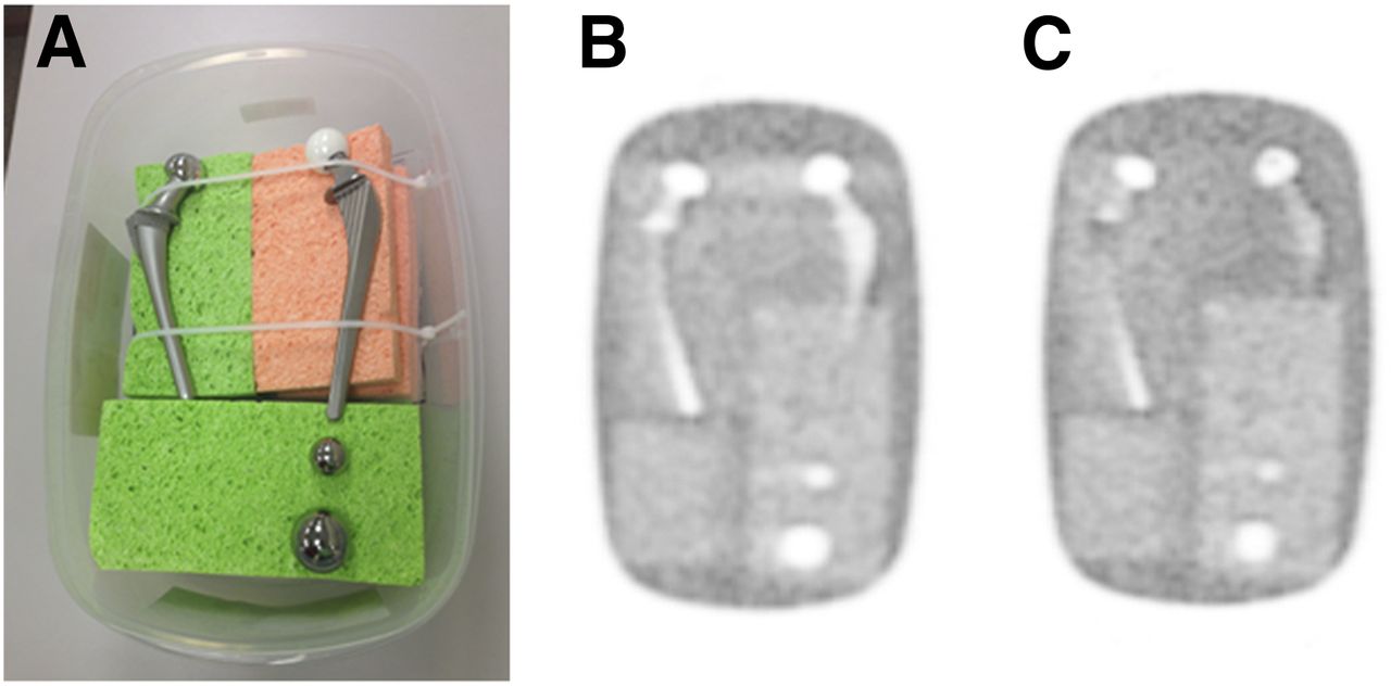
    (A) Phantom consisting of 2 types of hip prostheses in solution of 18F-FDG and water: a cobalt steel hip prosthesis and a Ti-Al-V hip prosthesis with ceramic head. Furthermore, phantom included 2 solid steel balls (2.5 and 3.8 cm in diameter). Phantom was scanned in mCT PET/CT scanner. Cold artifact caused by metal is present in standard PET reconstruction (B), whereas on iMAR PET reconstruction (C) these artifacts were not visible.

  • FIGURE 2.
    • Download figure
    • Open in new tab
    • Download powerpoint
    FIGURE 2.

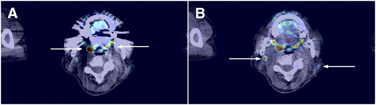
    Patient with uptake in palatine tonsils (arrows in A) and 18F-FDG–avid lymph nodes (arrows in B). Metal artifacts are visible on standard PET/CT reconstruction (A), whereas iMAR PET/CT reconstruction (B) shows less distortion.

  • FIGURE 3.
    • Download figure
    • Open in new tab
    • Download powerpoint
    FIGURE 3.

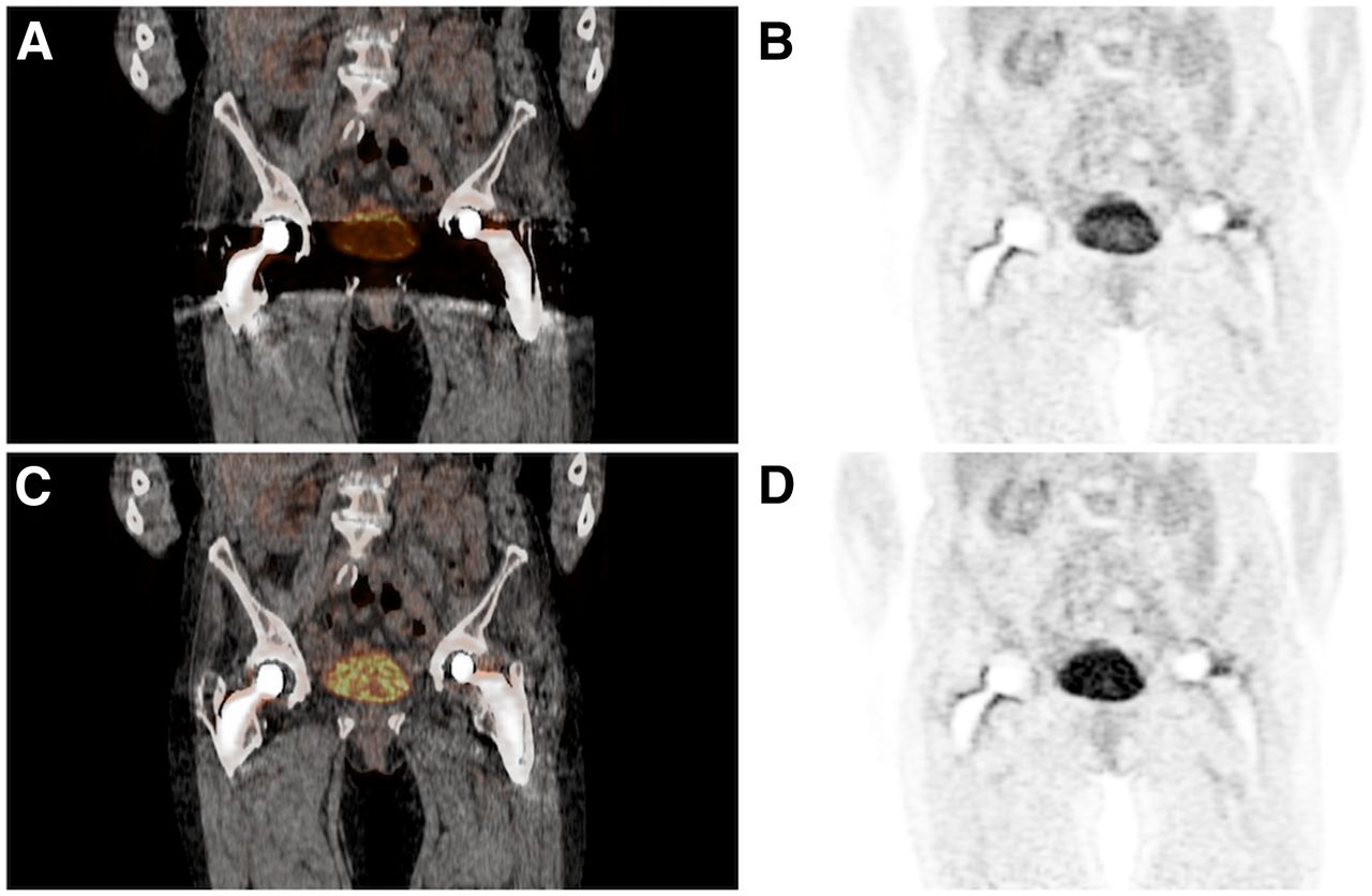
    Patient with possible malignant cyst in right ovary, which was not interpretable due to metal artifacts. Effect of metal artifact on CT is also visible on PET. Region between both hip implants show lower activity for standard PET/CT (A and B), which is clearly visible in bladder, whereas iMAR PET/CT (C and D) shows image closer to true distribution of 18F-FDG.

  • FIGURE 4.
    • Download figure
    • Open in new tab
    • Download powerpoint
    FIGURE 4.

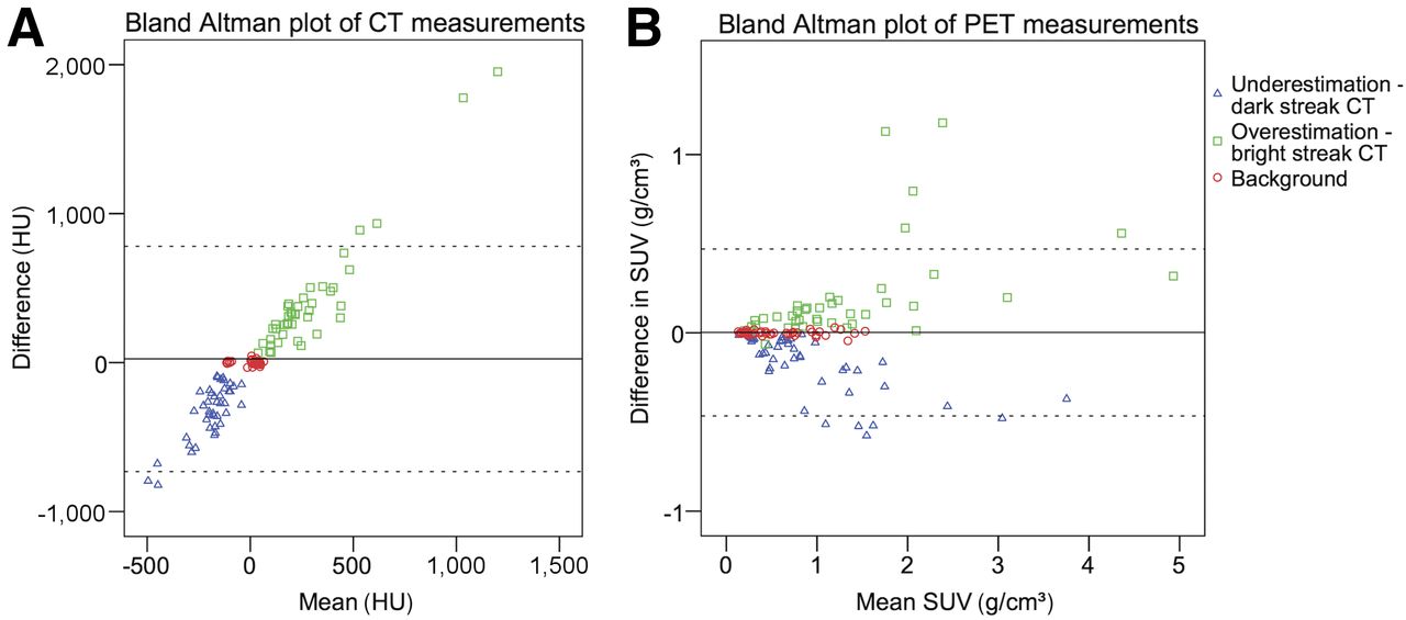
    Bland–Altman plots of ROI measurements, shown for HU measurements on CT (A) and SUVmean measurements for PET reconstructions (B). In the figure, difference between 2 measurements (standard − iMAR) is plotted against mean of these 2 measurements. Effect of algorithm not only is visible on CT image, but also influences PET image.

  • FIGURE 5.
    • Download figure
    • Open in new tab
    • Download powerpoint
    FIGURE 5.

    PET images of phantom when CT was misaligned by 3 mm (laterally in region of prosthetic femoral heads). (A) Normal, CT attenuation-based reconstruction. (B and C) One and 10 iterations of TOF-MLACF reconstruction.

Tables

  • Figures
  • Additional Files
    • View popup
    TABLE 1

    Summary of Patient Characteristics

    CharacteristicValue
    Sex
     Male12
     Female9
    Age (y)65.4 ± 8.6
    Weight (kg)80.8 ± 21.4
    Radiopharmaceutical
     18F-FDG14
     68Ga-PSMA7
    Type of prosthesis
     Dental implants6
     Hip prostheses12
     Shoulder prostheses1
     Screws in vertebrae2
    Indication PET/CT scan
     Staging/detection of disease16
     Radiotherapy planning1
     Therapy response monitoring4
    • Data are values, unless otherwise indicated.

    • View popup
    TABLE 2

    Results of Scoring of PET/CT Images by Nuclear Medicine Physicians (Reader 1/Reader 2)

    QuestionsDental implant (n = 6)Hip implant, 18F-FDG (n = 5)Hip implant, 68Ga-PSMA (n = 7)Shoulder implant (n = 1)Vertebra screw (n = 2)
    When normal CT was used, did metal artifacts affect your interpretation of the PET/CT image in a negative way?*5/64/56/70/10/1
    When iMAR CT was used, did metal artifacts affect your interpretation of the PET/CT image in a negative way?*0/00/00/00/00/1
    Was iMAR helpful?*5/65/57/71/10/1
    Was NAC PET helpful?*2/23/30/20/00/0
    Did the visual interpretation indicate that PET and CT were well aligned or misaligned? Answered with well aligned.6/45/37/71/12/2
    How much confidence do you have in your interpretation of the PET/CT image? (scale, 0%–100%; improvement of [%])30/3722/2841/3010/01/5
    Did the diagnosis/staging of the disease change when the iMAR CT was used?*1/02/11/10/00/0
    When normal CT was used, were more diagnostic tools needed because of the metal artifacts?*0/21/11/00/00/0
    • ↵* Answered with yes.

    • NAC = non–attenuation-corrected.

Additional Files

  • Figures
  • Tables
  • Supplemental Data

    Files in this Data Supplement:

    • Supplemental Data
PreviousNext
Back to top

In this issue

Journal of Nuclear Medicine: 58 (11)
Journal of Nuclear Medicine
Vol. 58, Issue 11
November 1, 2017
  • Table of Contents
  • Table of Contents (PDF)
  • About the Cover
  • Index by author
Print
Download PDF
Article Alerts
Sign In to Email Alerts with your Email Address
Email Article

Thank you for your interest in spreading the word on Journal of Nuclear Medicine.

NOTE: We only request your email address so that the person you are recommending the page to knows that you wanted them to see it, and that it is not junk mail. We do not capture any email address.

Enter multiple addresses on separate lines or separate them with commas.
Metal Artifact Reduction of CT Scans to Improve PET/CT
(Your Name) has sent you a message from Journal of Nuclear Medicine
(Your Name) thought you would like to see the Journal of Nuclear Medicine web site.
Citation Tools
Metal Artifact Reduction of CT Scans to Improve PET/CT
Charlotte S. van der Vos, Anne I.J. Arens, James J. Hamill, Christian Hofmann, Vladimir Y. Panin, Antoi P.W. Meeuwis, Eric P. Visser, Lioe-Fee de Geus-Oei
Journal of Nuclear Medicine Nov 2017, 58 (11) 1867-1872; DOI: 10.2967/jnumed.117.191171

Citation Manager Formats

  • BibTeX
  • Bookends
  • EasyBib
  • EndNote (tagged)
  • EndNote 8 (xml)
  • Medlars
  • Mendeley
  • Papers
  • RefWorks Tagged
  • Ref Manager
  • RIS
  • Zotero
Share
Metal Artifact Reduction of CT Scans to Improve PET/CT
Charlotte S. van der Vos, Anne I.J. Arens, James J. Hamill, Christian Hofmann, Vladimir Y. Panin, Antoi P.W. Meeuwis, Eric P. Visser, Lioe-Fee de Geus-Oei
Journal of Nuclear Medicine Nov 2017, 58 (11) 1867-1872; DOI: 10.2967/jnumed.117.191171
Twitter logo Facebook logo LinkedIn logo Mendeley logo
  • Tweet Widget
  • Facebook Like
  • Google Plus One
Bookmark this article

Jump to section

  • Article
    • Abstract
    • MATERIALS AND METHODS
    • RESULTS
    • DISCUSSION
    • CONCLUSION
    • DISCLOSURE
    • Acknowledgments
    • Footnotes
    • REFERENCES
  • Figures & Data
  • Supplemental
  • Info & Metrics
  • PDF

Related Articles

  • This Month in JNM
  • PubMed
  • Google Scholar

Cited By...

  • Hot Spot Imaging in Cardiovascular Diseases: An Information Statement from SNMMI, ASNC, and EANM
  • Google Scholar

More in this TOC Section

  • Investigating the Prospect of Cross Calibrating Bone Density Scanners across a Medical Enterprise
  • DIFFERENCES IN THYROID UPTAKE PERCENTAGE BASED ON COUNTING STATISTICS
  • Monte Carlo N-Particle Code Analysis of Multiple Gamma Camera Components from Varying Manufacturers
Show more Instrumentation

Similar Articles

Keywords

  • PET/CT
  • iterative metal artifact reduction
  • 18F-FDG
  • 68Ga-PSMA
  • TOF-MLACF
SNMMI

© 2025 SNMMI

Powered by HighWire