


Article Figures & Data
Figures
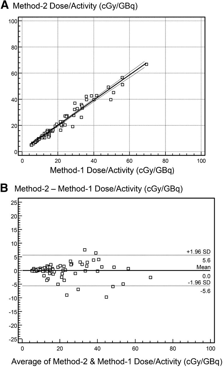
- FIGURE 1.
Linear regression (A) and Bland–Altman plot (B) for method 2 vs. method 1 dose.
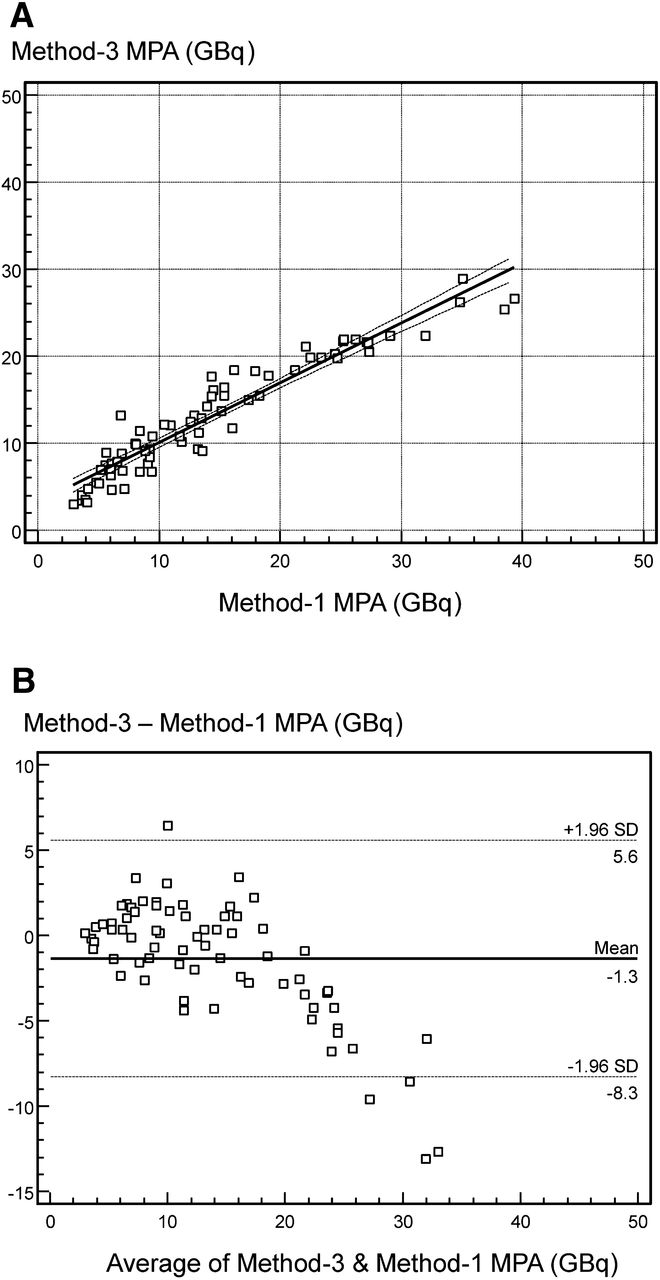
- FIGURE 2.
Linear regression (A) and Bland–Altman plot (B) for method 3 vs. method 1 dose.
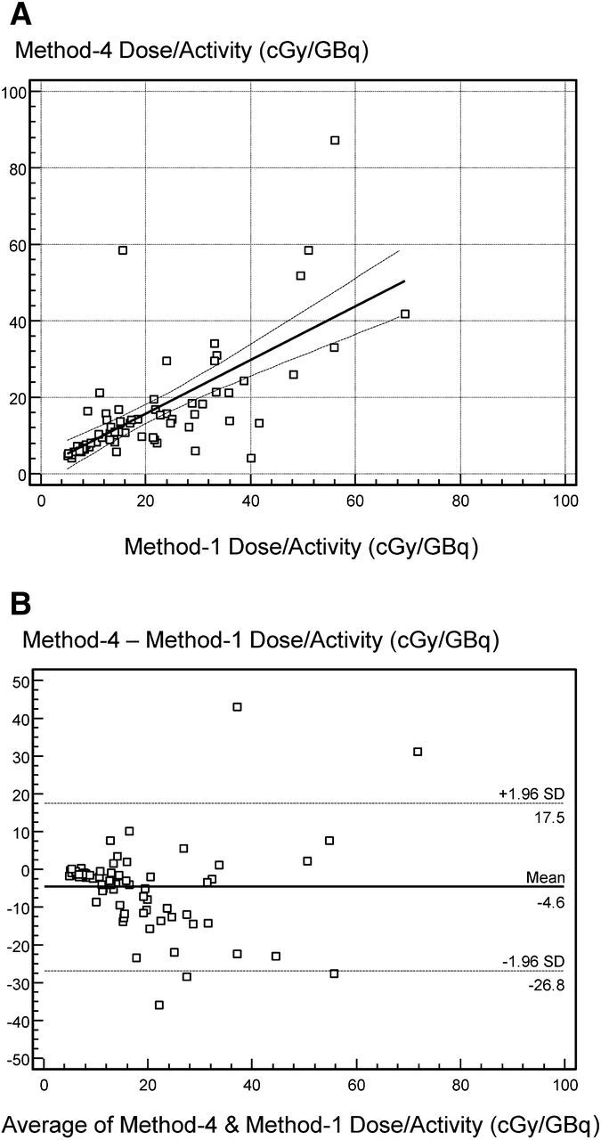
- FIGURE 3.
Linear regression (A) and Bland–Altman plot (B) for method 4 vs. method 1 dose.
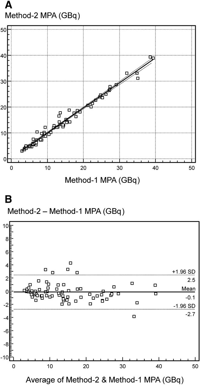
- FIGURE 4.
Linear regression (A) and Bland–Altman plots (B) for method 2 MPA vs. method 1.
- FIGURE 5.
Linear regression (A) and Bland–Altman plots (B) for method 3 MPA vs. method 1.
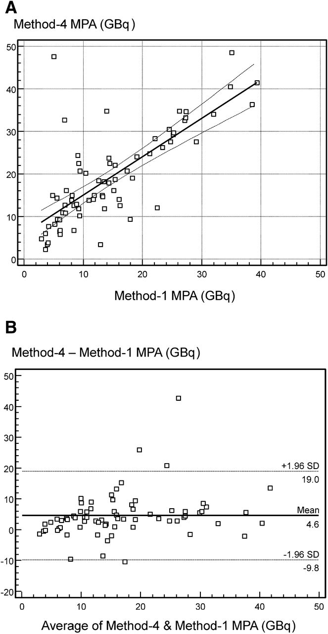
- FIGURE 6.
Linear regression (A) and Bland-Altman plots (B) for method 4 MPA vs. method 1.


Tables
- TABLE 1
Linear Regression and Bland–Altman Comparisons Versus Conventional Method 1 of Alternative Methods to Compute Total Blood Dose
Statistical measure Blood only, method 2 Camera only, method 3 48-h camera only, method 4 Regression R 0.98, P < 0.0001 0.94, *P < 0.0001 0.69, *P < 0.0001 Intercept 0.7 ± 0.6 cGy/GBq, P = 0.22 2.1 ± 1.0 cGy/GBq, *P = 0.05 1.6 ± 2.2 cGy/GBq, P = 0.49 Slope 0.97 ± 0.02, P < 0.0001 0.90 ± 0.04, *P < 0.0001 0.70 ± 0.09, *P < 0.0001 Bland–Altman R −0.07, P = 0.54 −0.12, P = 0.31 0.02, P = 0.84 Intercept 0.3 ± 0.6 cGy/GBq, P = 0.59 0.8 ± 1.1 cGy/GBq, *P = 0.44 −5. 0 ± 2.3 cGy/GBq, *P = 0.04 Slope −0.01 ± 0.02, P = 0.54 −0.04 ± 0.04, *P = 0.31 0.02 ± 0.10, *P = 0.84 ↵* P < 0.05 vs. method 2.
Statistical measure Blood only, method 2 Camera only, method 3 48-h camera only, method 4 Regression R 0.99, P < 0.0001 0.95, *P < 0.0001 0.75, *P < 0.0001 Intercept 0.4 ± 0.3 GBq, P = 0.20 3.2 ± 0.5 GBq, *P < 0.0001 6.1 ± 1.6 GBq,*P = 0.0001 Slope 0.96 ± 0.02, P < 0.0001 0.69 ± 0.03, *P < 0.0001 0.86 ± 0.09, P < 0.0001 Bland–Altman R −0.18, P = 0.14 −0.74, *P < 0.0001 0.26, *P = 0.03 Intercept 0.2 ± 0.3 GBq, P = 0.43 3.3 ± 0.6 GBq, *P < 0.0001 1.2 ± 1.7 GBq, *P = 0.48 Slope −0.03 ± 0.02, P = 0.14 −0.33 ± 0.04, *P < 0.0001 0.20 ± 0.09, *P = 0.03 ↵* P < 0.05 vs. method 2.
- TABLE 3
Comparison of Methods 2–4 Against Cases for Which MPA < 7.4 GBq by Conventional Method 1
Statistical measure Blood only, method 2 Camera only, method 3 48-h camera only, method 4 κ 0.86 (very good agreement) 0.70* (good agreement) 0.43* (moderate agreement) McNemar Δ 0.0%, P = 0.62 5.6%, P = 0.29 14.1%, *P = 0.02 Sensitivity 90% 70% 40%* Specificity 96% 96% 96% Accuracy 94% 89% 80%* Positive predictive value 90% 88% 80% Negative predictive value 96% 89% 80%* ↵* P < 0.05 vs. method 2.
- TABLE 4
Comparison of Dose Estimates of Methods 2–4 Against Conventional Method 1 Dose Estimates for Patients Grouped by Prior Treatments and Abnormal Renal Function
Patient group Conventional, method 1 Blood only, method 2 Camera only, method 3 48-h camera only, method 4 PT+ and AF− (n = 26) 12 ± 9 cGy/GBq 12 ± 9 cGy/GBq, P = 0.63 13 ± 8 cGy/GBq, *P = 0.03 13 ± 11 cGy/GBq, P = 0.76 PT− and AF− (n = 22) 22 ± 17 cGy/GBq 21 ± 16 cGy/GBq, P = 0.22 23 ± 17 cGy/GBq, P = 0.25 14 ± 12 cGy/GBq, *P = 0.002 PT− and AF+ (n = 17) 29 ± 9 cGy/GBq 30 ± 10 cGy/GBq, P = 0.54 27 ± 11 cGy/GBq, P = 0.17 20 ± 13 cGy/GBq, *P = 0.0008 PT+ and AF+ (n = 6) 34 ± 13 cGy/GBq 35 ± 11 cGy/GBq, P = 0.51 30 ± 14 cGy/GBq, P = 0.11 29 ± 29 cGy/GBq, P = 0.52 ↵* P < 0.05 vs. method 1.
PT+ = patients who had prior 131I treatment; AF− = patients with normal renal function; PT− = patients who did not have prior 131I treatment; AF+ = patients with abnormal renal function.
- TABLE 5
Comparison of MPA of Methods 2–4 Against Conventional Method 1 MPA for Patients Grouped by Prior Treatments and Abnormal Renal Function
Patient group Conventional, method 1 Blood only, method 2 Camera only, method 3 48-h camera only, method 4 PT+ and AF− (n = 26) 21.4 ± 9.3 GBq 20.9 ± 9.0 GBq, P = 0.14 18.0 ± 6.1 GBq, *P = 0.0003 23.4 ± 11.9 GBq, P = 0.08 PT− and AF− (n = 22) 13.9 ± 7.8 GBq 14.1 ± 7.7 GBq, P = 0.61 12.6 ± 6.2 GBq, *P = 0.03 21.0 ± 10.8 GBq, P = 0.002 PT− and AF+ (n = 17) 7.6 ± 2.4 GBq 7.6 ± 2.7 GBq, P = 0.92 8.5 ± 2.9 GBq, P = 0.09 13.3 ± 7.4 GBq, *P = 0.003 PT+ and AF+ (n = 6) 6.7 ± 2.7 GBq 6.5 ± 2.8 GBq, P = 0.29 7.6 ± 2.5 GBq, P = 0.12 10.6 ± 4.5 GBq, *P = 0.04 ↵* P < 0.05 vs. method 1.
PT+ = patients who had prior 131I treatment; AF− = patients with normal renal function; PT− = patients who did not have prior 131I treatment; AF+ = patients with abnormal renal function.


Jump to section
Related Articles
Cited By...
- No citing articles found.