
Main menu

  • Home
  • Content
    • Current
    • Ahead of print
    • Past Issues
    • JNM Supplement
    • SNMMI Annual Meeting Abstracts
    • Continuing Education
    • JNM Podcasts
  • Subscriptions
    • Subscribers
    • Institutional and Non-member
    • Rates
    • Journal Claims
    • Corporate & Special Sales
  • Authors
    • Submit to JNM
    • Information for Authors
    • Assignment of Copyright
    • AQARA requirements
  • Info
    • Reviewers
    • Permissions
    • Advertisers
  • About
    • About Us
    • Editorial Board
    • Contact Information
  • More
    • Alerts
    • Feedback
    • Help
    • SNMMI Journals
  • SNMMI
    • JNM
    • JNMT
    • SNMMI Journals
    • SNMMI

User menu

  • Subscribe
  • My alerts
  • Log in
  • My Cart

Search

  • Advanced search
Journal of Nuclear Medicine
  • SNMMI
    • JNM
    • JNMT
    • SNMMI Journals
    • SNMMI
  • Subscribe
  • My alerts
  • Log in
  • My Cart
Journal of Nuclear Medicine

Advanced Search

  • Home
  • Content
    • Current
    • Ahead of print
    • Past Issues
    • JNM Supplement
    • SNMMI Annual Meeting Abstracts
    • Continuing Education
    • JNM Podcasts
  • Subscriptions
    • Subscribers
    • Institutional and Non-member
    • Rates
    • Journal Claims
    • Corporate & Special Sales
  • Authors
    • Submit to JNM
    • Information for Authors
    • Assignment of Copyright
    • AQARA requirements
  • Info
    • Reviewers
    • Permissions
    • Advertisers
  • About
    • About Us
    • Editorial Board
    • Contact Information
  • More
    • Alerts
    • Feedback
    • Help
    • SNMMI Journals
  • View or Listen to JNM Podcast
  • Visit JNM on Facebook
  • Join JNM on LinkedIn
  • Follow JNM on Twitter
  • Subscribe to our RSS feeds
Research ArticleOncology

In Vivo Relationship Between Hypoxia and Angiogenesis in Human Glioblastoma: A Multimodal Imaging Study

Keven Ferreira da Ponte, David Hassanein Berro, Solène Collet, Jean-Marc Constans, Evelyne Emery, Samuel Valable and Jean-Sébastien Guillamo
Journal of Nuclear Medicine October 2017, 58 (10) 1574-1579; DOI: https://doi.org/10.2967/jnumed.116.188557
Keven Ferreira da Ponte
1Normandie University, UNICAEN, CEA, CNRS, ISTCT/CERVOxy Group, Caen, France
2Department of Neurosurgery, University Hospital of Caen, Caen, France
  • Find this author on Google Scholar
  • Find this author on PubMed
  • Search for this author on this site
David Hassanein Berro
1Normandie University, UNICAEN, CEA, CNRS, ISTCT/CERVOxy Group, Caen, France
2Department of Neurosurgery, University Hospital of Caen, Caen, France
  • Find this author on Google Scholar
  • Find this author on PubMed
  • Search for this author on this site
Solène Collet
1Normandie University, UNICAEN, CEA, CNRS, ISTCT/CERVOxy Group, Caen, France
  • Find this author on Google Scholar
  • Find this author on PubMed
  • Search for this author on this site
Jean-Marc Constans
1Normandie University, UNICAEN, CEA, CNRS, ISTCT/CERVOxy Group, Caen, France
3Department of Neuroradiology, University Hospital of Caen, Caen, France
  • Find this author on Google Scholar
  • Find this author on PubMed
  • Search for this author on this site
Evelyne Emery
2Department of Neurosurgery, University Hospital of Caen, Caen, France
4Normandie University, UNICAEN, INSERM, Inserm UMR-S U919, Caen, France
  • Find this author on Google Scholar
  • Find this author on PubMed
  • Search for this author on this site
Samuel Valable
1Normandie University, UNICAEN, CEA, CNRS, ISTCT/CERVOxy Group, Caen, France
  • Find this author on Google Scholar
  • Find this author on PubMed
  • Search for this author on this site
Jean-Sébastien Guillamo
1Normandie University, UNICAEN, CEA, CNRS, ISTCT/CERVOxy Group, Caen, France
5Department of Neurology, University Hospital of Caen, Caen, France; and
6Department of Neurology, University Hospital of Nimes, Nimes, France
  • Find this author on Google Scholar
  • Find this author on PubMed
  • Search for this author on this site
  • Article
  • Figures & Data
  • Info & Metrics
  • PDF
Loading

Article Figures & Data

Figures

  • Tables
  • FIGURE 1.
    • Download figure
    • Open in new tab
    • Download powerpoint
    FIGURE 1.

    Delimitation of different ROIs. T1Gd images (A) were used to calculate the following ROIs: classic tumor region (B, cyan), radiologic necrosis region (T1Gd central hyposignal; B, magenta), and, by subtraction, CE region (C). FLAIR images (D) were used to calculate the total tumor region (D, yellow) and exclusive FLAIR region (D, region between yellow and cyan). ROIs have been transposed on modalities (E and F) for calculating parameters. Then, a semiautomatic segmentation was performed for high-CBV (G) and –18F-FMISO uptake (H) volumes.

  • FIGURE 2.
    • Download figure
    • Open in new tab
    • Download powerpoint
    FIGURE 2.

    Right occipital glioblastoma. (A) MRI T1Gd sequence, illustrating typical appearance of glioblastoma with peripheral hypersignal reflecting extravasation of gadolinium, associated with central hyposignal (radiologic necrosis). (B) FLAIR sequence showing hyperintensity that far exceeds volume of CE. (C) 18F-FMISO PET image (T/B) revealing significant uptake within tumor. Yellow outline shows automatic thresholding by contralateral 95 centile, which yielded hypoxic tumor volume. (D) CBV map also shows widespread tumor vascularization. Cyan outline represents threshold by contralateral 95 centile, which has produced hypervascularized tumor volume. (E) Representation of different volumes on T1Gd sequence after coregistration of all modalities. White area represents CE compartment, and brown zone represents exclusive FLAIR region. (F–H) Automatic thresholding on same images of C, D, and E of volume of maximal tumor hypoxia (F) and volume of maximal tumor vascularization (G) by tumoral 95 centile (5% of most intense pixels).
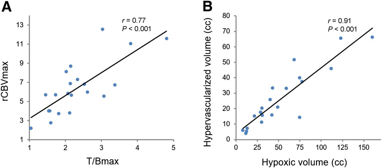
  • FIGURE 3.
    • Download figure
    • Open in new tab
    • Download powerpoint
    FIGURE 3.

    Scatterplot with linear regression lines illustrating relationship between uptake of 18F-FMISO and vascularization. (A) Correlation between maximum values of 18F-FMISO uptake expressed as T/B and rCBV in glioblastomas (classic tumor volume). (B) Relationship between hypoxic tumor volume (18F-FMISO uptake thresholded by contralateral 95 centile) and hypervascularized tumor volume (high CBV thresholded by contralateral 95 centile).
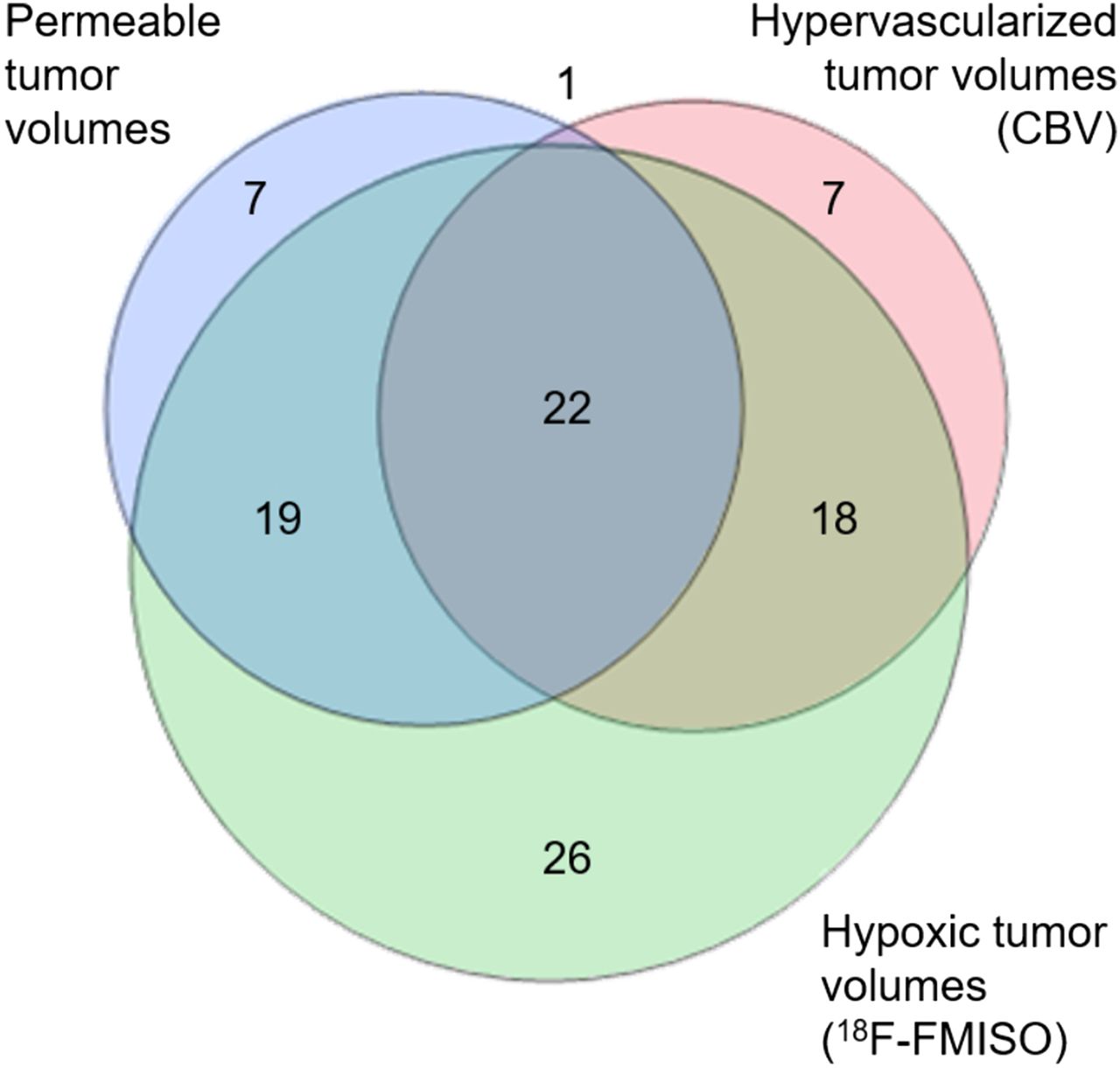
  • FIGURE 4.
    • Download figure
    • Open in new tab
    • Download powerpoint
    FIGURE 4.

    Venn diagram representing relationship between hypoxic (18F-FMISO uptake), hypervascularized (high CBV), and permeable (CE) tumor volumes of study population. Numbers and colored areas indicate proportions between different volumes and overlap rates (Table 2). There is greater volume of 18F-FMISO uptake, with large overlap with both CBV and permeable tumor volume. There is also overlap between hypervascularized and permeable tumor volumes, corresponding to almost 50% of these volumes.

Tables

  • Figures
    • View popup
    TABLE 1

    Correlation Between Different Imaging Parameters (18F-FMISO PET and MRI)*

    Total tumor regionsClassic tumor regions
    ParameterrPrP
    Maximal and mean value
     T/Bmax vs. rCBVmax0.610.002†0.77<0.001†
     T/Bmean vs. rCBVmean0.290.1850.380.079
    Volume
     Hypoxic tumor volume vs. hypervascularized tumor volume0.91<0.001†
     Hypoxic tumor volume vs. permeable tumor volume0.93<0.001†
     Hypervascularized tumor volume vs. permeable tumor volume0.88<0.001†
    • ↵* Pearson correlation coefficients (r) with corresponding P values.

    • ↵† P values are statistically significant.

    • Hypoxic tumor volume = 18F-FMISO uptake volume thresholded by contralateral 95 centile; hypervascularized tumor volume = high CBV thresholded by contralateral 95 centile; permeable tumor volume = volume of CE region.

    • View popup
    TABLE 2

    Mean Overlap Rate Between Hypoxic, Hypervascularized, and Permeable Tumor Volumes

    Recovery ofByMean %
    Hypoxic tumor volumeHypervascularized tumor volume48.8
    Permeable tumor volume44.3
    Volume of maximal tumor hypoxiaHypervascularized tumor volume68.0
    Permeable tumor volume60.8
    Volume of maximal tumor vascularization18.0
    Hypervascularized tumor volumeHypoxic tumor volume81.3
    Permeable tumor volume46.5
    • Hypoxic tumor volume =18F-FMISO uptake volume thresholded by contralateral 95 centile; hypervascularized tumor volume = high CBV thresholded by contralateral 95 centile; permeable tumor volume = volume of CE region; volume of maximal tumor hypoxia = 18F-FMISO uptake volume thresholded by tumoral 95 centile (most intense 5% pixels); volume of maximal tumor vascularization = high CBV thresholded by tumoral 95 centile.

PreviousNext
Back to top

In this issue

Journal of Nuclear Medicine: 58 (10)
Journal of Nuclear Medicine
Vol. 58, Issue 10
October 1, 2017
  • Table of Contents
  • Table of Contents (PDF)
  • About the Cover
  • Index by author
Print
Download PDF
Article Alerts
Sign In to Email Alerts with your Email Address
Email Article

Thank you for your interest in spreading the word on Journal of Nuclear Medicine.

NOTE: We only request your email address so that the person you are recommending the page to knows that you wanted them to see it, and that it is not junk mail. We do not capture any email address.

Enter multiple addresses on separate lines or separate them with commas.
In Vivo Relationship Between Hypoxia and Angiogenesis in Human Glioblastoma: A Multimodal Imaging Study
(Your Name) has sent you a message from Journal of Nuclear Medicine
(Your Name) thought you would like to see the Journal of Nuclear Medicine web site.
Citation Tools
In Vivo Relationship Between Hypoxia and Angiogenesis in Human Glioblastoma: A Multimodal Imaging Study
Keven Ferreira da Ponte, David Hassanein Berro, Solène Collet, Jean-Marc Constans, Evelyne Emery, Samuel Valable, Jean-Sébastien Guillamo
Journal of Nuclear Medicine Oct 2017, 58 (10) 1574-1579; DOI: 10.2967/jnumed.116.188557

Citation Manager Formats

  • BibTeX
  • Bookends
  • EasyBib
  • EndNote (tagged)
  • EndNote 8 (xml)
  • Medlars
  • Mendeley
  • Papers
  • RefWorks Tagged
  • Ref Manager
  • RIS
  • Zotero
Share
In Vivo Relationship Between Hypoxia and Angiogenesis in Human Glioblastoma: A Multimodal Imaging Study
Keven Ferreira da Ponte, David Hassanein Berro, Solène Collet, Jean-Marc Constans, Evelyne Emery, Samuel Valable, Jean-Sébastien Guillamo
Journal of Nuclear Medicine Oct 2017, 58 (10) 1574-1579; DOI: 10.2967/jnumed.116.188557
Twitter logo Facebook logo LinkedIn logo Mendeley logo
  • Tweet Widget
  • Facebook Like
  • Google Plus One
Bookmark this article

Jump to section

  • Article
    • Abstract
    • MATERIALS AND METHODS
    • RESULTS
    • DISCUSSION
    • CONCLUSION
    • DISCLOSURE
    • Acknowledgments
    • Footnotes
    • REFERENCES
  • Figures & Data
  • Info & Metrics
  • PDF

Related Articles

  • This Month in JNM
  • PubMed
  • Google Scholar

Cited By...

  • Simultaneous Mapping of Vasculature, Hypoxia, and Proliferation Using Dynamic Susceptibility Contrast MRI, 18F-FMISO PET, and 18F-FLT PET in Relation to Contrast Enhancement in Newly Diagnosed Glioblastoma
  • Monitoring Oxygenation Levels Deep in the Tumor Core: Noninvasive Imaging of Hypoxia, Now in Real-Time 3D
  • Association between Tumor Acidity and Hypervascularity in Human Gliomas Using pH-Weighted Amine Chemical Exchange Saturation Transfer Echo-Planar Imaging and Dynamic Susceptibility Contrast Perfusion MRI at 3T
  • Google Scholar

More in this TOC Section

Oncology

  • 18F-FDG PET/CT manifestations of three Cases of Female Desmoplastic Small Round Cell Tumor
  • 18F-FDG PET/CT imaging features of parotid lesions; Case based pictorial review and its multi-modality correlation.
  • Utility of Fluorine-18-fluorodeoxyglucose PET/CT in Uterine sarcoma patients.
Show more Oncology

Clinical

  • TauIQ: A Canonical Image Based Algorithm to Quantify Tau PET Scans
  • Dual PET Imaging in Bronchial Neuroendocrine Neoplasms: The NETPET Score as a Prognostic Biomarker
  • Addition of 131I-MIBG to PRRT (90Y-DOTATOC) for Personalized Treatment of Selected Patients with Neuroendocrine Tumors
Show more Clinical

Similar Articles

Keywords

  • Glioblastoma
  • hypoxia
  • 18F-FMISO PET
  • angiogenesis
  • MRI
SNMMI

© 2025 SNMMI

Powered by HighWire