
Main menu

  • Home
  • Content
    • Current
    • Ahead of print
    • Past Issues
    • JNM Supplement
    • SNMMI Annual Meeting Abstracts
    • Continuing Education
    • JNM Podcasts
  • Subscriptions
    • Subscribers
    • Institutional and Non-member
    • Rates
    • Journal Claims
    • Corporate & Special Sales
  • Authors
    • Submit to JNM
    • Information for Authors
    • Assignment of Copyright
    • AQARA requirements
  • Info
    • Reviewers
    • Permissions
    • Advertisers
  • About
    • About Us
    • Editorial Board
    • Contact Information
  • More
    • Alerts
    • Feedback
    • Help
    • SNMMI Journals
  • SNMMI
    • JNM
    • JNMT
    • SNMMI Journals
    • SNMMI

User menu

  • Subscribe
  • My alerts
  • Log in
  • My Cart

Search

  • Advanced search
Journal of Nuclear Medicine
  • SNMMI
    • JNM
    • JNMT
    • SNMMI Journals
    • SNMMI
  • Subscribe
  • My alerts
  • Log in
  • My Cart
Journal of Nuclear Medicine

Advanced Search

  • Home
  • Content
    • Current
    • Ahead of print
    • Past Issues
    • JNM Supplement
    • SNMMI Annual Meeting Abstracts
    • Continuing Education
    • JNM Podcasts
  • Subscriptions
    • Subscribers
    • Institutional and Non-member
    • Rates
    • Journal Claims
    • Corporate & Special Sales
  • Authors
    • Submit to JNM
    • Information for Authors
    • Assignment of Copyright
    • AQARA requirements
  • Info
    • Reviewers
    • Permissions
    • Advertisers
  • About
    • About Us
    • Editorial Board
    • Contact Information
  • More
    • Alerts
    • Feedback
    • Help
    • SNMMI Journals
  • View or Listen to JNM Podcast
  • Visit JNM on Facebook
  • Join JNM on LinkedIn
  • Follow JNM on Twitter
  • Subscribe to our RSS feeds
Meeting ReportInstrumentation & Data Analysis Track

A novel two-stage iterative vessel tracking algorithm for determining an image derived input function for PET

Artem Mikheev, Jean Logan, Yu-Shin Ding and Henry Rusinek
Journal of Nuclear Medicine May 2016, 57 (supplement 2) 1905;
Artem Mikheev
1Radiology New York University School of Medicine New York NY United States
  • Find this author on Google Scholar
  • Find this author on PubMed
  • Search for this author on this site
Jean Logan
1Radiology New York University School of Medicine New York NY United States
  • Find this author on Google Scholar
  • Find this author on PubMed
  • Search for this author on this site
Yu-Shin Ding
1Radiology New York University School of Medicine New York NY United States
  • Find this author on Google Scholar
  • Find this author on PubMed
  • Search for this author on this site
Henry Rusinek
1Radiology New York University School of Medicine New York NY United States
  • Find this author on Google Scholar
  • Find this author on PubMed
  • Search for this author on this site
  • Article
  • Figures & Data
  • Info & Metrics
Loading

Abstract

1905

Objectives In dynamic PET a plasma arterial input function (AIF) is required to quantify data with a compartment model. As this is cumbersome to acquire and causes discomfort to the subject, alternative methods based on an image derived input function (IDIF) have been explored. We report here a semi-automatic 2 stage algorithm for extracting the IDIF directly from dynamic PET images, without the need for a coregistered anatomical image.

Methods Stage I Given a seed region R, find a sphere of diameter D adjacent to R with the highest peak value of its time activity curve (TAC). Stage II (vessel tracking): Take a step size S in either direction along a set of discretized lines passing through center of the current sphere Si and construct the next sphere Si+1 of diameter D. Select the direction that produces a TAC with the closest match to Si . Continue until the specified maximum length is reached or tracking fails. The algorithm was implemented in the software FireVoxel (wp.nyu.edu/FireVoxel) and applied to FDG PET (Siemens HR+) data for n=9 subjects for which a direct invasive measure of AIF was available (see below). Parameter estimates of K1, k2, k3 and KI for the FDG model were made using the IDIF as it was derived and using a linear correction technique (for partial volume and whole blood to plasma) (Zhou et al, 2011) that requires a few measured plasma samples. An exponential fit to the IDIF was used for smoothing and the ratios of the IDIF to measured samples at 3 points were fit to a straight line after 10 minutes. The IDIF was corrected using this linear function to form IDIFcorr. Results are reported for the average (over subjects and 7 brain ROIs) of K1, K1/k2, k3 and KI, the influx constant using AIF (plasma from left radial artery collected on the Ole-Dich blood sampler with 5 sec samples around the peak, 6mCi dose), IDIF and IDIFcorr. To assess the ability of the two IDIFs to correctly rank subjects, differences in rank position of the 9 subjects between invasively sampled AIF and the IDIFs were calculated for each ROI.

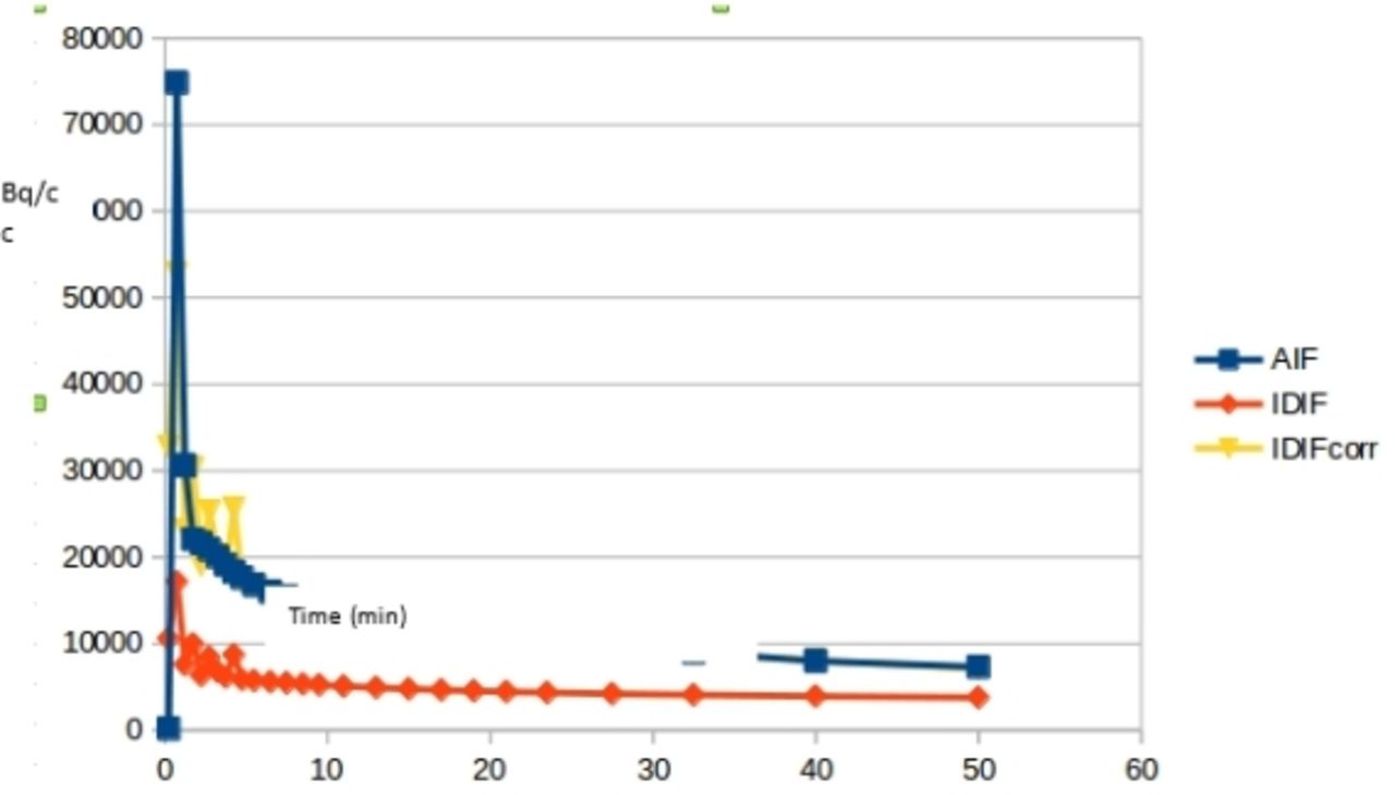
Results A typical AIF, IDIF and IDIFcorr are shown in the figure. Average model parameters were K1: 0.099 ± 0.013 (AIF), 0.16 ± 0.078 (IDIFcorr),0.284 ± .077 (IDIF);K1/k2: 0.58 ± 0.15 (AIF), 0.50 ± 0.14 (IDIFcorr), 1.40 ± 0.32 (IDIF); k3: .069 ± .013 (AIF), 0.074 ± 0.017 (IDIFcorr), 0.039 ± 0.007 (IDIF); KI: 0.0276 ± 0.005 (AIF), 0.0284 ± 0.0049 (IDIFcorr), 0.044 ± 0.0088 (IDIF). KI AIF vs KI IDIFcorr slope 1.05, int=-.005 and correlation .989 (ICC=0.97). KI AIF vs IDIF slope 1.4, int .0053 and correlation 0.73 (ICC=-.32). Average displacements from AIF rank among 9 subjects were using IDIFcorr K1 1.41, k3 1.62, KI 0.44 and using IDIF K1 2.63 k3 1.52 KI 2.02.

Conclusions Good agreement was found for KI for AIF and IDIFcorr. However due to underestimation of the AIF peak by the IDIF and IDIFcorr, K1 is higher for both. The underestimation of the peak is due to 30 second temporal sampling (compared to 5 sec for AIF) for first 5 min and low (with respect to vessel) spatial resolution of the HR+ PET camera which could not adequately capture AIF shape . However more important is the ability to correctly rank subjects and this is achieved using IDIFcorr for KI. For the IDIF the ranking was not as good. Better temporal sampling and spatial resolution will help in this respect. The combination of the automated IDIF and a calibration based on few venous plasma samples promises an adequate input function for dynamic FDG PET studies and may work for other PET tracers as well.

Figure
  • Download figure
  • Open in new tab
  • Download powerpoint
Previous
Back to top

In this issue

Journal of Nuclear Medicine
Vol. 57, Issue supplement 2
May 1, 2016
  • Table of Contents
  • Index by author
Article Alerts
Sign In to Email Alerts with your Email Address
Email Article

Thank you for your interest in spreading the word on Journal of Nuclear Medicine.

NOTE: We only request your email address so that the person you are recommending the page to knows that you wanted them to see it, and that it is not junk mail. We do not capture any email address.

Enter multiple addresses on separate lines or separate them with commas.
A novel two-stage iterative vessel tracking algorithm for determining an image derived input function for PET
(Your Name) has sent you a message from Journal of Nuclear Medicine
(Your Name) thought you would like to see the Journal of Nuclear Medicine web site.
Citation Tools
A novel two-stage iterative vessel tracking algorithm for determining an image derived input function for PET
Artem Mikheev, Jean Logan, Yu-Shin Ding, Henry Rusinek
Journal of Nuclear Medicine May 2016, 57 (supplement 2) 1905;

Citation Manager Formats

  • BibTeX
  • Bookends
  • EasyBib
  • EndNote (tagged)
  • EndNote 8 (xml)
  • Medlars
  • Mendeley
  • Papers
  • RefWorks Tagged
  • Ref Manager
  • RIS
  • Zotero
Share
A novel two-stage iterative vessel tracking algorithm for determining an image derived input function for PET
Artem Mikheev, Jean Logan, Yu-Shin Ding, Henry Rusinek
Journal of Nuclear Medicine May 2016, 57 (supplement 2) 1905;
Twitter logo Facebook logo LinkedIn logo Mendeley logo
  • Tweet Widget
  • Facebook Like
  • Google Plus One
Bookmark this article

Jump to section

  • Article
  • Figures & Data
  • Info & Metrics

Related Articles

  • No related articles found.
  • Google Scholar

Cited By...

  • No citing articles found.
  • Google Scholar

More in this TOC Section

Instrumentation & Data Analysis Track

  • Deep Learning Based Kidney Segmentation for Glomerular Filtration Rate Measurement Using Quantitative SPECT/CT
  • The Benefit of Time-of-Flight in Digital Photon Counting PET Imaging: Physics and Clinical Evaluation
  • Preclinical validation of a single-scan rest/stress imaging technique for 13NH3 cardiac perfusion studies
Show more Instrumentation & Data Analysis Track

MTA II: Data Analysis & Management Posters

  • Comparison of radiation dosimetry estimates for [11C]L-Deprenyl and [11C]Clorgyline across species and with D for H isotopic substitution.
  • Direct VOI-dedicated voxelwise Patlak estimation for quantitative dynamic imaging
  • SPECT/CT and MR image registration for 123I-FP-CIT imaging
Show more MTA II: Data Analysis & Management Posters

Similar Articles

SNMMI

© 2025 SNMMI

Powered by HighWire