


Article Figures & Data
Figures
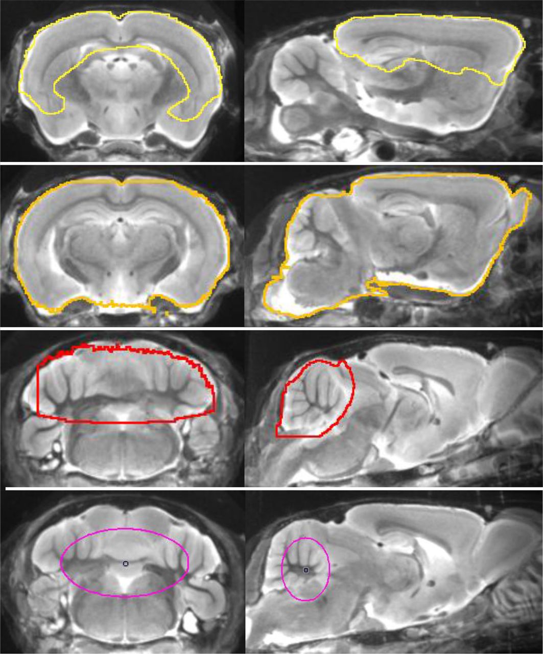
- FIGURE 1.
Definitions of forebrain (yellow), whole brain (orange), cerebellar (red), and white matter (purple) volumes of interest projected on mouse brain MRI atlas in coronal and sagittal slices.
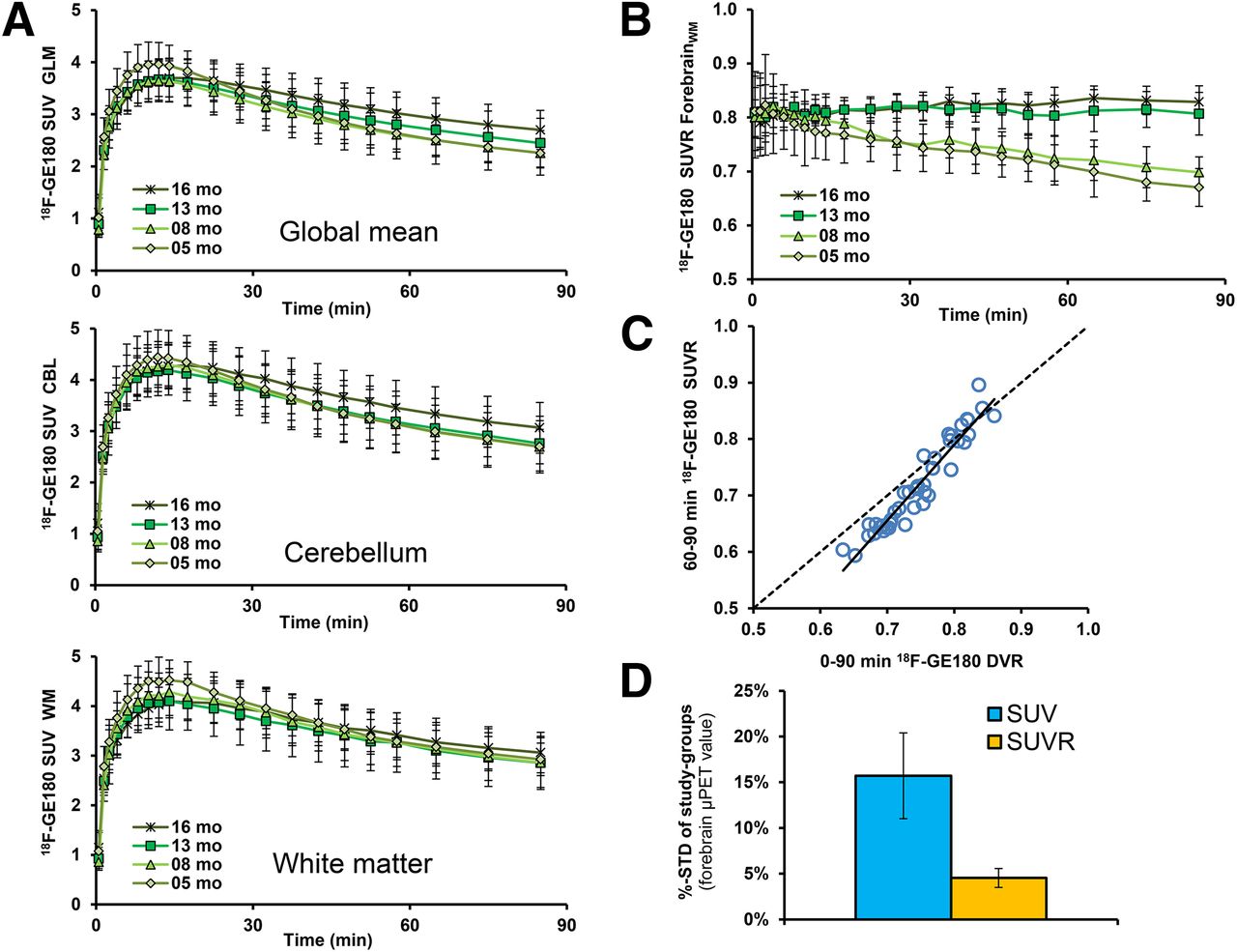
- FIGURE 2.
(A) Mean 18F-GE180 uptake reported as SUV for each of 3 reference regions as function of time after tracer administration in groups of PS2APP mice. Least evidence of pathology (i.e., stability between TG and WT) was detected for a white matter pseudo reference region. Error bars indicate SD for estimates in groups of 5–8 animals. (B) Target-to-reference ratios as functions of time after 18F-GE180 administration in groups of PS2APP mice. (C) Correlation of 18F-GE180 distribution volume ratio (DVR) calculated from 90-min dynamic small-animal PET recordings with corresponding SUVR (forebrain/white-matter) results from 60- to 90-min static frame. Dotted line represents a perfect line of identity (DVR = SUVR). (D) Stability of forebrain 18F-GE180 values after SUV calculation (blue) and pseudo reference tissue (orange) scaling as expressed by mean %-SD (± SD) in all 8 groups of TG and WT mice.
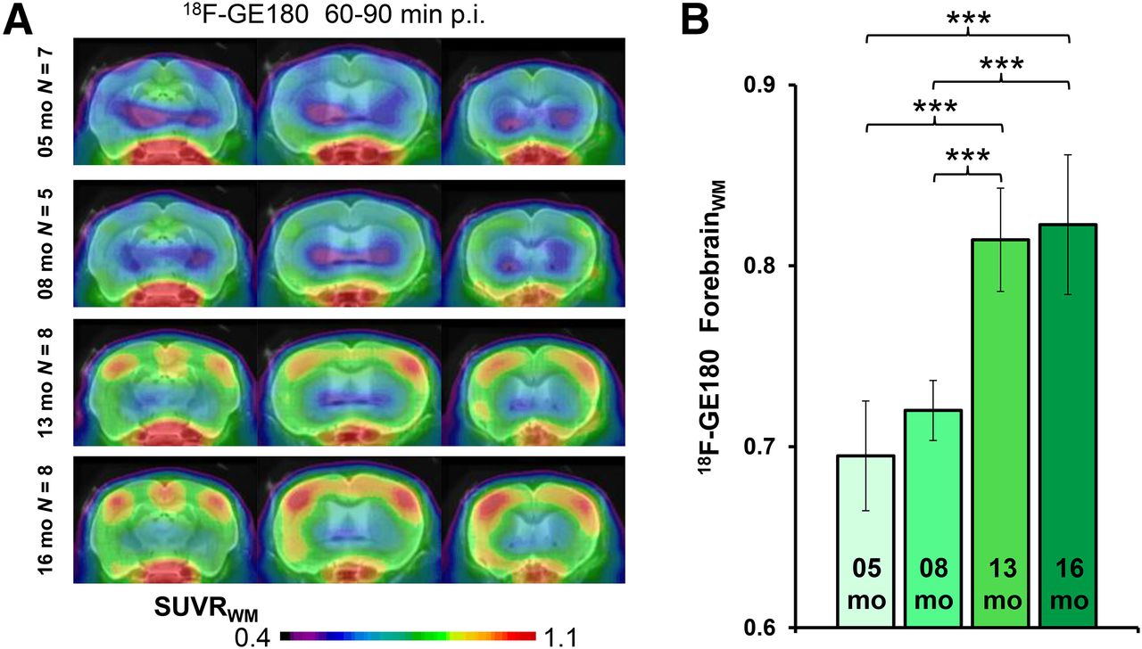
- FIGURE 3.
(A) Coronal planes of 18F-GE180 mean SUVR maps at different ages of PS2APP animals projected on an MRI mouse atlas (gray scale). (B) Mean (±SD) SUVR estimates for PS2APP animals at different ages. Significant differences between subgroups are marked by ***P < 0.001; 1-way ANOVA. p.i. = after injection.
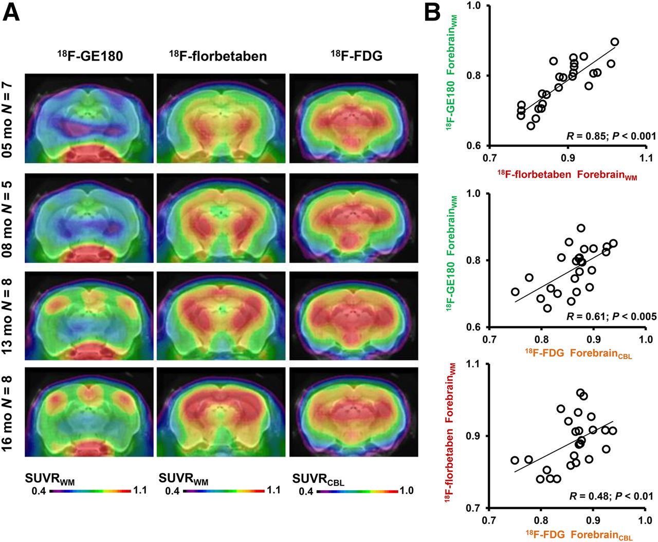
- FIGURE 4.
(A) Coronal planes of mean SUVR maps for all 3 different radiotracers at different ages of PS2APP animals, projected on an MRI mouse atlas (gray scale). (B) Correlations between the different forebrain radiotracer SUVR for all PS2APP mice.
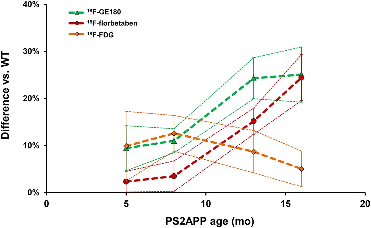
- FIGURE 5.
Life-course kinetics for PS2APP mice as expressed by %-difference (±SD) versus C57BL/6 controls for the 3 different radiotracers.
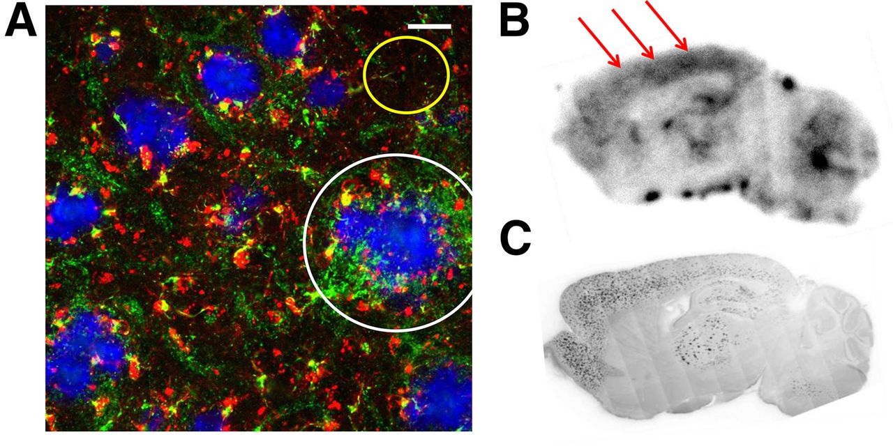
- FIGURE 6.
(A) Immunohistochemical costaining shows IBA-1– (red) and TSPO- (green) positive cells adjacent (white circle) to fibrillar amyloid plaques (blue) in the frontal cortex of a PS2APP mouse aged 22 mo. Yellow circle indicates low IBA-1 and TSPO staining between amyloid plaques. Scale bar represents 20 μm. (B) Sagittal plane for 18F-GE180 autoradiography ex vivo (60 min after injection) in a 22-mo-old PS2APP mouse indicates extensive radiotracer uptake in neocortex (red arrows), hippocampus, and thalamus, which is colocalized with fibrillar amyloid as detected in corresponding methoxy-X04 staining in these brain regions (C). No specific binding is observed in cerebellum.


Tables
Mouse model Age (mo) 18F-GE180 small-animal PET (n) 18F-GE180 small-animal PET (SUVRFBR/WM) 18F-florbetaben small-animal PET (n) 18F-florbetaben small-animal PET (SUVRFBR/WM) 18F-FDG small-animal PET (n) 18F-FDG small-animal PET (SUVRFBR/CBL) PS2APP 5 6 0.69 ± 0.03† 6 0.80 ± 0.02 6 0.85 ± 0.05‡ 8 5 0.72 ± 0.02‡ 4 0.82 ± 0.03 4 0.90 ± 0.05‡ 13 8 0.81 ± 0.03‡ 8 0.90 ± 0.02‡ 8 0.94 ± 0.02‡ 16 8 0.83 ± 0.04‡ 7 0.96 ± 0.03‡ 7 0.91 ± 0.03* C57BL/6 5 8 0.63 ± 0.01 8 0.79 ± 0.01 8 0.77 ± 0.03 8 8 0.65 ± 0.03 8 0.79 ± 0.01 8 0.80 ± 0.03 13 8 0.66 ± 0.02 8 0.78 ± 0.02 8 0.86 ± 0.04‡ 16 8 0.67 ± 0.02* 8 0.78 ± 0.02 8 0.86 ± 0.04‡ Groups of PS2APP and C57BL/6 mice are provided together with forebrain small-animal PET SUVRs for the 3 radiotracers. Significant differences in PS2APP mice versus age-matched C57BL/6 control or significant differences in C57BL/6 controls against their 5-mo-old littermates are marked by *P < 0.05; †P < 0.01; ‡P < 0.001; 1-way ANOVA.


Jump to section
Related Articles
Cited By...
- Early brain neuroinflammatory and metabolic changes identified by dual tracer microPET imaging in mice with acute liver injury
- Remote Neuroinflammation in Newly Diagnosed Glioblastoma Correlates with Unfavorable Clinical Outcome
- Hippocampal purinergic P2X7 receptor level is increased in Alzheimers disease patients, and associated with amyloid and tau pathologies
- Western diet increases brain metabolism and adaptive immune responses in a mouse model of amyloidosis
- Deciphering sources of PET signals in the tumor microenvironment of glioblastoma at cellular resolution
- Tracking Innate Immune Activation in a Mouse Model of Parkinsons Disease Using TREM1 and TSPO PET Tracers
- Chronic PPAR{gamma} Stimulation Shifts Amyloidosis to Higher Fibrillarity but Improves Cognition
- Loss of TREM2 reduces hyperactivation of progranulin deficient microglia but not lysosomal pathology
- Pre-therapeutic Microglia Activation and Sex Determine Therapy Effects of Chronic Immunomodulation
- In vivo Assessment of Neuroinflammation in 4-Repeat Tauopathies
- Asymmetry of Fibrillar Plaque Burden in Amyloid Mouse Models
- Increased Neurite Orientation-Dispersion and Density in the TgCRND8 Mouse Model of Amyloidosis: Inverse Relation with Functional Connectome Clustering and Modulation by Interleukin-6
- Longitudinal PET Monitoring of Amyloidosis and Microglial Activation in a Second-Generation Amyloid-{beta} Mouse Model
- Opposite microglial activation stages upon loss of PGRN or TREM2 result in reduced cerebral glucose metabolism
- Early and Longitudinal Microglial Activation but Not Amyloid Accumulation Predicts Cognitive Outcome in PS2APP Mice
- Neuroinflammation Appears Early on PET Imaging and Then Plateaus in a Mouse Model of Alzheimer Disease
- Time Courses of Cortical Glucose Metabolism and Microglial Activity Across the Life Span of Wild-Type Mice: A PET Study
- Evaluation of Small-Animal PET Outcome Measures to Detect Disease Modification Induced by BACE Inhibition in a Transgenic Mouse Model of Alzheimer Disease
- The FTD-like syndrome causing TREM2 T66M mutation impairs microglia function, brain perfusion, and glucose metabolism