


Article Figures & Data
Figures
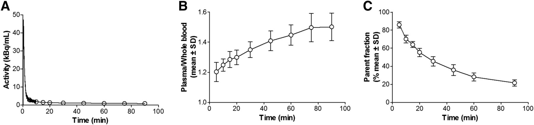
- FIGURE 1.
(A) Typical whole-blood activity curve from 1 AD patient. (B) Plasma–to–whole-blood ratio (mean ± SD) for all 9 subjects. (C) Fraction of unmetabolized (S)-18F-THK5117 (mean ± SD) for all 9 subjects.
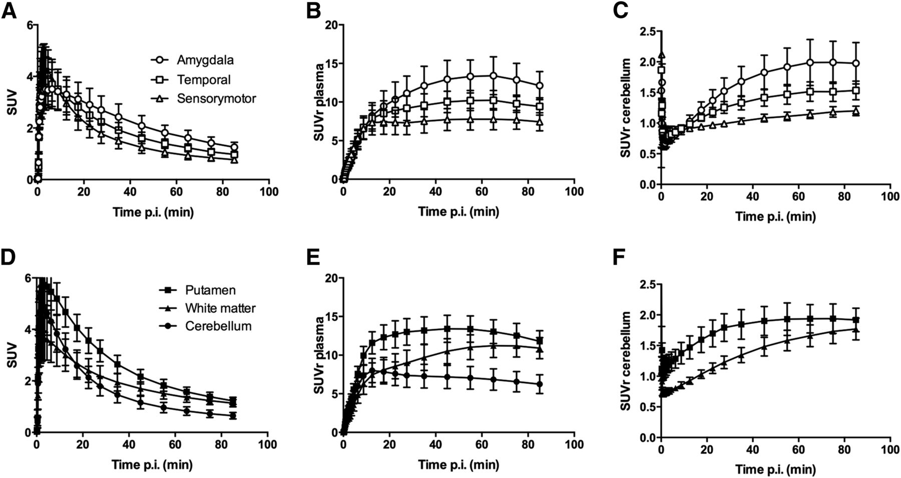
- FIGURE 2.
Time–activity curves (A and D), tissue–to–unmetabolized (S)-18F-THK5117 in plasma ratio (B and E), and tissue-to-cerebellum ratio (C and F) in regions with high, intermediate, and low tau binding (A–C) as well as cerebellum, putamen, and white matter (D–F). Mean for all 5 AD subjects. p.i. = after injection.
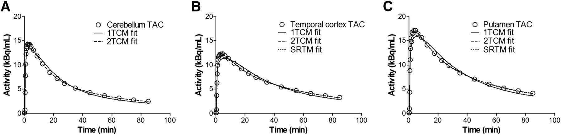
- FIGURE 3.
Typical time–activity curves (TACs) for cerebellum (A), temporal cortex (B), and putamen (C), from 1 AD patient, with fits of 1TCM, 2TCM, and SRTM. Observe that 2TCM and SRTM fits overlap in B and C.
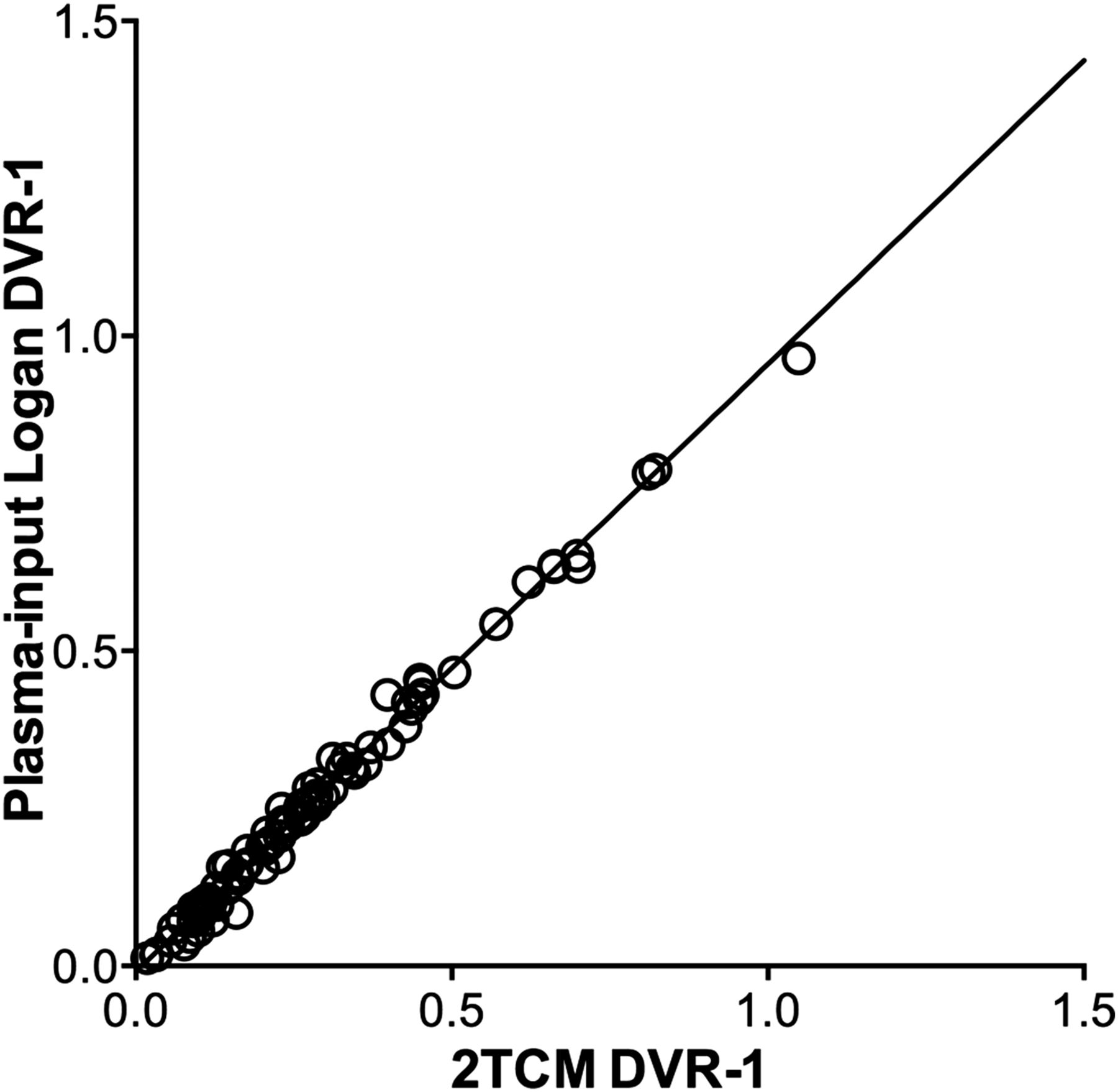
- FIGURE 4.
2TCM DVR-1 values versus plasma-input Logan DVR-1 values. Line is orthogonal regression with slope 0.96 (confidence interval, 0.94–0.98) and intercept −0.01 (confidence interval, −0.01–0.00). Correlation coefficient (R2) between 2TCM DVR-1 and plasma-input Logan DVR-1 was 0.99.
- FIGURE 5.
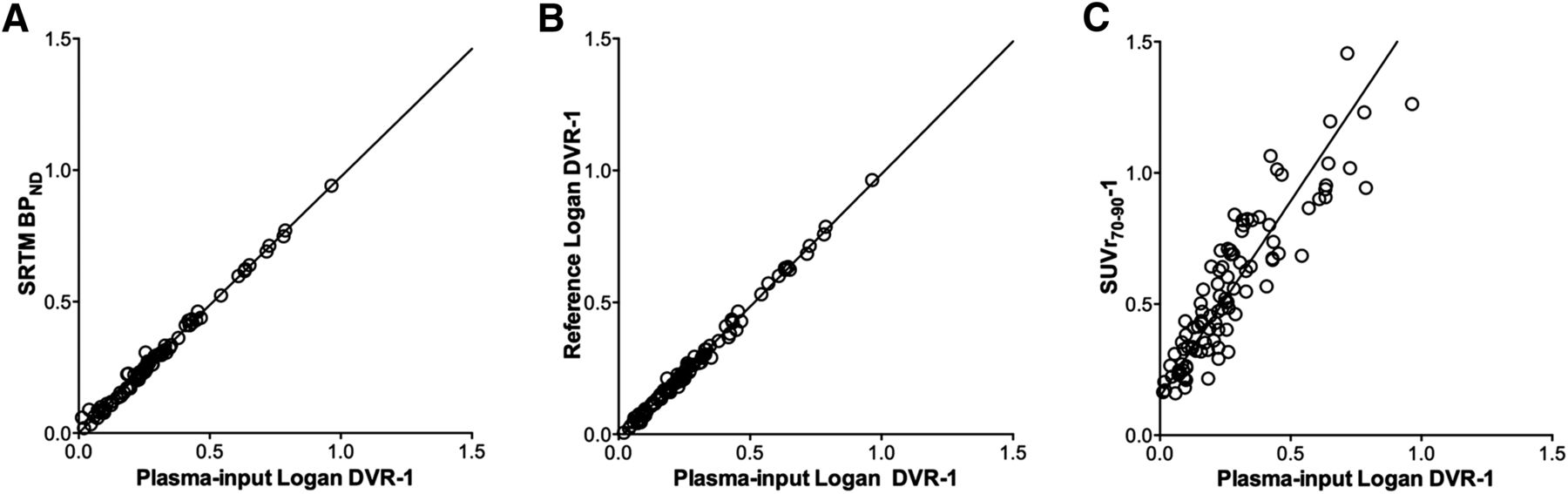
Plasma-input Logan DVR-1 values versus SRTM BPND (A), reference Logan DVR-1 (B), and SUVr-1 values (C). Lines are orthogonal regressions. Slopes and correlation coefficients are given in Table 1.
- FIGURE 6.
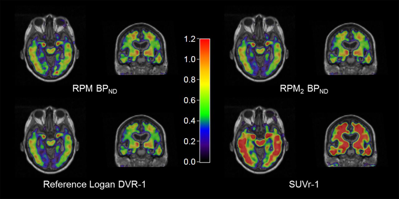
Parametric (S)-18F-THK5117 images of RPM and RPM2 BPND, reference Logan DVR-1, and SUVr70–90-1, from 1 AD patient.
- FIGURE 7.
Relationship between SRTM BPND and SUVr70–90-1 values in whole-brain white matter. Line is orthogonal regression.


Tables
Patient Sex Age (y) Amyloid-β (11C-PIB) Mini-Mental State Examination Years of education AD1 F 70 Pos 25 16 AD2 M 58 Pos 25 11 AD3 F 74 Pos 23 8 AD4 F 57 Pos 23 17 AD5 F 67 Pos 23 14 MCI1 M 79 Neg 27 18 MCI2 F 77 Pos 30 8 MCI3 F 74 Pos 23 12 MCI4 M 65 Pos 30 18 Pos = positive; Neg = negative.
- TABLE 2
Regression Parameters of Reference Tissue Methods Relative to Plasma-Input Logan DVR-1 Values
Model Slope R2 SRTM BPND 0.97 (0.96–0.99) 0.99 Reference Logan DVR-1 1.00 (0.99–1.02) 1.00 SUVr70–90-1 1.49 (1.36–1.62) 0.84 Data in parentheses are confidence intervals.
- TABLE 3
Regression Parameters of Parametric Methods Relative to VOI-Based SRTM BPND Values
Model Slope R2 RPM BPND 0.96 (0.95–0.98) 0.99 RPM2 BPND 1.08 (1.05–1.11) 0.98 Reference Logan DVR-1 1.03 (1.01–1.05) 0.99 SUVr70–90-1 1.55 (1.38–1.72) 0.80 Data in parentheses are confidence intervals.
BPND = 0.80 BPND = 0.32 Model Bias (%) COV (%) Bias (%) COV (%) 2TCM DVR-1 0.1 3.7 0.1 8.7 Plasma Logan DVR-1 −3.0 2.7 −4.3 6.0 SRTM BPND −4.3 2.9 −13.9 6.5 RPM BPND −3.7 2.8 −12.7 6.3 Reference Logan DVR-1 −3.0 0.8 −1.2 0.7 SUVr70–90-1 114.4 3.8 147.2 7.6 Slope R2 Bias Model 40 min 60 min 40 min 60 min 40 min 60 min SRTM BPND 1.06 (1.00 to 1.12) 1.01 (0.98 to 1.03) 0.95 0.99 −0.01 (−0.10 to 0.08) −0.01 (−0.05 to 0.03) Reference Logan DVR-1 0.95 (0.91 to 0.99) 0.99 (0.97 to 1.01) 0.95 0.99 −0.10 (−0.19 to −0.01) −0.03 (−0.07 to 0.01) SUVr-1 0.78 (0.68 to 0.87) 0.97 (0.92 to 1.03) 0.72 0.92 −0.27 (−0.57 to 0.04) −0.08 (−0.24 to 0.08) Data in parentheses are confidence intervals.
Supplemental Data
Files in this Data Supplement:


Jump to section
Related Articles
Cited By...
- In vivo characterization and quantification of neurofibrillary tau PET radioligand [18F]MK-6240 in humans from Alzheimers disease dementia to young controls
- In Vivo Characterization and Quantification of Neurofibrillary Tau PET Radioligand 18F-MK-6240 in Humans from Alzheimer Disease Dementia to Young Controls
- Evaluation of 18F-RO-948 PET for Quantitative Assessment of Tau Accumulation in the Human Brain
- Cerebrospinal Fluid Clearance in Alzheimer Disease Measured with Dynamic PET
- In Vivo Comparison of Tau Radioligands 18F-THK-5351 and 18F-THK-5317