


Article Figures & Data
Figures
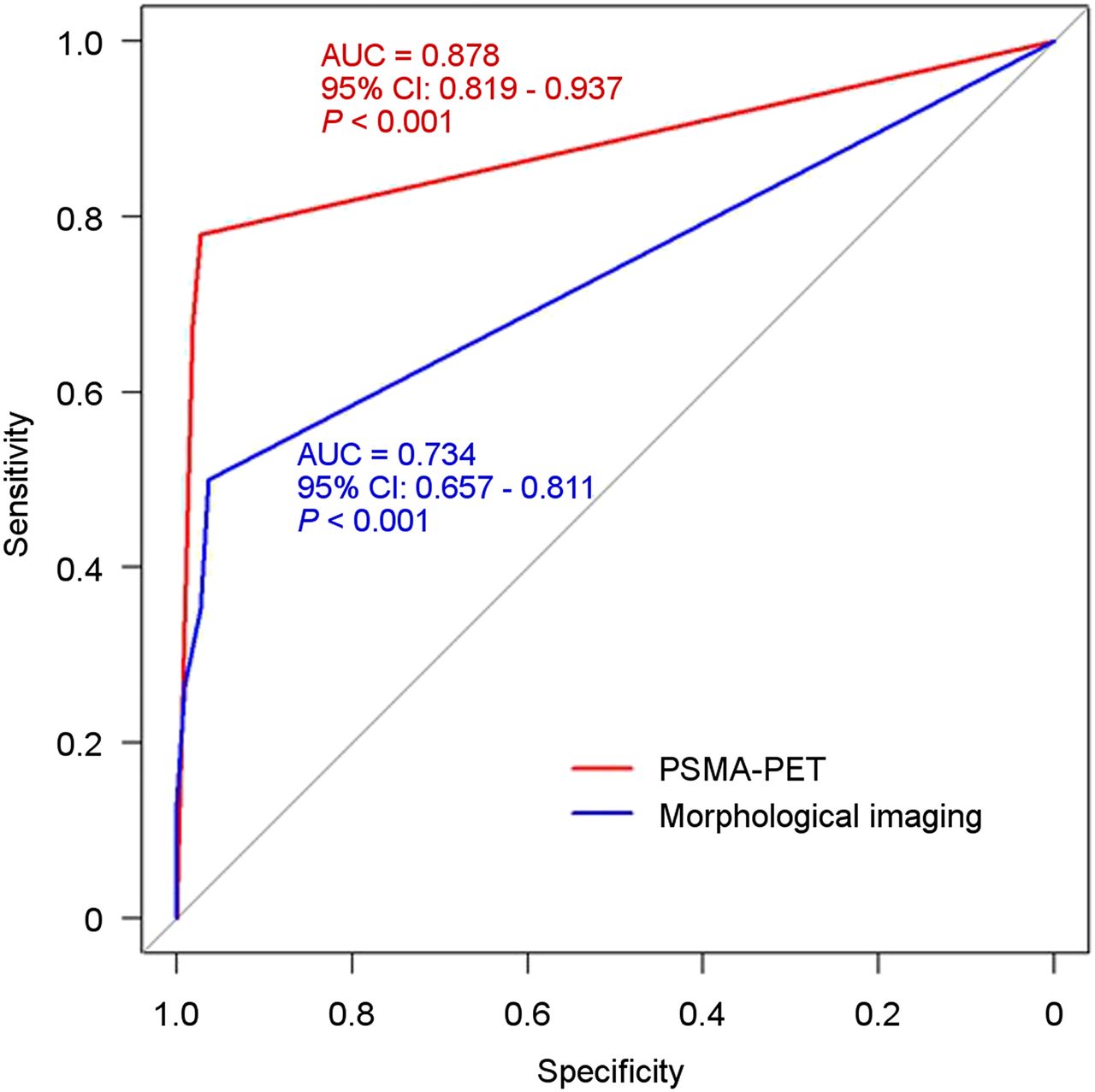
- FIGURE 1.
ROC curves for 68Ga-PSMA HBED-CC PET imaging (red) and morphologic imaging (CT/MR) (blue) for detection of LNM on a field-based analysis (P values: comparison to AUC = 0.5).
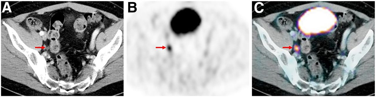
- FIGURE 2.
Example of a 55-y-old patient with biochemical recurrence after radical prostatectomy (Gleason score, 7; PSA level at PET examination, 0.77 ng/mL) and a correctly classified LNM by 68Ga-PSMA HBED-CC PET imaging: a 6-mm LN is visible in right obturator fossa on CT imaging (A, arrow) that shows intense, focal and thus suspicious tracer uptake on 68Ga-PSMA HBED-CC PET (B) and PET/CT fusion imaging (C). Salvage lymphadenectomy with histologic evaluation confirmed a single LNM.
- FIGURE 3.
68Ga-PSMA HBED-CC PET imaging of a 55-y-old patient with recurrent PC (Gleason score, 8; PSA level at PET examination, 5.1 ng/mL). Patient presented with 2 correctly classified LNM (5 and 6 mm) behind left common iliac artery with intense, focal uptake on 68Ga-PSMA HBED-CC PET/CT fusion (B, arrow) and on maximum-intensity-projection images (C, arrow). However, paraaortal/interaortocaval LN field was negative on 68Ga-PSMA HBED-CC PET, showing no increased uptake either on maximum-intensity-projection images (C) or on axial 68Ga-PSMA HBED-CC PET/CT (E). In conventional CT imaging, only small, unsuspicious LNs could be found (D, arrow). Histopathology revealed overall 9 further LNM in this anatomic field (paraaortic).


Tables
- TABLE 1
Total Number and Location of Resected LN Fields and Histopathologically Proven LNM
LN fields No. of resected fields No. of histopathologically proven LNM % of histopathologically proven LNM Level 1 right common iliac vessel 18 6 33.3 Level 2 left common iliac vessel 22 7 31.8 Level 3 right internal iliac vessel 16 4 25 Level 4 left internal iliac vessel 20 9 45 Level 5 right external iliac vessel 17 6 35.3 Level 6 left external iliac vessel 19 9 47.4 Level 7 right obturator fossa 18 7 38.9 Level 8 left obturator fossa 18 3 16.7 Level 9 presacral 13 9 69.2 Level 10 others 18 8 44.4 Histology: LNM Results Positive Negative Diagnostic accuracy 68Ga-PSMA HBED-CC PET rating Positive 53 3 PPV, 94.6% Negative 15 108 NPV, 87.8% Total 68 111 179 Sensitivity, 77.9% Specificity, 97.3% Accuracy, 89.9% Morphologic rating (CT/MR) Positive 18 1 PPV, 94.7% Negative 49 110 NPV, 69.2% Total 67 111 178 Sensitivity, 26.9% Specificity, 99.1% Accuracy, 71.9% PPV = positive predictive value; NPV = negative predictive value.
Histology: LNM Results Positive Negative Diagnostic accuracy 68Ga-PSMA HBED-CC PET rating Positive 42 3 PPV, 93.3% Negative 0 3 NPV, 100% Total 42 6 48 Sensitivity, 100% Specificity, 50% Accuracy, 93.8% Morphologic rating (CT/MR) Positive 14 1 PPV, 93.3% Negative 27 5 NPV, 84.3% Total 41 6 47 Sensitivity, 34.1% Specificity, 83.3% Accuracy, 40.4% PPV = positive predictive value; NPV = negative predictive value.
Supplemental Data
Files in this Data Supplement:


Jump to section
Related Articles
Cited By...
- Validation of 18F-rhPSMA-7 and 18F-rhPSMA-7.3 PET Imaging Results with Histopathology from Salvage Surgery in Patients with Biochemical Recurrence of Prostate Cancer
- Detection Efficacy of 18F-rhPSMA-7.3 PET/CT and Impact on Management in Patients with Biochemical Recurrence of Prostate Cancer After Radical Prostatectomy and Before Potential Salvage Treatment
- Matched-Pair Comparison of 68Ga-PSMA-11 and 18F-rhPSMA-7 PET/CT in Patients with Primary and Biochemical Recurrence of Prostate Cancer: Frequency of Non-Tumor-Related Uptake and Tumor Positivity
- Imaging the Distribution of Gastrin-Releasing Peptide Receptors in Cancer
- 18F-rhPSMA-7 PET for the Detection of Biochemical Recurrence of Prostate Cancer After Radical Prostatectomy
- Evaluation of an Automated Module Synthesis and a Sterile Cold Kit-Based Preparation of 68Ga-PSMA-11 in Patients with Prostate Cancer
- Head-to-Head Comparison of 68Ga-PSMA-11 with 18F-PSMA-1007 PET/CT in Staging Prostate Cancer Using Histopathology and Immunohistochemical Analysis as a Reference Standard
- Can the Injected Dose Be Reduced in 68Ga-PSMA-11 PET/CT While Maintaining High Image Quality for Lesion Detection?
- Matched-Pair Comparison of 68Ga-PSMA-11 PET/CT and 18F-PSMA-1007 PET/CT: Frequency of Pitfalls and Detection Efficacy in Biochemical Recurrence After Radical Prostatectomy
- Detection Rate of 18F-Choline PET/CT and 68Ga-PSMA-HBED-CC PET/CT for Prostate Cancer Lymph Node Metastases with Direct Link from PET to Histopathology: Dependence on the Size of Tumor Deposits in Lymph Nodes
- Metaanalysis of 68Ga-PSMA-11 PET Accuracy for the Detection of Prostate Cancer Validated by Histopathology
- 68Gallium-labelled PSMA-PET/CT as a diagnostic and clinical decision-making tool in Asian prostate cancer patients following prostatectomy
- 68Ga-PSMA-HBED-CC Uptake in Cervical, Celiac, and Sacral Ganglia as an Important Pitfall in Prostate Cancer PET Imaging
- Effects of Fasting on 18F-DCFPyL Uptake in Prostate Cancer Lesions and Tissues with Known High Physiologic Uptake
- Reply: Comparison of 68Ga-PSMA-11 and 18F-Fluciclovine PET/CT in a Case Series of 10 Patients with Prostate Cancer Recurrence: Prospective Trial Is on Its Way
- Prostate Cancer Molecular Imaging Standardized Evaluation (PROMISE): Proposed miTNM Classification for the Interpretation of PSMA-Ligand PET/CT
- Impact of 68Ga-PSMA-11 PET/CT on the Management of Prostate Cancer Patients with Biochemical Recurrence
- 68Ga-PSMA-11 PET/CT Mapping of Prostate Cancer Biochemical Recurrence After Radical Prostatectomy in 270 Patients with a PSA Level of Less Than 1.0 ng/mL: Impact on Salvage Radiotherapy Planning
- PSMA-targeted polyinosine/polycytosine vector induces prostate tumor regression and invokes an antitumor immune response in mice
- Impact of 68Ga-PSMA-11 PET on Management in Patients with Biochemically Recurrent Prostate Cancer
- Seduction by Sensitivity: Reality, Illusion, or Delusion? The Challenge of Assessing Outcomes after PSMA Imaging Selection of Patients for Treatment
- 68Ga-PSMA PET/CT and Volumetric Morphology of PET-Positive Lymph Nodes Stratified by Tumor Differentiation of Prostate Cancer
- Initial Experience with Volumetric 68Ga-PSMA I&T PET/CT for Assessment of Whole-Body Tumor Burden as a Quantitative Imaging Biomarker in Patients with Prostate Cancer
- 18F-DCFPyL PET/CT in the Detection of Prostate Cancer at 60 and 120 Minutes: Detection Rate, Image Quality, Activity Kinetics, and Biodistribution
- PSMA Ligands for PET Imaging of Prostate Cancer
- Detection Efficacy of Hybrid 68Ga-PSMA Ligand PET/CT in Prostate Cancer Patients with Biochemical Recurrence After Primary Radiation Therapy Defined by Phoenix Criteria
- The Clinical Impact of Additional Late PET/CT Imaging with 68Ga-PSMA-11 (HBED-CC) in the Diagnosis of Prostate Cancer
- 68Ga-PSMA Ligand PET/CT-based Radiotherapy for Lymph Node Relapse of Prostate Cancer After Primary Therapy Delays Initiation of Systemic Therapy
- PET-Guided Stereotactic Irradiation of Prostate Cancer Lymph Node Metastases