


Article Figures & Data
Figures
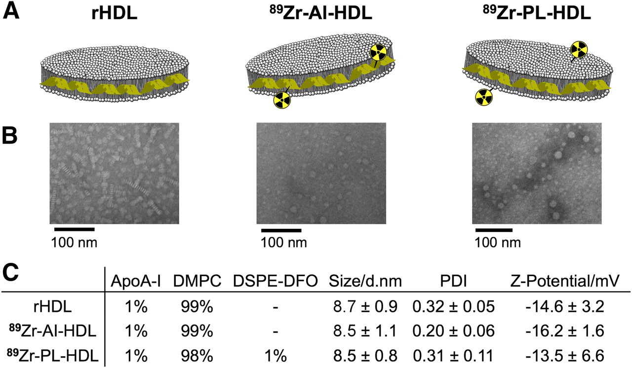
- FIGURE 1.
Structure and composition of rHDL and 89Zr-HDL nanotracers. (A) Schematic of rHDL (left), 89Zr-AI-HDL (middle), and 89Zr-PL-HDL (right). (B) Transmission electron microscopy images of rHDL (left), Zr-AI-HDL (middle), and Zr-PL-HDL (right). (C) Composition (in mol %), size, polydispersity index (PDI), and surface charge of rHDL, 89Zr-AI-HDL, and 89Zr-PL-HDL. Data are presented as mean ± SD (n ≥ 3).
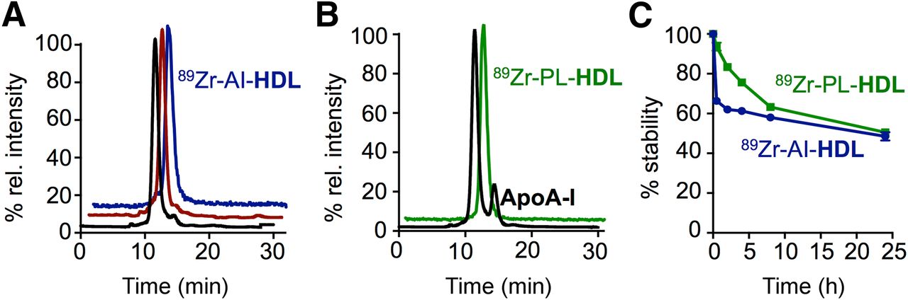
- FIGURE 2.
Radiosynthesis and in vitro stability of 89Zr-HDL nanotracers. Size-exclusion chromatograms showing coelution of plain rHDL (black trace), DFO-apoA-I@rHDL (red trace), and 89Zr-AI-HDL (blue, radioactive trace) (A) and coelution of 1% DSPE-DFO@rHDL (black trace) and 89Zr-PL-HDL (green, radioactive trace) (B). (C) In vitro serum stability of 89Zr-HDL nanotracers at 37°C.
- FIGURE 3.
Pharmacokinetics and biodistribution of 89Zr-HDL nanotracers. (A) Blood time–activity curve for 89Zr-AI-HDL and 89Zr-PL-HDL (n = 3). (B) Radioactivity distribution in selected tissues of 89Zr-AI-HDL (blue) and 89Zr-PL-HDL (green) in mice bearing orthotopic breast cancer tumors, expressed as %ID/g ± SD (n ≥ 3).
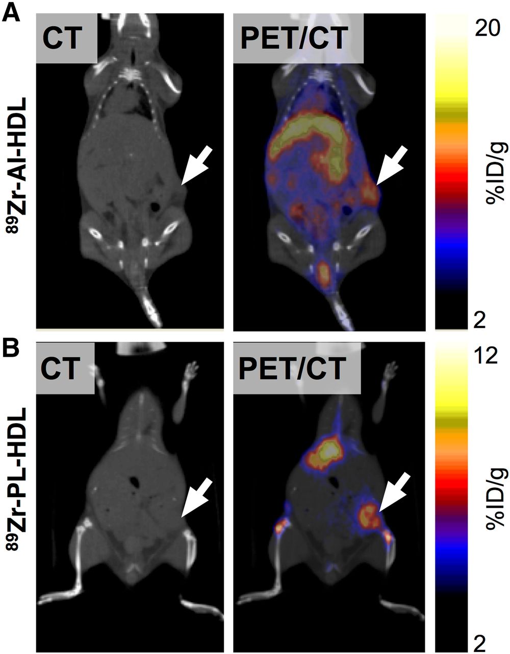
- FIGURE 4.
Accumulation of 89Zr-HDL nanotracers in tumor tissues can be visualized by in vivo PET imaging. CT (left) and PET/CT fusion (right) images of 89Zr-AI-HDL (A) and 89Zr-PL-HDL (B) obtained at 24 h after injection in mice bearing orthotopic 4T1 tumors (indicated by arrows).
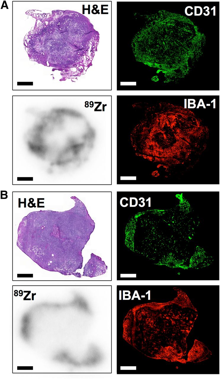
- FIGURE 5.
89Zr-HDL nanotracers accumulate in TAM-rich areas. Ex vivo histologic analysis of tumor sections at 24 h after administration of HDL nanotracers, showing hematoxylin and eosin (H&E) staining (top left), immunofluorescence for CD31 (top right) and IBA-1 (bottom right), and autoradiography (bottom left) for 89Zr-AI-HDL (A) and 89Zr-PL-HDL (B). Scale bar = 2 mm.
- FIGURE 6.
Both DiO@Zr-PL-HDL and DiO@Zr-AI-HDL preferentially target tumor-associated macrophages. 4T1 cell–induced orthotopic breast tumors were used to isolate single cells. (A) Representative DiO levels in 5 immune cells, namely TAMs, monocyte-derived cells (Mo-derived cells), monocytes, dendritic cells (DCs), and T cells. (B) Representative DiO levels in ECs and tumor cells (4T1). Cells from a phosphate-buffered saline–injected mouse served as controls (gray histograms to left). (C) Quantification of DiO levels presented as mean fluorescence intensity (MFI). Importantly, no statistical significance was found when comparing DiO levels of same cell type from 2 HDL formulations. Statistics were calculated with 2-tailed Student t test with unequal variance by comparing with TAM from same group. **P < 0.01. ***P < 0.001.


Additional Files
Supplemental Data
Files in this Data Supplement:


Jump to section
Related Articles
Cited By...
- 89Zr-Labeled High-Density Lipoprotein Nanoparticle PET Imaging Reveals Tumor Uptake in Patients with Esophageal Cancer
- A modular approach toward producing nanotherapeutics targeting the innate immune system
- An 89Zr-HDL PET Tracer Monitors Response to a CSF1R Inhibitor
- Imaging-assisted nanoimmunotherapy for atherosclerosis in multiple species
- An Overview of PET Radiochemistry, Part 2: Radiometals
- Magnetic Resonance Imaging of Tumor-Associated Macrophages: Clinical Translation
- Combined PET/DCE-MRI in a Rabbit Model of Atherosclerosis: Integrated Quantification of Plaque Inflammation, Permeability, and Burden During Treatment With a Leukotriene A4 Hydrolase Inhibitor
- In Vivo Imaging of Pro- and Antitumoral Cellular Components of the Tumor Microenvironment
- Immune cell screening of a nanoparticle library improves atherosclerosis therapy
- In Vivo PET Imaging of HDL in Multiple Atherosclerosis Models