


Article Figures & Data
Figures
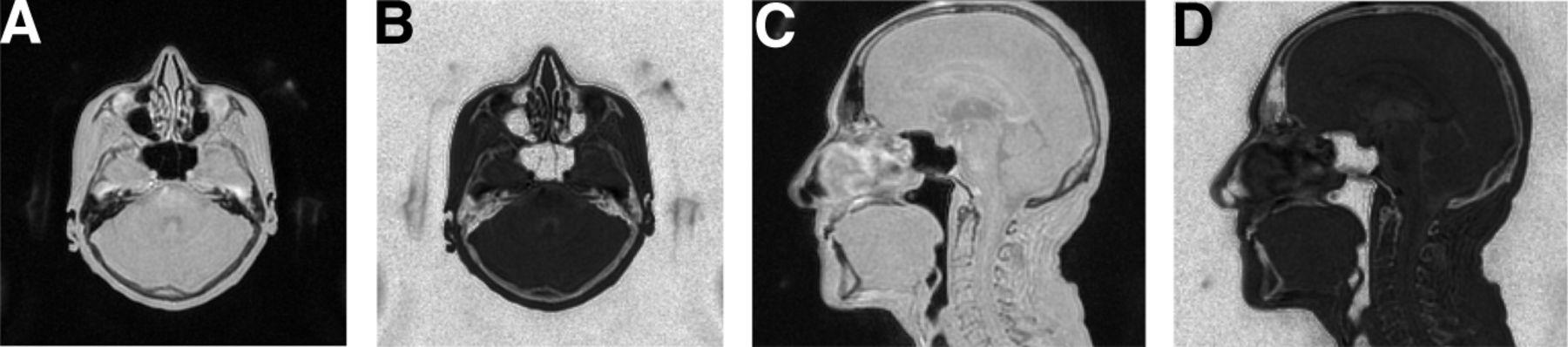
- FIGURE 1.
Axial (A and B) and sagittal views (C and D) of reconstructed ZTE dataset. Logarithmic intensity rescaling (B and D) was applied to enhance bone tissue and facilitate its separation from background and internal air cavities.
- FIGURE 2.
(A) Typical intensity histogram of ZTE dataset after logarithmic rescaling. Gaussian fitting result for soft-tissue and background peaks is indicated by dashed lines. Expected intensity range of bone tissue used in segmentation is indicated by dotted lines. (B) Axial view of corresponding ZTE volume. (C) Soft tissue (blue) and bone mask (green) obtained with segmentation.
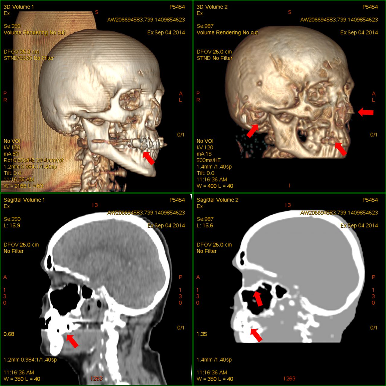
- FIGURE 3.
Volume rendering (top) and sagittal views (bottom) of CT dataset (left) and corresponding segmented ZTE dataset (right). Dental artifacts on both images, minor misclassification of cartilage and auditory canal air, and oversegmentation around sinusal cavities exist.
- FIGURE 4.
Jaccard distance between CT and ZTE bone masks, as function of CT threshold value. Each line in plot corresponds to 1 of the patients.
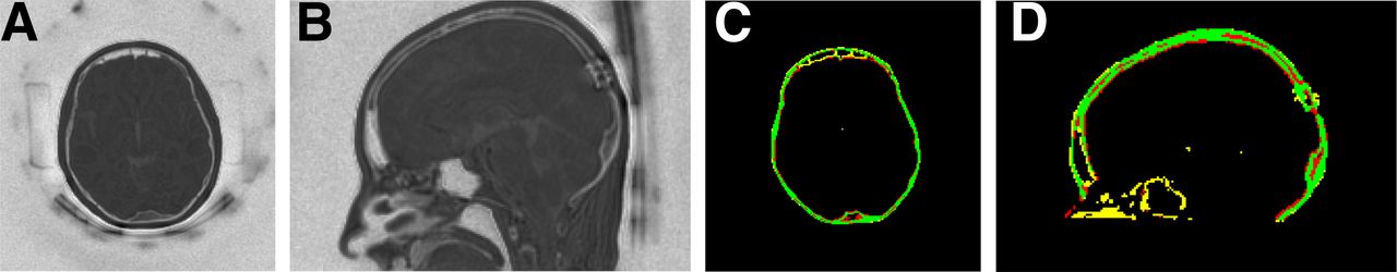
- FIGURE 5.
Logarithmic rescaling of ZTE dataset (A and B) and corresponding map of agreement between ZTE and CT bone masks (C and D). Green pixels indicate true-positive bone identification, red pixels indicate false-negative (missed bone), and yellow pixels indicate false-positive. Residual misregistration can be perceived in, for example, occipital region. Notice surgical bone alteration.


Tables
Distance Patient Whole Cranium 1 59% 46% 2 50% 37% 3 53% 38% 4 51% 40% 5 46% 33% 6 38% 32% 7 57% 36% 8 54% 39% 9 63% 40% 10 58% 40% 11 46% 41% 12 53% 43% 13 50% 38% 14 52% 49% 15 46% 40% Mean 52% 39% Maximum 63% 49% Minimum 38% 32% SD 6% 4% Calvaria Cerebrospinal fluid space Patient Frontal Parietal Temporal Occipital Inner ear Mastoid Orbit Third ventricle Fourth ventricle Choroid plexus of lateral ventricle (calcification) Pineal gland (calcification) Artificial alteration 1 1 0 1 1 2 1 1 1 1 1 0 NA 2 1 1 1 0 2 1 0 0 0 1 0 1 3 0 1 1 1 2 1 1 2 0 1 1 1 4 0 1 1 1 1 1 0 2 0 1 0 1 5 1 1 0 1 1 1 2 2 0 1 0 0 6 1 1 1 1 1 1 1 1 0 0 0 2 7 2 0 1 1 2 1 1 2 1 0 0 0 8 2 1 1 1 2 1 0 0 0 0 1 1 9 1 1 1 1 2 1 1 0 1 0 0 NA 10 1 1 1 1 2 1 1 2 0 1 0 NA 11 1 2 2 2 2 1 1 0 0 1 0 2 12 1 1 1 1 1 1 0 0 0 0 0 NA 13 1 0 1 1 2 1 0 2 0 1 0 1 14 2 2 2 1 2 1 1 2 0 1 0 2 15 1 0 1 1 2 1 0 2 2 1 0 1 Mean 1.1 0.9 1.1 1.0 1.7 1.0 0.7 1.2 0.3 0.7 0.1 1.1 SD 0.6 0.6 0.5 0.4 0.5 0.0 0.6 0.9 0.6 0.5 0.4 0.7 1 = minor issue; 0 = no issue; 2 = major issue; NA = not applicable.
Supplemental Data
Files in this Data Supplement:


Jump to section
Related Articles
Cited By...
- Enhancing the Diagnostic Accuracy of Amyloid PET: The Impact of MR-Guided PET Reconstruction
- Measuring 3D Cochlear Duct Length on MRI: Is It Accurate and Reliable?
- Zero TE MRI for Craniofacial Bone Imaging
- Clinical Feasibility of Zero TE Skull MRI in Patients with Head Trauma in Comparison with CT: A Single-Center Study
- The Effect of Susceptibility Artifacts Related to Metallic Implants on Adjacent-Lesion Assessment in Simultaneous TOF PET/MR
- Clinical Evaluation of Zero-Echo-Time Attenuation Correction for Brain 18F-FDG PET/MRI: Comparison with Atlas Attenuation Correction
- MRI-Based Attenuation Correction for PET/MRI Using Multiphase Level-Set Method
- Clinical Assessment of Emission- and Segmentation-Based MR-Guided Attenuation Correction in Whole-Body Time-of-Flight PET/MR Imaging