
Main menu

  • Home
  • Content
    • Current
    • Ahead of print
    • Past Issues
    • JNM Supplement
    • SNMMI Annual Meeting Abstracts
    • Continuing Education
    • JNM Podcasts
  • Subscriptions
    • Subscribers
    • Institutional and Non-member
    • Rates
    • Journal Claims
    • Corporate & Special Sales
  • Authors
    • Submit to JNM
    • Information for Authors
    • Assignment of Copyright
    • AQARA requirements
  • Info
    • Reviewers
    • Permissions
    • Advertisers
  • About
    • About Us
    • Editorial Board
    • Contact Information
  • More
    • Alerts
    • Feedback
    • Help
    • SNMMI Journals
  • SNMMI
    • JNM
    • JNMT
    • SNMMI Journals
    • SNMMI

User menu

  • Subscribe
  • My alerts
  • Log in
  • My Cart

Search

  • Advanced search
Journal of Nuclear Medicine
  • SNMMI
    • JNM
    • JNMT
    • SNMMI Journals
    • SNMMI
  • Subscribe
  • My alerts
  • Log in
  • My Cart
Journal of Nuclear Medicine

Advanced Search

  • Home
  • Content
    • Current
    • Ahead of print
    • Past Issues
    • JNM Supplement
    • SNMMI Annual Meeting Abstracts
    • Continuing Education
    • JNM Podcasts
  • Subscriptions
    • Subscribers
    • Institutional and Non-member
    • Rates
    • Journal Claims
    • Corporate & Special Sales
  • Authors
    • Submit to JNM
    • Information for Authors
    • Assignment of Copyright
    • AQARA requirements
  • Info
    • Reviewers
    • Permissions
    • Advertisers
  • About
    • About Us
    • Editorial Board
    • Contact Information
  • More
    • Alerts
    • Feedback
    • Help
    • SNMMI Journals
  • View or Listen to JNM Podcast
  • Visit JNM on Facebook
  • Join JNM on LinkedIn
  • Follow JNM on Twitter
  • Subscribe to our RSS feeds
Research ArticleBasic Science Investigations

α-Imaging Confirmed Efficient Targeting of CD45-Positive Cells After 211At-Radioimmunotherapy for Hematopoietic Cell Transplantation

Sofia H.L. Frost, Brian W. Miller, Tom A. Bäck, Erlinda B. Santos, Donald K. Hamlin, Sue E. Knoblaugh, Shani L. Frayo, Aimee L. Kenoyer, Rainer Storb, Oliver W. Press, D. Scott Wilbur, John M. Pagel and Brenda M. Sandmaier
Journal of Nuclear Medicine November 2015, 56 (11) 1766-1773; DOI: https://doi.org/10.2967/jnumed.115.162388
Sofia H.L. Frost
1Clinical Research Division, Fred Hutchinson Cancer Research Center, Seattle, Washington
  • Find this author on Google Scholar
  • Find this author on PubMed
  • Search for this author on this site
Brian W. Miller
2Pacific Northwest National Laboratory, Richland, Washington
3College of Optical Sciences, University of Arizona, Tucson, Arizona
  • Find this author on Google Scholar
  • Find this author on PubMed
  • Search for this author on this site
Tom A. Bäck
4Sahlgrenska Academy, University of Gothenburg, Gothenburg, Sweden
  • Find this author on Google Scholar
  • Find this author on PubMed
  • Search for this author on this site
Erlinda B. Santos
1Clinical Research Division, Fred Hutchinson Cancer Research Center, Seattle, Washington
  • Find this author on Google Scholar
  • Find this author on PubMed
  • Search for this author on this site
Donald K. Hamlin
5Department of Radiation Oncology, University of Washington, Seattle, Washington
  • Find this author on Google Scholar
  • Find this author on PubMed
  • Search for this author on this site
Sue E. Knoblaugh
6Comparative Medicine Shared Resource, Fred Hutchinson Cancer Research Center, Seattle, Washington; and
  • Find this author on Google Scholar
  • Find this author on PubMed
  • Search for this author on this site
Shani L. Frayo
1Clinical Research Division, Fred Hutchinson Cancer Research Center, Seattle, Washington
  • Find this author on Google Scholar
  • Find this author on PubMed
  • Search for this author on this site
Aimee L. Kenoyer
1Clinical Research Division, Fred Hutchinson Cancer Research Center, Seattle, Washington
  • Find this author on Google Scholar
  • Find this author on PubMed
  • Search for this author on this site
Rainer Storb
1Clinical Research Division, Fred Hutchinson Cancer Research Center, Seattle, Washington
7Department of Medicine, University of Washington, Seattle, Washington
  • Find this author on Google Scholar
  • Find this author on PubMed
  • Search for this author on this site
Oliver W. Press
1Clinical Research Division, Fred Hutchinson Cancer Research Center, Seattle, Washington
7Department of Medicine, University of Washington, Seattle, Washington
  • Find this author on Google Scholar
  • Find this author on PubMed
  • Search for this author on this site
D. Scott Wilbur
5Department of Radiation Oncology, University of Washington, Seattle, Washington
  • Find this author on Google Scholar
  • Find this author on PubMed
  • Search for this author on this site
John M. Pagel
1Clinical Research Division, Fred Hutchinson Cancer Research Center, Seattle, Washington
7Department of Medicine, University of Washington, Seattle, Washington
  • Find this author on Google Scholar
  • Find this author on PubMed
  • Search for this author on this site
Brenda M. Sandmaier
1Clinical Research Division, Fred Hutchinson Cancer Research Center, Seattle, Washington
7Department of Medicine, University of Washington, Seattle, Washington
  • Find this author on Google Scholar
  • Find this author on PubMed
  • Search for this author on this site
  • Article
  • Figures & Data
  • Supplemental
  • Info & Metrics
  • PDF
Loading

Article Figures & Data

Figures

  • Tables
  • Additional Files
  • FIGURE 1.
    • Download figure
    • Open in new tab
    • Download powerpoint
    FIGURE 1.

    Biodistribution and clearance of 211At-anti-CD45 mAb. (A) Radioactive content expressed as %IA/g ± SD (n = 3) in selected tissues from 2 dogs euthanized 19–22 h after injection. (B) Retention of 211At in blood for all treated dogs, determined by radioactivity measurements of repeated blood samples (%IA/g ± SD, 0.75 mg/kg, n = 5; 1.00 mg/kg, n = 3). p.i. = after injection.

  • FIGURE 2.
    • Download figure
    • Open in new tab
    • Download powerpoint
    FIGURE 2.

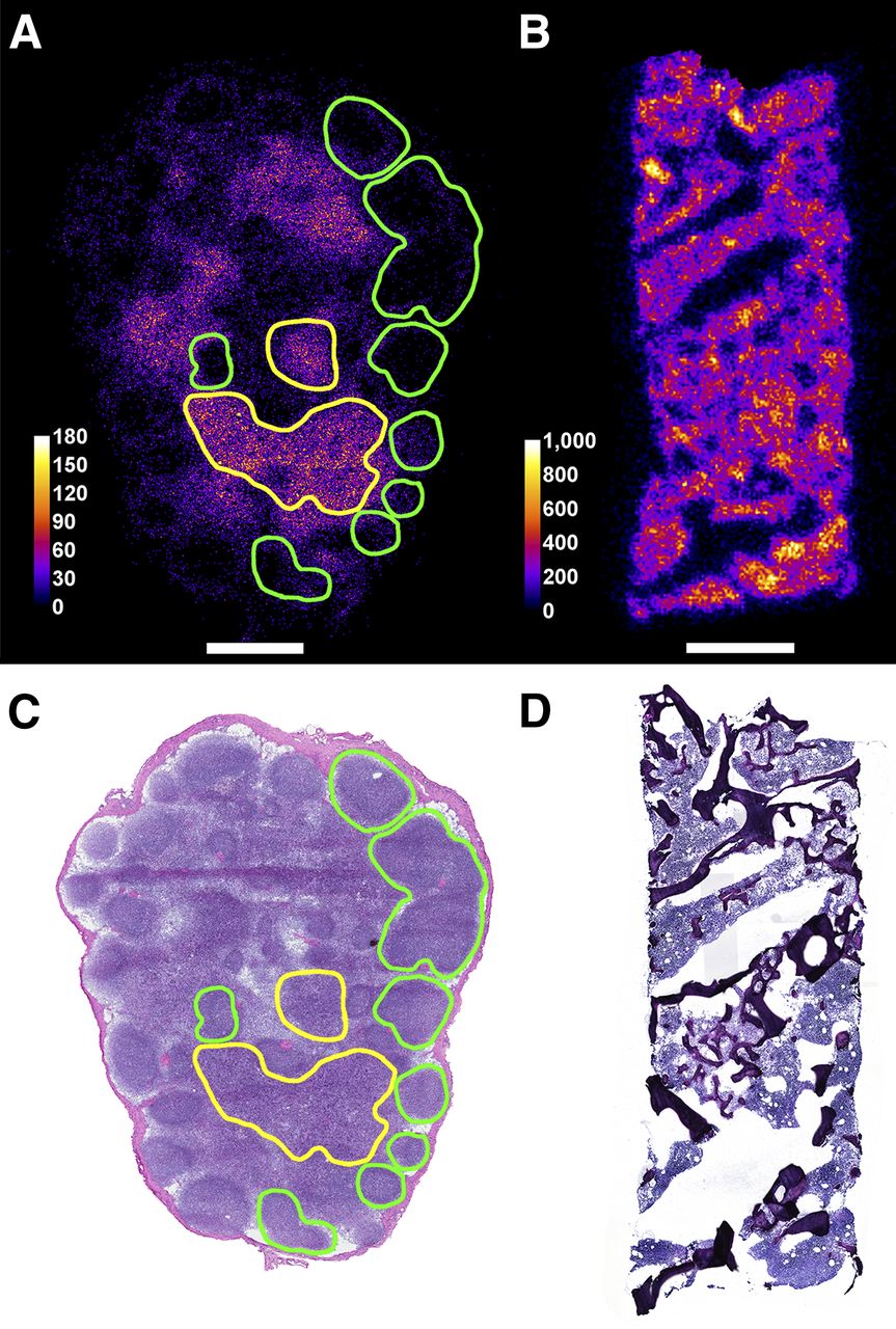
    Distribution of 211At in cryosectioned tissue samples imaged using iQID camera (A and B), and corresponding H&E-stained sections (C and D). A and C show lymph node sections from dog H638, biopsied 19 h after injection; B and D show sections of bone marrow core from dog H632, biopsied 2 h after injection. Green and yellow lines indicate lymph node follicles and paracortex (including visibly apoptotic areas), respectively, identified in H&E section. Overlaid with the α-image, outlined regions coincide well with low- and high-activity areas. Horizontal scale bars indicate 1 mm; color bars show 211At activity in microbecquerels.

  • FIGURE 3.
    • Download figure
    • Open in new tab
    • Download powerpoint
    FIGURE 3.

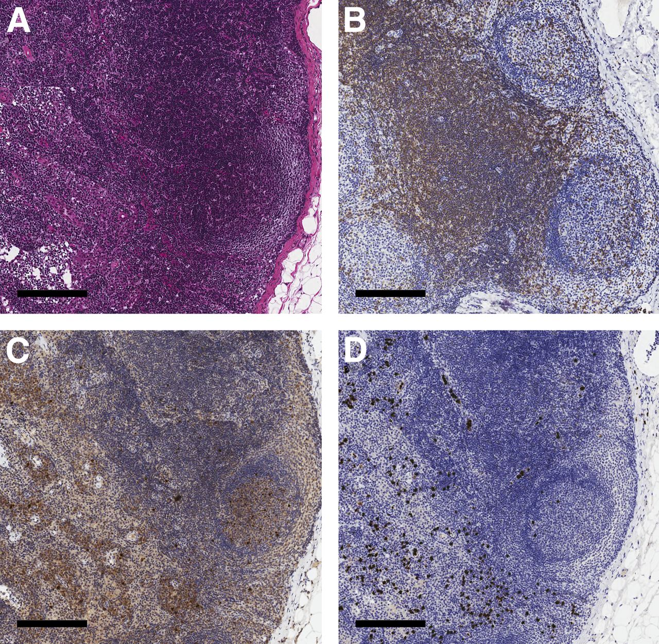
    Immunohistochemical staining of a formalin-fixed, paraffin-embedded lymph node from dog H629, biopsied 2 h after 211At-B10-CA12.10C12 injection. (A) H&E-stained section, including representative areas of cortex, paracortex, and medulla. B–D show staining for T cells (CD3), apoptosis (cleaved caspase-3), and macrophages (MAC 387), respectively, in corresponding areas of serially cut sections from the same lymph node. Scale bar represents 200 μm.

  • FIGURE 4.
    • Download figure
    • Open in new tab
    • Download powerpoint
    FIGURE 4.

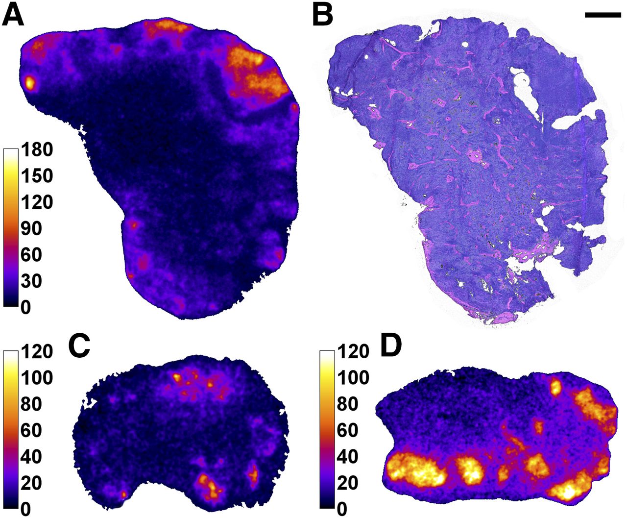
    Dose rate images derived by 3-dimensional dosimetry using α-camera imaging. (A) Dose rate distribution at biopsy for dog H638 4 h after injection. (B) Corresponding H&E-stained section. (C and D) Dose rate distribution for dog H689 2 and 19 h after injection, respectively. Color bars express dose rate in mGy/h; scale bar (top right) indicates 1 mm.

Tables

  • Figures
  • Additional Files
    • View popup
    TABLE 1

    Dogs Treated with 211At-Anti-CD45 Radioimmunotherapy

    Dog no.Age (mo)Weight (kg)mAb dose* (mg/kg)211At activity (MBq/kg)Specific 211At activity (MBq/mg mAb)Autologous transplantSample time (h after injection)Transfusions (d after HCT)Survival (wk)
    H573127.70.7514.619.5No22——
    H58599.40.7527.636.7No19——
    H5432012.80.7511.515.3Yes197, 11, 1948†
    H68978.70.7513.918.5Yes2, 19732†
    H5222211.80.7513.918.6Yes2846†
    H62989.51.0014.614.6Yes2, 196, 8, 1220‡
    H6381012.31.0018.418.4Yes4, 196, 10, 14, 16, 19, 2232†
    H632108.41.0018.718.7Yes2, 197, 932†
    • ↵* Including 0.05 mg/kg of unlabeled mAb.

    • ↵† End of study.

    • ↵‡ Severe ascites.

    • View popup
    TABLE 2

    Mean Fluorescence Intensity after Staining of CD45+ Cells (Gated on Lymphocytes)

    Bone marrow coreLymph node
    FMFCD45FFMFCD45F
    Dog no.2–4 h19–22 h2–4 h19–22 h2–4 h19–22 h2–4 h19–22 h
    H573*—7,245—1,292—1,288—7,500
    H585*—1,009—213—402—7,027
    H543*—17,931—669—2,358—5,257
    H689*28,17210,0825531,8412,1781,01112,1336,020
    H522*10,432—82—505—6,935—
    H629†17,9561,000109657582,7746,0416,025
    H638†37,17632,8312205481,7801,05715,11313,754
    H632†24,29838,8861555641,5174,47814,11217,067
    • ↵* mAb dose, 0.75 mg/kg.

    • ↵† mAb dose, 1.00 mg/kg.

    • View popup
    TABLE 3

    Percentage of Stained CD45+ Cells (Gated on Lymphocytes)

    Bone marrow coreLymph node
    FMFCD45FFMFCD45F
    Dog no.2–4 h19–22 h2–4 h19–22 h2–4 h19–22 h2–4 h19–22 h
    H573*—44.7—51.0—9.9—97.0
    H585*—13.7—6.1—2.7—98.2
    H543*—94.2—15.2—17.2—82.4
    H689*95.881.711.787.88.010.093.796.8
    H522*91.4—0.4—4.2—97.0—
    H629†95.65.20.10.85.424.494.188.3
    H638†97.292.80.85.36.86.896.897.2
    H632†78.993.60.57.56.235.897.997.9
    • ↵* mAb dose, 0.75 mg/kg.

    • ↵† mAb dose, 1.00 mg/kg.

    • View popup
    TABLE 4

    Mean Absorbed Dose to Blood After Injection of 211At-Anti-CD45 mAb

    Dog no.211At activity (MBq/kg)Injected 211At activity (A) (MBq)Absorbed dose to blood (D) (Gy)D/A (Gy/MBq)
    H573*14.61123.30.030
    H585*27.62595.70.022
    H543*11.51472.20.015
    H689*13.91201.60.013
    H522*13.91652.50.015
    H629†14.61392.70.020
    H638†18.42273.00.013
    H632†18.71573.40.022
    • ↵* mAb dose, 0.75 mg/kg.

    • ↵† mAb dose, 1.00 mg/kg.

Additional Files

  • Figures
  • Tables
  • Supplemental Data

    Files in this Data Supplement:

    • Supplemental Data
PreviousNext
Back to top

In this issue

Journal of Nuclear Medicine: 56 (11)
Journal of Nuclear Medicine
Vol. 56, Issue 11
November 1, 2015
  • Table of Contents
  • Table of Contents (PDF)
  • About the Cover
  • Index by author
Print
Download PDF
Article Alerts
Sign In to Email Alerts with your Email Address
Email Article

Thank you for your interest in spreading the word on Journal of Nuclear Medicine.

NOTE: We only request your email address so that the person you are recommending the page to knows that you wanted them to see it, and that it is not junk mail. We do not capture any email address.

Enter multiple addresses on separate lines or separate them with commas.
α-Imaging Confirmed Efficient Targeting of CD45-Positive Cells After 211At-Radioimmunotherapy for Hematopoietic Cell Transplantation
(Your Name) has sent you a message from Journal of Nuclear Medicine
(Your Name) thought you would like to see the Journal of Nuclear Medicine web site.
Citation Tools
α-Imaging Confirmed Efficient Targeting of CD45-Positive Cells After 211At-Radioimmunotherapy for Hematopoietic Cell Transplantation
Sofia H.L. Frost, Brian W. Miller, Tom A. Bäck, Erlinda B. Santos, Donald K. Hamlin, Sue E. Knoblaugh, Shani L. Frayo, Aimee L. Kenoyer, Rainer Storb, Oliver W. Press, D. Scott Wilbur, John M. Pagel, Brenda M. Sandmaier
Journal of Nuclear Medicine Nov 2015, 56 (11) 1766-1773; DOI: 10.2967/jnumed.115.162388

Citation Manager Formats

  • BibTeX
  • Bookends
  • EasyBib
  • EndNote (tagged)
  • EndNote 8 (xml)
  • Medlars
  • Mendeley
  • Papers
  • RefWorks Tagged
  • Ref Manager
  • RIS
  • Zotero
Share
α-Imaging Confirmed Efficient Targeting of CD45-Positive Cells After 211At-Radioimmunotherapy for Hematopoietic Cell Transplantation
Sofia H.L. Frost, Brian W. Miller, Tom A. Bäck, Erlinda B. Santos, Donald K. Hamlin, Sue E. Knoblaugh, Shani L. Frayo, Aimee L. Kenoyer, Rainer Storb, Oliver W. Press, D. Scott Wilbur, John M. Pagel, Brenda M. Sandmaier
Journal of Nuclear Medicine Nov 2015, 56 (11) 1766-1773; DOI: 10.2967/jnumed.115.162388
Twitter logo Facebook logo LinkedIn logo Mendeley logo
  • Tweet Widget
  • Facebook Like
  • Google Plus One
Bookmark this article

Jump to section

  • Article
    • Abstract
    • MATERIALS AND METHODS
    • RESULTS
    • DISCUSSION
    • CONCLUSION
    • DISCLOSURE
    • Acknowledgments
    • Footnotes
    • REFERENCES
  • Figures & Data
  • Supplemental
  • Info & Metrics
  • PDF

Related Articles

  • This Month in JNM
  • PubMed
  • Google Scholar

Cited By...

  • The bone marrow niche and hematopoietic system are distinctly remodeled by CD45-targeted astatine-211 radioimmunotherapy
  • 211At-Labeled Anti-CD45 Antibody as a Nonmyeloablative Conditioning for Canine DLA-Haploidentical Stem Cell Transplantation
  • Impact of Donor Type in Patients with AML Given Allogeneic Hematopoietic Cell Transplantation After Low-Dose TBI-Based Regimen
  • Cure of Human Ovarian Carcinoma Solid Xenografts by Fractionated {alpha}-Radioimmunotherapy with 211At-MX35-F(ab')2: Influence of Absorbed Tumor Dose and Effect on Long-Term Survival
  • Google Scholar

More in this TOC Section

  • Tumor-Specific Binding of Radiolabeled PEGylated GIRLRG Peptide: A Novel Agent for Targeting Cancers
  • PET/MRI of Hypoxic Atherosclerosis Using 64Cu-ATSM in a Rabbit Model
  • Tumor Uptake of Anti-CD20 Fabs Depends on Tumor Perfusion
Show more Basic Science Investigations

Similar Articles

Keywords

  • radioimmunotherapy
  • 211At
  • Alpha imaging
  • CD45
  • lymphoma
SNMMI

© 2025 SNMMI

Powered by HighWire