
Main menu

  • Home
  • Content
    • Current
    • Ahead of print
    • Past Issues
    • JNM Supplement
    • SNMMI Annual Meeting Abstracts
    • Continuing Education
    • JNM Podcasts
  • Subscriptions
    • Subscribers
    • Institutional and Non-member
    • Rates
    • Journal Claims
    • Corporate & Special Sales
  • Authors
    • Submit to JNM
    • Information for Authors
    • Assignment of Copyright
    • AQARA requirements
  • Info
    • Reviewers
    • Permissions
    • Advertisers
  • About
    • About Us
    • Editorial Board
    • Contact Information
  • More
    • Alerts
    • Feedback
    • Help
    • SNMMI Journals
  • SNMMI
    • JNM
    • JNMT
    • SNMMI Journals
    • SNMMI

User menu

  • Subscribe
  • My alerts
  • Log in
  • My Cart

Search

  • Advanced search
Journal of Nuclear Medicine
  • SNMMI
    • JNM
    • JNMT
    • SNMMI Journals
    • SNMMI
  • Subscribe
  • My alerts
  • Log in
  • My Cart
Journal of Nuclear Medicine

Advanced Search

  • Home
  • Content
    • Current
    • Ahead of print
    • Past Issues
    • JNM Supplement
    • SNMMI Annual Meeting Abstracts
    • Continuing Education
    • JNM Podcasts
  • Subscriptions
    • Subscribers
    • Institutional and Non-member
    • Rates
    • Journal Claims
    • Corporate & Special Sales
  • Authors
    • Submit to JNM
    • Information for Authors
    • Assignment of Copyright
    • AQARA requirements
  • Info
    • Reviewers
    • Permissions
    • Advertisers
  • About
    • About Us
    • Editorial Board
    • Contact Information
  • More
    • Alerts
    • Feedback
    • Help
    • SNMMI Journals
  • View or Listen to JNM Podcast
  • Visit JNM on Facebook
  • Join JNM on LinkedIn
  • Follow JNM on Twitter
  • Subscribe to our RSS feeds
Research ArticleBasic Science Investigations

Variability of Proliferation and Diffusion in Different Lung Cancer Models as Measured by 3′-Deoxy-3′-18F-Fluorothymidine PET and Diffusion-Weighted MR Imaging

Sonja Schelhaas, Lydia Wachsmuth, Thomas Viel, Davina J. Honess, Kathrin Heinzmann, Donna-Michelle Smith, Sven Hermann, Stefan Wagner, Michael T. Kuhlmann, Carsten Müller-Tidow, Klaus Kopka, Otmar Schober, Michael Schäfers, Richard Schneider, Eric O. Aboagye, John Griffiths, Cornelius Faber and Andreas H. Jacobs
Journal of Nuclear Medicine June 2014, 55 (6) 983-988; DOI: https://doi.org/10.2967/jnumed.113.133348
Sonja Schelhaas
1European Institute for Molecular Imaging (EIMI), Westfälische Wilhelms-Universität (WWU) Münster, Münster, Germany
  • Find this author on Google Scholar
  • Find this author on PubMed
  • Search for this author on this site
Lydia Wachsmuth
2Department of Clinical Radiology, University Hospital of Münster, Münster, Germany
  • Find this author on Google Scholar
  • Find this author on PubMed
  • Search for this author on this site
Thomas Viel
1European Institute for Molecular Imaging (EIMI), Westfälische Wilhelms-Universität (WWU) Münster, Münster, Germany
  • Find this author on Google Scholar
  • Find this author on PubMed
  • Search for this author on this site
Davina J. Honess
3Cancer Research United Kingdom Cambridge Institute, Cambridge, United Kingdom
  • Find this author on Google Scholar
  • Find this author on PubMed
  • Search for this author on this site
Kathrin Heinzmann
3Cancer Research United Kingdom Cambridge Institute, Cambridge, United Kingdom
  • Find this author on Google Scholar
  • Find this author on PubMed
  • Search for this author on this site
Donna-Michelle Smith
3Cancer Research United Kingdom Cambridge Institute, Cambridge, United Kingdom
  • Find this author on Google Scholar
  • Find this author on PubMed
  • Search for this author on this site
Sven Hermann
1European Institute for Molecular Imaging (EIMI), Westfälische Wilhelms-Universität (WWU) Münster, Münster, Germany
  • Find this author on Google Scholar
  • Find this author on PubMed
  • Search for this author on this site
Stefan Wagner
4Department of Nuclear Medicine, University Hospital of Münster, Münster, Germany
  • Find this author on Google Scholar
  • Find this author on PubMed
  • Search for this author on this site
Michael T. Kuhlmann
1European Institute for Molecular Imaging (EIMI), Westfälische Wilhelms-Universität (WWU) Münster, Münster, Germany
  • Find this author on Google Scholar
  • Find this author on PubMed
  • Search for this author on this site
Carsten Müller-Tidow
5Department of Hematology and Oncology, University Hospital of Münster, Münster, Germany
  • Find this author on Google Scholar
  • Find this author on PubMed
  • Search for this author on this site
Klaus Kopka
4Department of Nuclear Medicine, University Hospital of Münster, Münster, Germany
  • Find this author on Google Scholar
  • Find this author on PubMed
  • Search for this author on this site
Otmar Schober
1European Institute for Molecular Imaging (EIMI), Westfälische Wilhelms-Universität (WWU) Münster, Münster, Germany
4Department of Nuclear Medicine, University Hospital of Münster, Münster, Germany
  • Find this author on Google Scholar
  • Find this author on PubMed
  • Search for this author on this site
Michael Schäfers
1European Institute for Molecular Imaging (EIMI), Westfälische Wilhelms-Universität (WWU) Münster, Münster, Germany
4Department of Nuclear Medicine, University Hospital of Münster, Münster, Germany
  • Find this author on Google Scholar
  • Find this author on PubMed
  • Search for this author on this site
Richard Schneider
6Merck Serono, Darmstadt, Germany
  • Find this author on Google Scholar
  • Find this author on PubMed
  • Search for this author on this site
Eric O. Aboagye
7Comprehensive Cancer Imaging Centre, Imperial College London, London, United Kingdom; and
  • Find this author on Google Scholar
  • Find this author on PubMed
  • Search for this author on this site
John Griffiths
3Cancer Research United Kingdom Cambridge Institute, Cambridge, United Kingdom
  • Find this author on Google Scholar
  • Find this author on PubMed
  • Search for this author on this site
Cornelius Faber
2Department of Clinical Radiology, University Hospital of Münster, Münster, Germany
  • Find this author on Google Scholar
  • Find this author on PubMed
  • Search for this author on this site
Andreas H. Jacobs
1European Institute for Molecular Imaging (EIMI), Westfälische Wilhelms-Universität (WWU) Münster, Münster, Germany
8Department of Geriatric Medicine, Johanniter Hospital, Bonn, Germany
  • Find this author on Google Scholar
  • Find this author on PubMed
  • Search for this author on this site
  • Article
  • Figures & Data
  • Supplemental
  • Info & Metrics
  • PDF
Loading

Article Figures & Data

Figures

  • Additional Files
  • FIGURE 1.
    • Download figure
    • Open in new tab
    • Download powerpoint
    FIGURE 1.

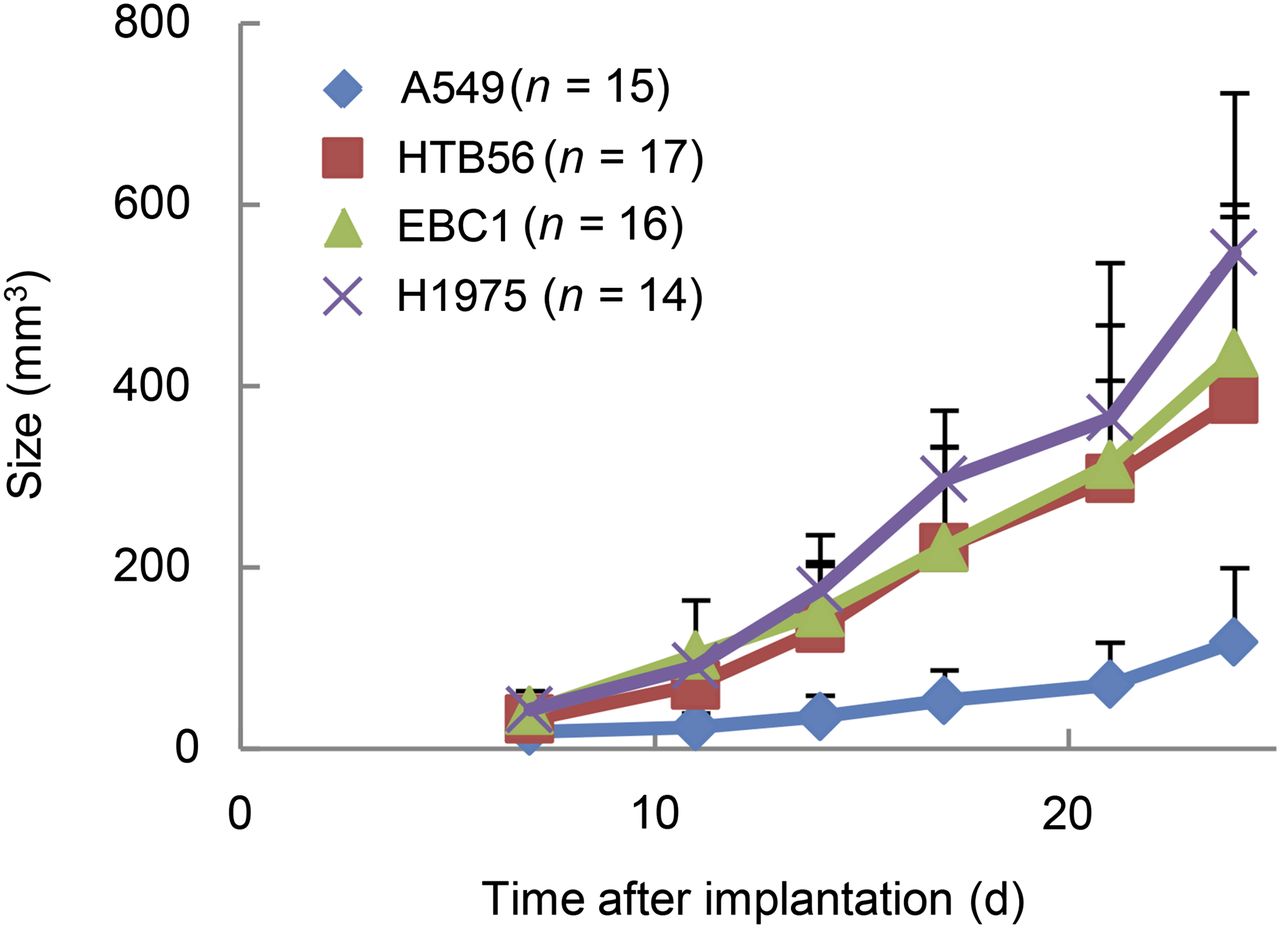
    NSCLC xenografts differ with respect to growth. Tumor size was determined by caliper measurements. n = number of tumors.

  • FIGURE 2.
    • Download figure
    • Open in new tab
    • Download powerpoint
    FIGURE 2.

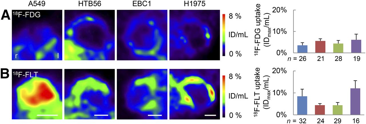
    PET imaging of lung cancer xenografts reveals pronounced 18F-FLT uptake in A549 and H1975 xenografts. Transverse slices of 18F-FDG (A) and 18F-FLT (B) PET images at biggest tumor diameter of representative tumors about 4 wk after implantation are shown. Maximum radiotracer uptake of whole tumors was determined. Some tumors were measured several times during their growth (∼2 and 4 wk after implantation). However, no influence of imaging time point was detected, and data were combined in analysis. Blue = A549, red = HTB56, green = EBC1, purple = H1975. n = number of analyses per cell line. Scale bars = 5 mm.

  • FIGURE 3.
    • Download figure
    • Open in new tab
    • Download powerpoint
    FIGURE 3.

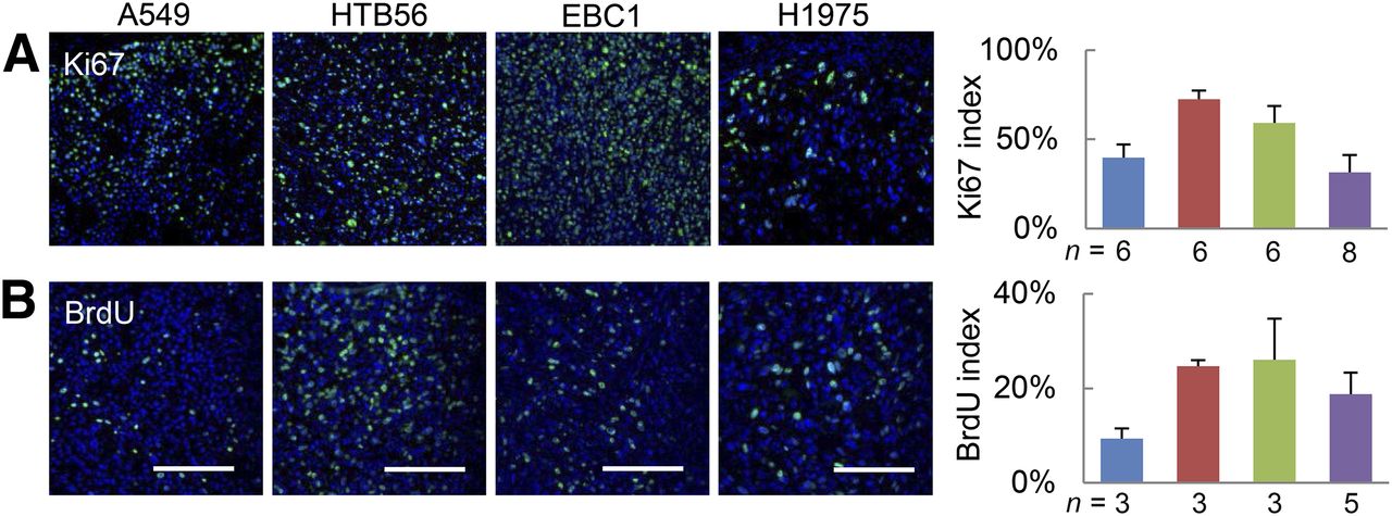
    Proliferation as determined by histologic markers does not positively correlate with 18F-FLT uptake. Histologic sections were probed for Ki67 (A) and BrdU (B). Percentage of specifically stained nuclei was quantified in viable tumor regions. Blue = A549, red = HTB56, green = EBC1, purple = H1975. n = number of tumors analyzed (1 section per tumor). Scale bars = 100 μm.

  • FIGURE 4.
    • Download figure
    • Open in new tab
    • Download powerpoint
    FIGURE 4.

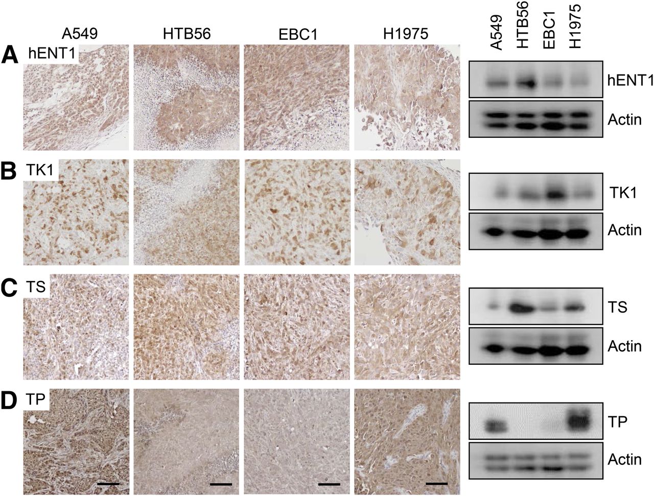
    Expression of thymidine metabolism proteins does not account for variations in 18F-FLT uptake, except for TP. Tumor homogenates were analyzed by Western blot for expression of hENT1 (A), TK1 (B), TS (C), or TP (D). Five different xenografts per cell line were examined by this method, and representative blot is shown. Same proteins were also detected by immunohistochemistry. Sections of 4 tumors per cell line were analyzed, and representative figures are depicted here. Scale bars = 100 μm.

  • FIGURE 5.
    • Download figure
    • Open in new tab
    • Download powerpoint
    FIGURE 5.

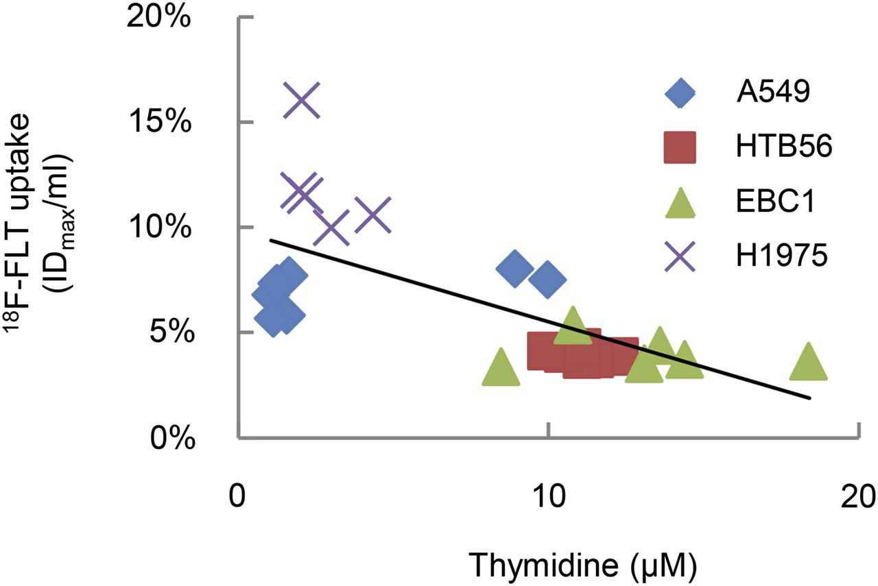
    Tumor thymidine negatively correlates with 18F-FLT uptake in lung cancer xenografts. Thymidine levels in tumor homogenates were determined by thymidine-specific liquid chromatography–mass spectrometry. Correlation coefficient = −0.682. P < 0.005.
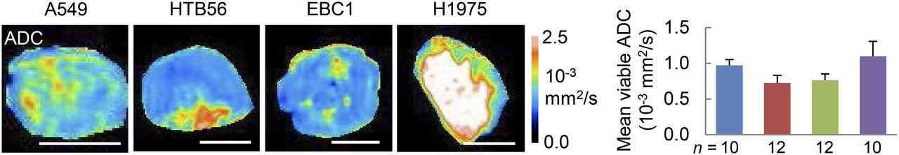
  • FIGURE 6.
    • Download figure
    • Open in new tab
    • Download powerpoint
    FIGURE 6.

    ADC differs between analyzed xenografts. Transverse slices of ADC images are depicted here, and respective T2w images can be found in Supplemental Figure 2B. Viable tumor regions were defined on these T2w images, and mean ADC was quantified within 1 representative ROI in this area. Blue = A549, red = HTB56, green = EBC1, purple = H1975. n = number of analyzed tumors per cell line. Scale bars = 5 mm.

  • FIGURE 7.
    • Download figure
    • Open in new tab
    • Download powerpoint
    FIGURE 7.

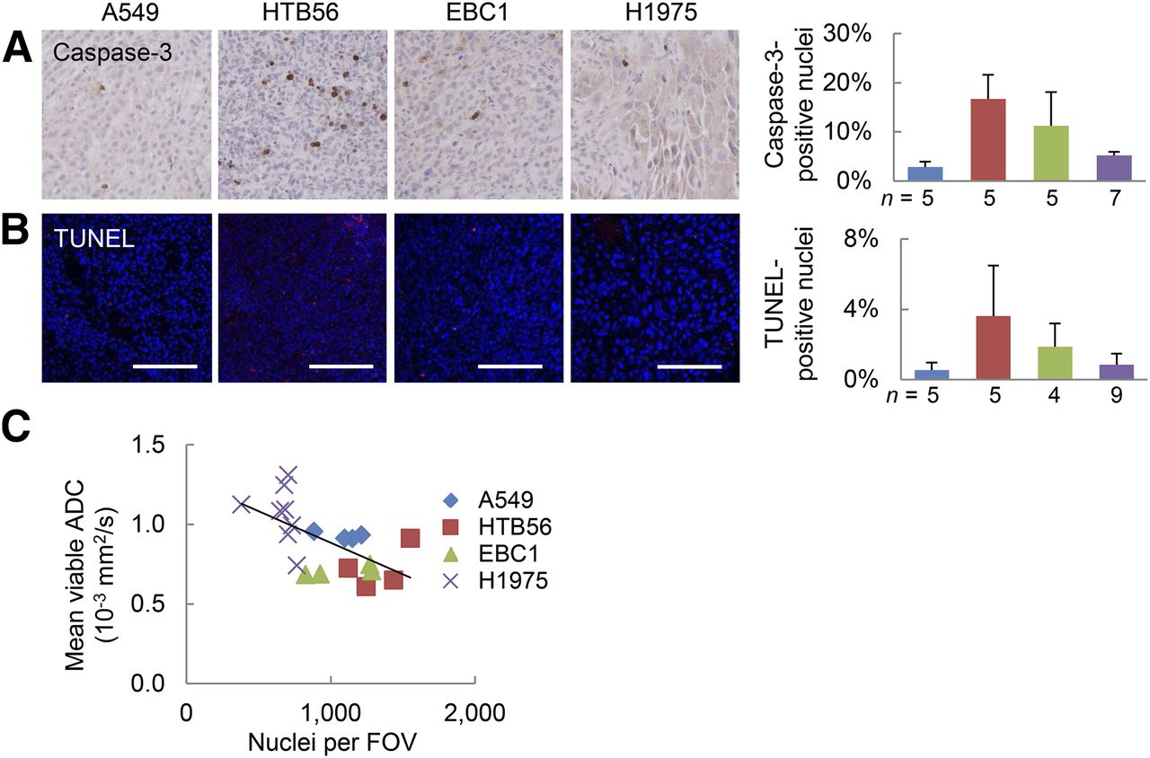
    ADC in various lung cancer xenografts is not related to cell death but negatively correlates with cell density. Histologic sections were analyzed for active caspase-3 (A) and TUNEL (B). Representative images of viable tumor regions and respective quantifications are shown here. Scale bars = 100 μm. (C) Number of 4′,6-diamidino-2-phenylindole (DAPI)–stained nuclei per field of view (field of view [FOV], 20× resolution; 580 × 460 μm) was determined as measure for cellular density. Transverse sections at biggest tumor diameter were investigated to directly relate findings to respective MR slices. Correlation coefficient = −0.61; P < 0.005; n = number of tumors analyzed. blue = A549, red = HTB56, green = EBC1, purple = H1975.

Additional Files

  • Figures
  • Supplemental Data

    Files in this Data Supplement:

    • Supplemental Data
PreviousNext
Back to top

In this issue

Journal of Nuclear Medicine: 55 (6)
Journal of Nuclear Medicine
Vol. 55, Issue 6
June 1, 2014
  • Table of Contents
  • Table of Contents (PDF)
  • About the Cover
  • Index by author
Print
Download PDF
Article Alerts
Sign In to Email Alerts with your Email Address
Email Article

Thank you for your interest in spreading the word on Journal of Nuclear Medicine.

NOTE: We only request your email address so that the person you are recommending the page to knows that you wanted them to see it, and that it is not junk mail. We do not capture any email address.

Enter multiple addresses on separate lines or separate them with commas.
Variability of Proliferation and Diffusion in Different Lung Cancer Models as Measured by 3′-Deoxy-3′-18F-Fluorothymidine PET and Diffusion-Weighted MR Imaging
(Your Name) has sent you a message from Journal of Nuclear Medicine
(Your Name) thought you would like to see the Journal of Nuclear Medicine web site.
Citation Tools
Variability of Proliferation and Diffusion in Different Lung Cancer Models as Measured by 3′-Deoxy-3′-18F-Fluorothymidine PET and Diffusion-Weighted MR Imaging
Sonja Schelhaas, Lydia Wachsmuth, Thomas Viel, Davina J. Honess, Kathrin Heinzmann, Donna-Michelle Smith, Sven Hermann, Stefan Wagner, Michael T. Kuhlmann, Carsten Müller-Tidow, Klaus Kopka, Otmar Schober, Michael Schäfers, Richard Schneider, Eric O. Aboagye, John Griffiths, Cornelius Faber, Andreas H. Jacobs
Journal of Nuclear Medicine Jun 2014, 55 (6) 983-988; DOI: 10.2967/jnumed.113.133348

Citation Manager Formats

  • BibTeX
  • Bookends
  • EasyBib
  • EndNote (tagged)
  • EndNote 8 (xml)
  • Medlars
  • Mendeley
  • Papers
  • RefWorks Tagged
  • Ref Manager
  • RIS
  • Zotero
Share
Variability of Proliferation and Diffusion in Different Lung Cancer Models as Measured by 3′-Deoxy-3′-18F-Fluorothymidine PET and Diffusion-Weighted MR Imaging
Sonja Schelhaas, Lydia Wachsmuth, Thomas Viel, Davina J. Honess, Kathrin Heinzmann, Donna-Michelle Smith, Sven Hermann, Stefan Wagner, Michael T. Kuhlmann, Carsten Müller-Tidow, Klaus Kopka, Otmar Schober, Michael Schäfers, Richard Schneider, Eric O. Aboagye, John Griffiths, Cornelius Faber, Andreas H. Jacobs
Journal of Nuclear Medicine Jun 2014, 55 (6) 983-988; DOI: 10.2967/jnumed.113.133348
Twitter logo Facebook logo LinkedIn logo Mendeley logo
  • Tweet Widget
  • Facebook Like
  • Google Plus One
Bookmark this article

Jump to section

  • Article
    • Abstract
    • MATERIALS AND METHODS
    • RESULTS
    • DISCUSSION
    • CONCLUSION
    • DISCLOSURE
    • Acknowledgments
    • Footnotes
    • REFERENCES
  • Figures & Data
  • Supplemental
  • Info & Metrics
  • PDF

Related Articles

  • This Month in JNM
  • PubMed
  • Google Scholar

Cited By...

  • Thymidine Metabolism as a Confounding Factor for 3'-Deoxy-3'-18F-Fluorothymidine Uptake After Therapy in a Colorectal Cancer Model
  • Preclinical Evidence That 3'-Deoxy-3'-[18F]Fluorothymidine PET Can Visualize Recovery of Hematopoiesis after Gemcitabine Chemotherapy
  • Gemcitabine Mechanism of Action Confounds Early Assessment of Treatment Response by 3'-Deoxy-3'-[18F]Fluorothymidine in Preclinical Models of Lung Cancer
  • Google Scholar

More in this TOC Section

  • 11C-Methionine PET of Myocardial Inflammation in a Rat Model of Experimental Autoimmune Myocarditis
  • Counting Rate Characteristics and Image Distortion in Preclinical PET Imaging During Radiopharmaceutical Therapy
  • Design and Fabrication of Kidney Phantoms for Internal Radiation Dosimetry Using 3D Printing Technology
Show more Basic Science Investigations

Similar Articles

Keywords

  • 18F-FLT PET
  • DW MRI
  • Molecular imaging
  • lung cancer
SNMMI

© 2025 SNMMI

Powered by HighWire