
Main menu

  • Home
  • Content
    • Current
    • Ahead of print
    • Past Issues
    • JNM Supplement
    • SNMMI Annual Meeting Abstracts
    • Continuing Education
    • JNM Podcasts
  • Subscriptions
    • Subscribers
    • Institutional and Non-member
    • Rates
    • Journal Claims
    • Corporate & Special Sales
  • Authors
    • Submit to JNM
    • Information for Authors
    • Assignment of Copyright
    • AQARA requirements
  • Info
    • Reviewers
    • Permissions
    • Advertisers
  • About
    • About Us
    • Editorial Board
    • Contact Information
  • More
    • Alerts
    • Feedback
    • Help
    • SNMMI Journals
  • SNMMI
    • JNM
    • JNMT
    • SNMMI Journals
    • SNMMI

User menu

  • Subscribe
  • My alerts
  • Log in
  • My Cart

Search

  • Advanced search
Journal of Nuclear Medicine
  • SNMMI
    • JNM
    • JNMT
    • SNMMI Journals
    • SNMMI
  • Subscribe
  • My alerts
  • Log in
  • My Cart
Journal of Nuclear Medicine

Advanced Search

  • Home
  • Content
    • Current
    • Ahead of print
    • Past Issues
    • JNM Supplement
    • SNMMI Annual Meeting Abstracts
    • Continuing Education
    • JNM Podcasts
  • Subscriptions
    • Subscribers
    • Institutional and Non-member
    • Rates
    • Journal Claims
    • Corporate & Special Sales
  • Authors
    • Submit to JNM
    • Information for Authors
    • Assignment of Copyright
    • AQARA requirements
  • Info
    • Reviewers
    • Permissions
    • Advertisers
  • About
    • About Us
    • Editorial Board
    • Contact Information
  • More
    • Alerts
    • Feedback
    • Help
    • SNMMI Journals
  • View or Listen to JNM Podcast
  • Visit JNM on Facebook
  • Join JNM on LinkedIn
  • Follow JNM on Twitter
  • Subscribe to our RSS feeds
Research ArticleBasic Science Investigations

Functional Imaging of Oxidative Stress with a Novel PET Imaging Agent, 18F-5-Fluoro-l-Aminosuberic Acid

Jack M. Webster, Christine A. Morton, Bruce F. Johnson, Hua Yang, Michael J. Rishel, Brian D. Lee, Qing Miao, Chittari Pabba, Donald T. Yapp and Paul Schaffer
Journal of Nuclear Medicine April 2014, 55 (4) 657-664; DOI: https://doi.org/10.2967/jnumed.113.126664
Jack M. Webster
1Diagnostics and Biomedical Technologies, GE Global Research, Niskayuna, New York
  • Find this author on Google Scholar
  • Find this author on PubMed
  • Search for this author on this site
Christine A. Morton
1Diagnostics and Biomedical Technologies, GE Global Research, Niskayuna, New York
  • Find this author on Google Scholar
  • Find this author on PubMed
  • Search for this author on this site
Bruce F. Johnson
1Diagnostics and Biomedical Technologies, GE Global Research, Niskayuna, New York
  • Find this author on Google Scholar
  • Find this author on PubMed
  • Search for this author on this site
Hua Yang
2Nuclear Medicine Division, TRIUMF, Vancouver, British Columbia, Canada
  • Find this author on Google Scholar
  • Find this author on PubMed
  • Search for this author on this site
Michael J. Rishel
1Diagnostics and Biomedical Technologies, GE Global Research, Niskayuna, New York
  • Find this author on Google Scholar
  • Find this author on PubMed
  • Search for this author on this site
Brian D. Lee
1Diagnostics and Biomedical Technologies, GE Global Research, Niskayuna, New York
  • Find this author on Google Scholar
  • Find this author on PubMed
  • Search for this author on this site
Qing Miao
2Nuclear Medicine Division, TRIUMF, Vancouver, British Columbia, Canada
  • Find this author on Google Scholar
  • Find this author on PubMed
  • Search for this author on this site
Chittari Pabba
1Diagnostics and Biomedical Technologies, GE Global Research, Niskayuna, New York
  • Find this author on Google Scholar
  • Find this author on PubMed
  • Search for this author on this site
Donald T. Yapp
3Division of Pharmaceutics and Biopharmaceutics, Faculty of Pharmaceutical Sciences, University of British Columbia, Vancouver, British Columbia, Canada; and
4Experimental Therapeutics, BC Cancer Agency, Vancouver, British Columbia, Canada
  • Find this author on Google Scholar
  • Find this author on PubMed
  • Search for this author on this site
Paul Schaffer
2Nuclear Medicine Division, TRIUMF, Vancouver, British Columbia, Canada
  • Find this author on Google Scholar
  • Find this author on PubMed
  • Search for this author on this site
  • Article
  • Figures & Data
  • Supplemental
  • Info & Metrics
  • PDF
Loading

Article Figures & Data

Figures

  • Tables
  • Additional Files
  • FIGURE 1.
    • Download figure
    • Open in new tab
    • Download powerpoint
    FIGURE 1.

    Cellular mechanism of xCT expression induced by oxidative stress and promoting increased glutathione synthesis.

  • FIGURE 2.
    • Download figure
    • Open in new tab
    • Download powerpoint
    FIGURE 2.

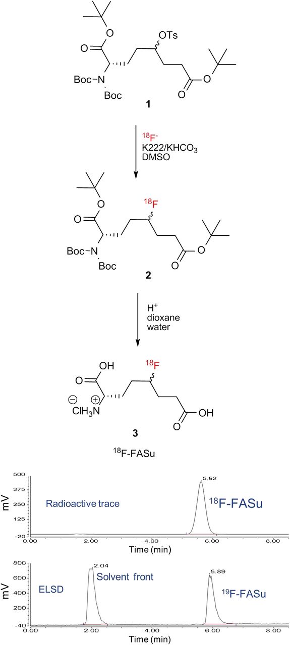
    2-step radiosynthesis and identification of 18F-FASu. HPLC of final product with γ detector demonstrates more than 98% radiochemical purity. HPLC of 19F-FASu standard with evaporative light scattering detector (ELSD) shows that differences in retention time of peaks matches 0.27-min (16-s) delay between the 2 detectors.

  • FIGURE 3.
    • Download figure
    • Open in new tab
    • Download powerpoint
    FIGURE 3.

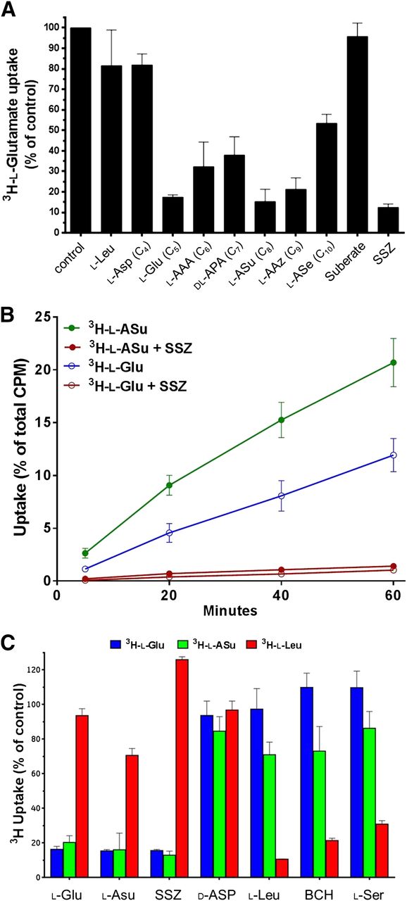
    In vitro system xc- inhibition, uptake, and specificity studies in diethylmaleate-treated SKOV-3 cells. (A) Competitive inhibition of test agents on 3H-l-Glu uptake during 30-min incubation. l-Asp, l-Glu, l-aminoadipic acid, dl-aminopimelic acid, l-ASu, l-AAz, and l-aminosebacic acid are all anionic amino acids from 4 to 10 carbons (C4–C10) in length, respectively. Values are normalized to uptake of 3H-l-Glu in phosphate-buffered saline alone (mean ± SD, n = 3). (B) Comparison of direct cell uptake of 3H-l-ASu and 3H-l-Glu. 3H-l-ASu and 3H-l-Glu were matched for specific activity and radiation dose per sample. Uptake values are expressed as percentage of total activity used per well (mean ± SD, n = 3). (C) Comparison of 3H-l-Glu, 3H-l-ASu, and 3H-l-Leu uptake in presence of selected amino acid transporter inhibitors for 30 min. 3H-l-Glu, 3H-l-ASu, and 3H-l-Leu were matched for specific activity and radiation dose per sample. Values are normalized to uptake of each agent in phosphate-buffered saline alone (mean ± SD, n = 3). SSZ = sulfasalazine.

  • FIGURE 4.
    • Download figure
    • Open in new tab
    • Download powerpoint
    FIGURE 4.

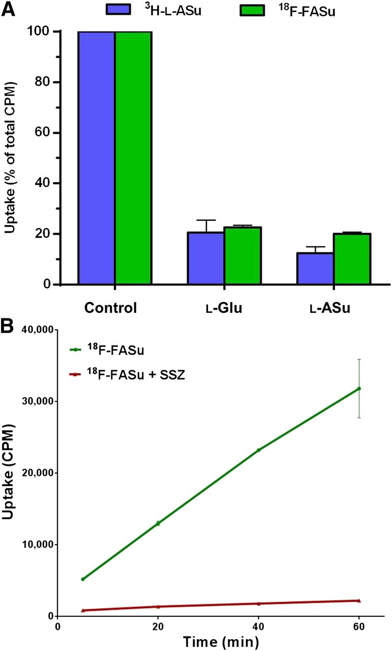
    Uptake of HPLC-purified 18F-FASu in EL4 and SKOV-3 cells in vitro. (A) EL4 cells were evaluated for uptake of 3H-l-ASu and 18F-FASu in absence or presence of 1 mM l-Glu or l-ASu for 30 min. Uptake and inhibition profiles were similar for both radiolabeled compounds. Values are normalized to uptake of each agent in phosphate-buffered-saline-alone control (mean ± SD, n = 4). (B) 18F-FASu uptake in SKOV-3 cells in absence and presence of 0.5 mM sulfasalazine was evaluated with 5-, 20-, 40-, and 60-min incubations. Uptake values are raw CPM values per sample (mean ± SD, n = 3). SSZ = sulfasalazine.

  • FIGURE 5.
    • Download figure
    • Open in new tab
    • Download powerpoint
    FIGURE 5.

    In vivo biodistribution in CD-1 nude mice bearing EL4 xenograft tumors with HPLC-purified 18F-FASu. (A) 18F-FASu retention in clearance organs at 5, 30, 60, 120, and 240 min after injection are shown in %ID/organ. (B) 18F-FASu retention in tumor, blood, muscle, bone, brain, and liver. Data are %ID/g (mean ± SD, n ≥ 4).
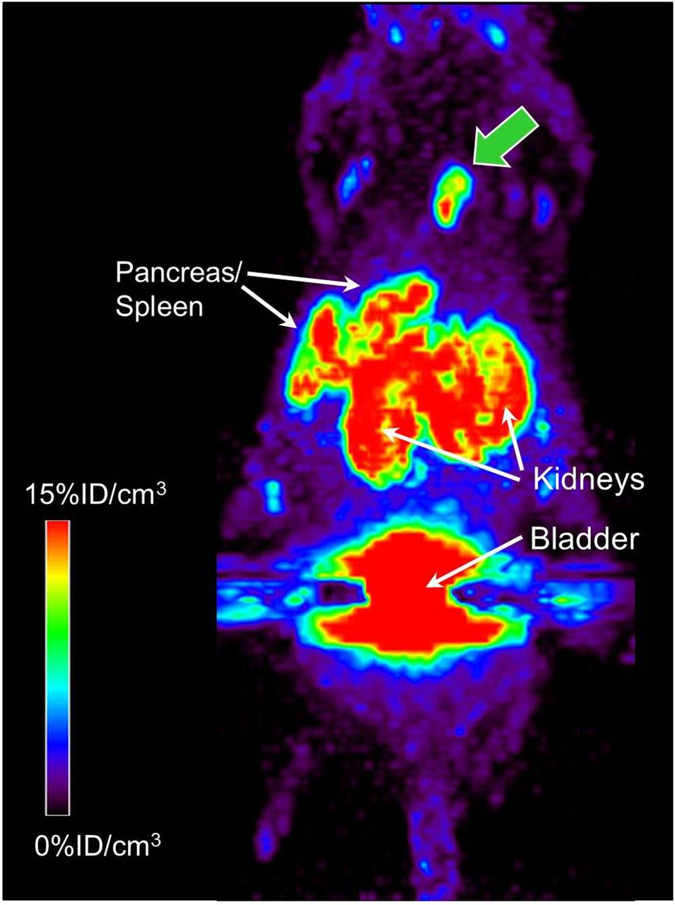
  • FIGURE 6.
    • Download figure
    • Open in new tab
    • Download powerpoint
    FIGURE 6.

    18F-FASu PET imaging in CD-1 nude mice bearing SKOV-3 xenograft tumors 1 h after injection. Maximum-intensity-projection image of 1 of 2 SKOV-3 tumor–bearing nude mice imaged in this study; tumor is indicated by green arrow. 18F-FASu was HPLC-purified. On necropsy, it was determined that weight of this tumor was 31 mg.
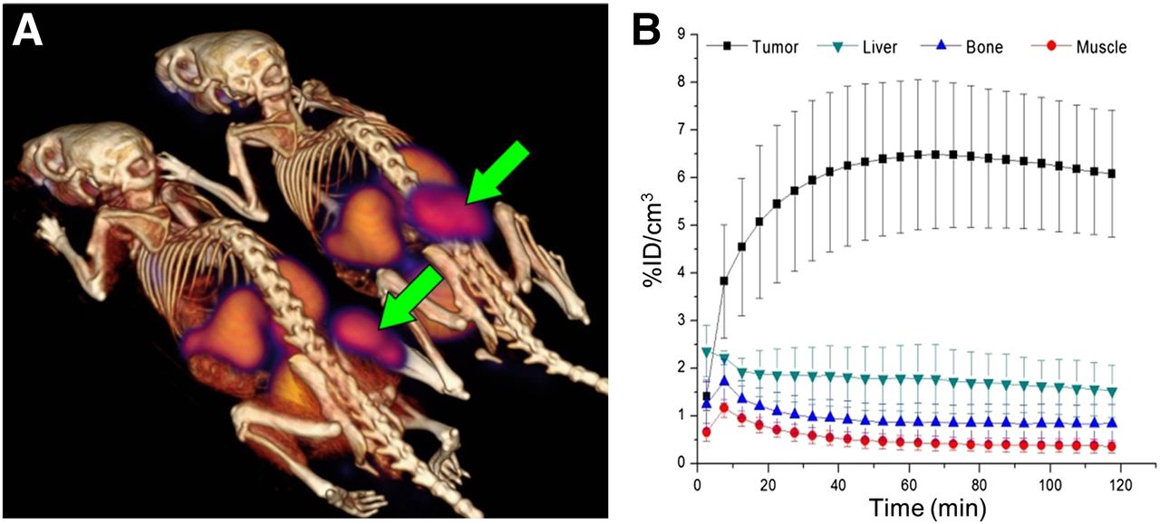
  • FIGURE 7.
    • Download figure
    • Open in new tab
    • Download powerpoint
    FIGURE 7.

    In vivo 18F-FASu dynamic PET imaging in Rag2 M mice bearing EL4 xenograft tumor. 18F-FASu was SepPak-purified. (A) PET/CT image summed over 110–120 min after injection. Tumors are indicated by arrows. (B) Region-of-interest analysis of 18F-FASu uptake in tumor, liver, bone, and muscle (mean ± SD, n = 4).

Tables

  • Figures
  • Additional Files
    • View popup
    TABLE 1

    Biodistribution of 18F-FASu and 18F-FDG in Mice Bearing Xenograft Tumors

    SKOV-3 tumors
    Organ or tissueEL4 tumors, 18F-FASu18F-FDG18F-FASu
    Blood0.49 ± 0.120.58 ± 0.300.72 ± 0.30
    Tumor5.81 ± 1.261.55 ± 0.968.08 ± 2.03
    Muscle0.25 ± 0.164.92 ± 2.260.35 ± 0.17
    Liver1.24 ± 0.180.74 ± 0.150.86 ± 0.08
    Kidney10.11 ± 2.581.04 ± 0.1615.36 ± 5.63
    Spleen4.98 ± 1.281.73 ± 0.596.74 ± 1.02
    Heart0.33 ± 0.0954.11 ± 29.000.32 ± 0.07
    Lungs2.00 ± 0.383.61 ± 1.681.95 ± 0.97
    Brain0.11 ± 0.037.76 ± 2.770.09 ± 0.02
    Bone0.85 ± 0.263.58 ± 1.740.85 ± 0.53
    Pancreas23.34 ± 5.731.49 ± 0.5421.19 ± 6.61
    Fat0.29 ± 0.220.59 ± 0.360.62 ± 0.67
    Tumor-to-blood12.01 ± 1.582.61 ± 0.2712.08 ± 0.4.39
    Tumor-to-muscle28.16 ± 10.390.33 ± 0.1329.59 ± 19.44
    Tumor size1,140 ± 29672.60 ± 37.6571.60 ± 39.09
    • Uptake values are %ID/g of tissue at 1 h after tracer injection; tumor size is in mg, mean ± SD (n ≥ 5).

Additional Files

  • Figures
  • Tables
  • Supplemental Data

    Files in this Data Supplement:

    • Supplemental Data
PreviousNext
Back to top

In this issue

Journal of Nuclear Medicine: 55 (4)
Journal of Nuclear Medicine
Vol. 55, Issue 4
April 1, 2014
  • Table of Contents
  • Table of Contents (PDF)
  • About the Cover
  • Index by author
Print
Download PDF
Article Alerts
Sign In to Email Alerts with your Email Address
Email Article

Thank you for your interest in spreading the word on Journal of Nuclear Medicine.

NOTE: We only request your email address so that the person you are recommending the page to knows that you wanted them to see it, and that it is not junk mail. We do not capture any email address.

Enter multiple addresses on separate lines or separate them with commas.
Functional Imaging of Oxidative Stress with a Novel PET Imaging Agent, 18F-5-Fluoro-l-Aminosuberic Acid
(Your Name) has sent you a message from Journal of Nuclear Medicine
(Your Name) thought you would like to see the Journal of Nuclear Medicine web site.
Citation Tools
Functional Imaging of Oxidative Stress with a Novel PET Imaging Agent, 18F-5-Fluoro-l-Aminosuberic Acid
Jack M. Webster, Christine A. Morton, Bruce F. Johnson, Hua Yang, Michael J. Rishel, Brian D. Lee, Qing Miao, Chittari Pabba, Donald T. Yapp, Paul Schaffer
Journal of Nuclear Medicine Apr 2014, 55 (4) 657-664; DOI: 10.2967/jnumed.113.126664

Citation Manager Formats

  • BibTeX
  • Bookends
  • EasyBib
  • EndNote (tagged)
  • EndNote 8 (xml)
  • Medlars
  • Mendeley
  • Papers
  • RefWorks Tagged
  • Ref Manager
  • RIS
  • Zotero
Share
Functional Imaging of Oxidative Stress with a Novel PET Imaging Agent, 18F-5-Fluoro-l-Aminosuberic Acid
Jack M. Webster, Christine A. Morton, Bruce F. Johnson, Hua Yang, Michael J. Rishel, Brian D. Lee, Qing Miao, Chittari Pabba, Donald T. Yapp, Paul Schaffer
Journal of Nuclear Medicine Apr 2014, 55 (4) 657-664; DOI: 10.2967/jnumed.113.126664
Twitter logo Facebook logo LinkedIn logo Mendeley logo
  • Tweet Widget
  • Facebook Like
  • Google Plus One
Bookmark this article

Jump to section

  • Article
    • Abstract
    • MATERIALS AND METHODS
    • RESULTS
    • DISCUSSION
    • CONCLUSION
    • DISCLOSURE
    • Acknowledgments
    • Footnotes
    • REFERENCES
  • Figures & Data
  • Supplemental
  • Info & Metrics
  • PDF

Related Articles

  • This Month in JNM
  • PubMed
  • Google Scholar

Cited By...

  • Comparative Evaluation of [18F]5-Fluoroaminosuberic Acid and (4S)-4-3-[18F]fluoropropyl)-L-Glutamate as System Formula-Targeting Radiopharmaceuticals
  • Comparative Evaluation of [18F]5-Fluoroaminosuberic Acid and (4S)-4-3-[18F]fluoropropyl)-L-Glutamate as System Formula-Targeting Radiopharmaceuticals
  • Latest Advances in Imaging Oxidative Stress in Cancer
  • Clinical Evaluation of (4S)-4-(3-[18F]Fluoropropyl)-L-glutamate (18F-FSPG) for PET/CT Imaging in Patients with Newly Diagnosed and Recurrent Prostate Cancer
  • The Characterization of 18F-hGTS13 for Molecular Imaging of xC- Transporter Activity with PET
  • Cystine/glutamate antiporter xCT (SLC7A11) facilitates oncogenic RAS transformation by preserving intracellular redox balance
  • Assessment of Tumor Redox Status through (S)-4-(3-[18F]fluoropropyl)-L-Glutamic Acid PET Imaging of System xc- Activity
  • 18F-5-Fluoroaminosuberic Acid as a Potential Tracer to Gauge Oxidative Stress in Breast Cancer Models
  • A Continuously Infused Microfluidic Radioassay System for the Characterization of Cellular Pharmacokinetics
  • Google Scholar

More in this TOC Section

  • 11C-Methionine PET of Myocardial Inflammation in a Rat Model of Experimental Autoimmune Myocarditis
  • Counting Rate Characteristics and Image Distortion in Preclinical PET Imaging During Radiopharmaceutical Therapy
  • Design and Fabrication of Kidney Phantoms for Internal Radiation Dosimetry Using 3D Printing Technology
Show more Basic Science Investigations

Similar Articles

Keywords

  • oxidative stress
  • PET
  • functional imaging
  • tumor imaging
  • 18F
SNMMI

© 2025 SNMMI

Powered by HighWire