


Abstract
Pancreatic ductal adenocarcinoma (PDAC) is a lethal disease. Late detection of then nonresectable or metastasized tumors emphasizes the need for novel imaging approaches. Here, we report on so far nonexploited potentials of αvβ3 integrin–targeted molecular imaging technologies for detection of PDAC using genetically engineered mouse models. Methods: Immunohistochemistry and Western blot were used for characterization of αvβ3 expression in murine and human PDAC. We applied IntegriSense 680 fluorescence molecular tomography, intraoperative fluorescence imaging, and 68Ga-NODAGA-RGD PET for αvβ3 integrin molecular in vivo imaging of spontaneous PDAC occurring in Ptf1a+/Cre;Kras+/LSL-G12D;p53LoxP/LoxP mice. (NODAGA is 1,4,7-triazacyclononane-1,4-bis[acetic acid]-7-[2-glutaric acid] and RGD is arginine-glycine-aspartic acid.) Results: αvβ3 integrin is expressed in tumor cells of human and murine PDAC. IntegriSense fluorescence molecular tomography and 68Ga-NODAGA-RGD PET enabled faithful visualization of PDAC. Furthermore, intraoperative optical imaging with IntegriSense 680 allowed good delineation of tumor borders. Conclusion: Imaging approaches targeting αvβ3 integrin expand the potential of molecular imaging for identification of αvβ3-positive PDAC with potential implications in early detection, fluorescence-guided surgery, and therapy monitoring.
- integrin αvβ3
- positron emission tomography
- fluorescence molecular tomography
- pancreatic cancer
- genetically engineered mice
The devastating status of pancreatic ductal adenocarcinoma (PDAC) as the fourth most lethal human malignancy results from the late detection of often metastasized disease and resistance to chemotherapies (1). Thus, new imaging methods for detection, improved surgical resection, and clinically relevant therapy response monitoring are key aims to increase the survival in PDAC patients.
Clinically, PDAC diagnosis and determination of resectability are typically performed by imaging modalities such as MR imaging and CT, which provide information regarding tumor size, location, and morphology (2). Molecular imaging approaches that provide clinically meaningful additional information, such as subtypes or tumor biology, are not well established. Molecular imaging using 18F-FDG and 3′-deoxy-3′-18F-fluorothymidine PET has been recently characterized in humans, demonstrating high sensitivity for PDAC detection but low specificity with high rates of false-positive findings of inflammatory lesions or pseudomasses (3,4).
Among molecular imaging approaches, fluorescence-based optical methods are especially appealing because of their low-cost, high sensitivity and spatial resolution and absence of ionizing radiation (5). As such, fluorescence-guided surgery is increasingly used in operation rooms in which it allows precise visualization of targeted tissue and its margins during surgery (6,7).
Genetically engineered mouse models (GEMM) that faithfully recapitulate molecular and morphologic features of human PDAC have already been used successfully for translational diagnostic and therapeutic approaches including molecular imaging. Recent studies in GEMM reported on identification of cathepsins and Claudin 4 as promising molecular targets for in vivo detection of PDAC using fluorescence-based imaging technologies. These studies suggested that molecular imaging of suitable targets may allow identification of PDAC and precursor lesions and may thus be suitable for early detection methods (8–11). However, the tissue volume in humans limits some of the approaches reported because of signal attenuation.
In this paper, we identify αvβ3 integrin as a candidate target for visualization of PDAC. Integrin αvβ3 belongs to the integrin family of cell surface receptors expressed on activated endothelial cells of new sprouting vessels in the tumor but also on tumor cells in various cancers including PDAC (12,13). αvβ3 integrin has been implicated as a key player in tumor angiogenesis and metastasis and was shown to be especially relevant for the metastatic spread of PDAC to lymph nodes (14,15). Molecular imaging of αvβ3 integrin by RGD (H-Arg-Gly-Asp-NH2; arginine-glycine-aspartic acid)–labeled PET tracers has been introduced into clinical practice and is used for visualization of angiogenesis in many cancers (13,16,17). However, its potential for PDAC imaging has not been addressed before.
Here, we report on the molecular imaging of αvβ3 integrin as a feasible and reliable approach for detection of PDAC and its high value for PDAC translational research. We report on robust αvβ3 expression in endogenous murine PDAC and in a subpopulation of human PDAC and demonstrate that detection of murine PDAC is achievable in vivo using αvβ3 integrin–targeted optical and PET imaging approaches.
MATERIALS AND METHODS
Animals
Animal strains and used mouse models have been previously described (18–20). Genetically engineered mice with pancreas-specific activation of oncogenic KrasG12D and additional pancreas-specific deletion of p53 for development of invasive PDAC (Ptf1a+/Cre;Kras+/LSL-G12D;p53LoxP/LoxP; referred to as CKP hereafter) were used. Animals were 5–7 wk old. All experiments were performed according to the guidelines of the local Animal Use and Care Committees.
Target Validation
Immunohistochemistry, immunofluorescence, and Western blot were performed on murine and human tissue samples according to standard protocols. A description of procedures and antibodies is given in the supplemental material (supplemental materials are available at http://jnm.snmjournals.org).
PET and Biodistribution Studies
One day before the PET scans, PDAC presence was confirmed via T2-weighted (T2w) MR imaging. Tumor-bearing CKP animals were injected with 68Ga-labeled NODAGA (1,4,7-triazacyclononane-1,4-bis[acetic acid]-7-[2-glutaric acid])-conjugated RGD peptide and scanned on a small-animal PET/CT scanner. Details on 68Ga labeling of NODAGA-RGD are provided in the supplemental material. The small-animal PET and biodistribution studies were performed by tail-vein injection of 7–12 MBq of 68Ga-NODAGA-RGD under isoflurane anesthesia. Static small-animal PET/CT scans were obtained using a combined PET/CT scanner (Inveon; Siemens). The scan was started 75 min after tracer injection (scan duration, 15 min). Image data were corrected for normalization, dead time, and decay. Images were reconstructed using a 2- and 3-dimensional ordered-subsets expectation maximum algorithm without scatter and attenuation correction. For anatomic information, axial PET images were fused to axial T2w MR images of corresponding animals, as the nonenhanced CT image from the PET/CT scanner was insufficient to distinguish tumor from surrounding soft-tissue structures because of low soft-tissue contrast. Images were analyzed using the Inveon Research Workplace (Siemens). For biodistribution experiments, animals were injected with 68Ga-NODAGA-RGD and euthanatized, and blood and all internal organs were collected and wet-weighed 75 min after injection. The radioactivity was measured using a γ counter (Wallac Wizard, PerkinElmer). The results are presented as percentage injected dose per gram of tissue.
Optical Imaging Procedures
IntegriSense 680 (PerkinElmer), a commercially available small nonpeptide molecule labeled with near-infrared fluorochrome that specifically binds to αvβ3 integrin, was dissolved in phosphate-buffered saline, administered via tail vein bolus injection (2 nmol), and allowed to circulate for 24 h before all optical imaging experiments.
Fluorescence Molecular Tomography (FMT)
Mice were imaged in vivo using the FMT–CT (FMT-XCT) hybrid modality at Helmholtz Zentrum München (21,22). FMT-XCT operates in 360° full-ration geometry, where the mouse is illuminated by a scanning 670-nm laser at equispaced 24 gantry locations. Fluorescence and intrinsic images are obtained for every source position using a cooled charge-coupled device in transillumination mode. Optical and x-ray modules are located on perpendicular axes, which are both perpendicular to the animal axial direction. The system was mathematically modeled using a finite element method for modeling light propagation in tissue assuming homogeneous optical absorption and scattering coefficients of 0.2 and 15 cm−1 for the mouse tissue. The inverse problem was regularized using Tikhonov regularization and solved using the least-squares algorithm method. The FMT and CT acquisitions lasted 34 and 23 min, respectively, during which the mouse was kept under isoflurane-induced anesthesia.
Intraoperative Optical Imaging
At 24 h after injection of IntegriSense 680, a surgical incision exposed the abdominal cavity to the intraoperative imaging setup described previously (23), which captured color, intrinsic, and fluorescence images of the exposed pancreas and its surroundings, simulating an intraoperative imaging situation.
Ex Vivo Fluorescence Cryoslicing
For whole-mount cryoslicing, animals used for FMT-XCT and intraoperative imaging experiments were sacrificed and whole-mount-frozen after the experiment, cryosliced, and imaged with a cryoslicer modality (24). Axial cut color and fluorescence images were captured every 250 μm and then used to volumetrically visualize pancreas or tumor and surrounding tissues and to compare the fluorescence images with the corresponding T2w MR imaging axial images under the same geometry.
Competitive Studies
To eliminate the possibility of nonspecific deposition of 68Ga-NODAGA-RGD in PDAC, around 400 μg of cilengitide, a selective αvβ3 inhibitor (25), were intravenously injected into animals 10–15 min before injections of 68Ga-NODAGA-RGD, and biodistribution was performed as described.
For the competitive study, IntegriSense 680 (2 nmol) was intravenously injected into CKP animals with PDAC and allowed to circulate and occupy αvβ3 integrin sites for 24 h. Subsequently, 68Ga-NODAGA-RGD was injected into the same animals and biodistribution performed as described.
RESULTS
αvβ3 Expression in Human PDAC
To investigate αvβ3 integrin expression in human PDAC, we performed immunohistochemistry for the integrin β3 subunit on 77 human PDAC specimens. Immunohistochemistry for β3 integrin identified 24 tumors as β3-positive (β+) (31%) with staining within neoplastic, cancer cells. β3 staining was of cytoplasmic or membranous localization, and expression levels in tumor cells varied from low to high (Fig. 1A). Correlation between tumor grading and β3+ positivity was not observed (data not shown).

Expression analysis of αvβ3 integrin. (A) Human PDAC with membranous/cytoplasmatic and absent expression of integrin β3. Scale bar: 50 μm. Table summarizes β3 expression results for 77 human PDAC samples. (B) Immunohistochemistry for β3 integrin demonstrating no expression in wild-type pancreatic tissue and high expression in PDAC in CKP mice. Scale bars: 50 μm. (C) Immunohistochemistry for CK19, CD31, and integrin β3 on consecutive paraffin sections of murine PDAC demonstrating partial colocalization of CK19 and β3 integrin and different expression patterns of CD31 and β3 integrin. Scale bars: 50 μm. (D) Western blot analysis demonstrating higher αv and β3 expression in PDAC from CKP animals than healthy tissue from wild-type mice. Hsp90 served as loading control. (E) αv and β3 expression in primary CKP murine cancer cell lines demonstrated by Western blot analysis. Hsp90 served as loading control. WT = wild-type.
αvβ3 Expression in Murine PDAC
We next analyzed expression of αv and β3 integrin subunits in the pancreas of wild-type mice and PDAC of CKP mice. As demonstrated by immunohistochemistry, in wild-type pancreas β3 expression was limited to occasional blood vessels whereas no staining was observed in the acinar or ductal compartments (Fig. 1B). In PDAC of CKP mice, a dramatic increase in staining signal was observed. β3+ cells were numerous and scattered throughout the tumor, with expression in invasive ductal cancer cells and the surrounding stroma (Fig. 1B). Staining for cytokeratin 19 (CK19), an epithelial marker of cancer cells, and β3 on consecutive paraffin sections (Fig. 1C) supported β3 expression in cancer cells because a similar expression pattern of β3 and CK19 was observed. Immunohistochemistry staining on consecutive paraffin sections for β3 and CD31, a blood vessel marker, demonstrated that most β3+ cells did not belong to tumor vessels (Fig. 1C). Western blot analysis demonstrated an increased expression of both αv and β3 protein subunits in PDAC, compared with wild-type pancreas (Fig. 1D). Furthermore, Western blot analysis of primary cancer cell lines isolated from tumors of CKP mice revealed that both αv and β3 were expressed in all 8 tumor cell lines isolated from different animals, albeit at different levels (Fig. 1E).
αvβ3-Targeted FMT and PET
Motivated by high αvβ3 expression, we implemented 2 different αvβ3-targeted molecular imaging methods and compared their efficiencies and benefits for PDAC detection: PET with 68Ga-NODAGA-RGD probe as an approach in clinical use and FMT-XCT with IntegriSense 680 probe as a novel strategy for imaging PDAC.
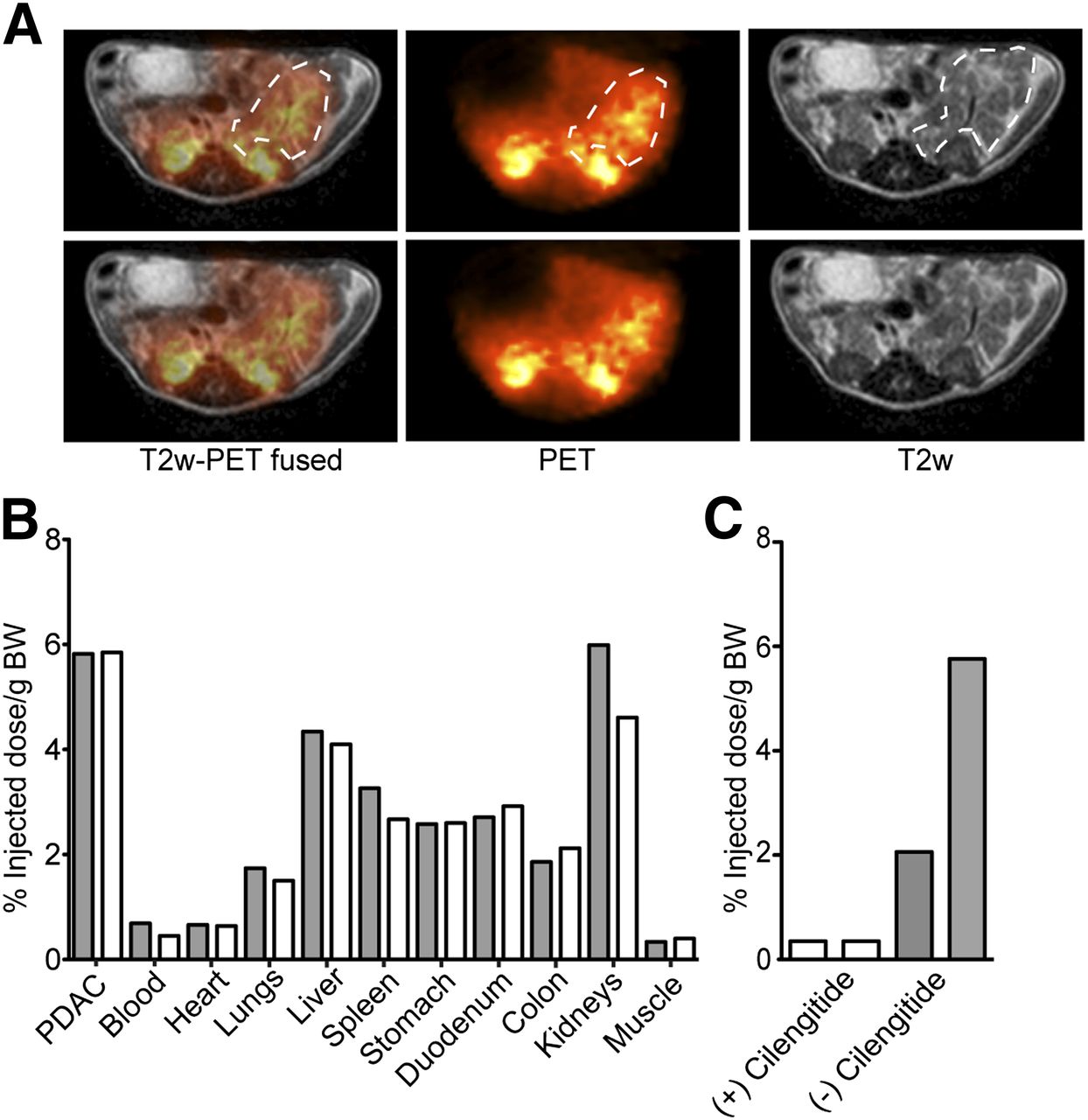
Positive PET signals were clearly detectable in PDAC (Fig. 2A), although delineation of cancer borders on PET images was not good because of the intrinsically low spatial resolution. To validate PET results, a biodistribution assay was performed. High accumulation of 68Ga-NODAGA-RGD in PDAC was detected (Fig. 2B). Competitive studies with αvβ3 inhibitor cilengitide showed that the amount of radioactivity accumulated in PDAC after cilengitide injections was dramatically lower than radioactivity measured in PDACs without cilengitide injections (Fig. 2C).

68Ga-NODAGA-RGD PET detects PDAC in vivo. (A) Fused and single axial T2w MR and 68Ga-NODAGA-RGD PET images of PDAC. Positive PET signal in orange is present in tumor and kidneys. In images, tumor is delineated with white dotted line (upper) and without delineation for readers' comfort (lower). Kidneys show positive PET signals due to clearance of 68Ga-NODAGA-RGD through kidneys. (B) Biodistribution of 68Ga-NODAGA-RGD demonstrating accumulation of radioactivity in PDAC in CKP animals. Results from 2 animals are presented (gray bar, animal 1; white bar, animal 2). (C) Preinjection of cilengitide decreases uptake of 68Ga-NODAGA-RGD into murine PDAC in CKP animals. Every bar represents PDAC of 1 animal. BW = body weight.
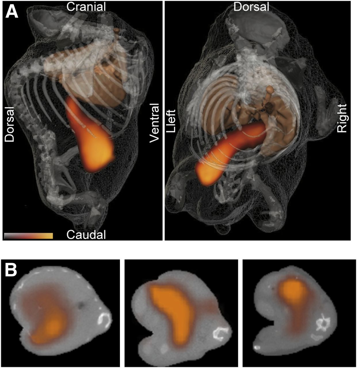
In animals bearing PDAC, we showcased a previously undocumented capability of FMT-XCT in 3-dimensionally resolving strong fluorescence signals localized specifically in pancreatic tumor (Fig. 3; Supplemental Video 1). Strong fluorescence signals were detected in PDAC, demonstrating the superiority of FMT-XCT to PET in this case.

In vivo imaging using FMT-XCT. (A) Three-dimensional representations of reconstructed fluorescence signal along with anatomic data; segmented skeleton and lungs (brown isosurface) are shown from side and top views, respectively. Bottom left is gated color map with orange color for high signal values, as detected in PDAC. (B) Reconstructed fluorescence overlaid as pseudo-orange transparency on corresponding CT slices in 3 different positions in mouse body.
Intraoperative Imaging
Because of high PDAC fluorescence in FMT-XCT experiments, we hypothesized that intraoperative detection of PDAC via IntegriSense fluorescence would be possible. As previously published in wild-type animals (24), no fluorescence was detectable from the surface of the healthy pancreas, suggesting no specific accumulation of IntegriSense in normal pancreatic tissue (Fig. 4A). In CKP animals, PDACs were remarkably well visualized, showing strong and specific fluorescence signals from tissue surface (Fig. 4A). Fluorescence contrast to the adjacent tissues and thus demarcation of tumor borders was conspicuous. High near-infrared fluorescence signals were detected only at the tumor surface, indicating specific IntegriSense accumulation in the tumor. Examination of other exposed organs showed some fluorescence signals arising primarily from the gut, stomach, fat pads, urinary bladder, and ovaries in females. The liver showed no increased signals.
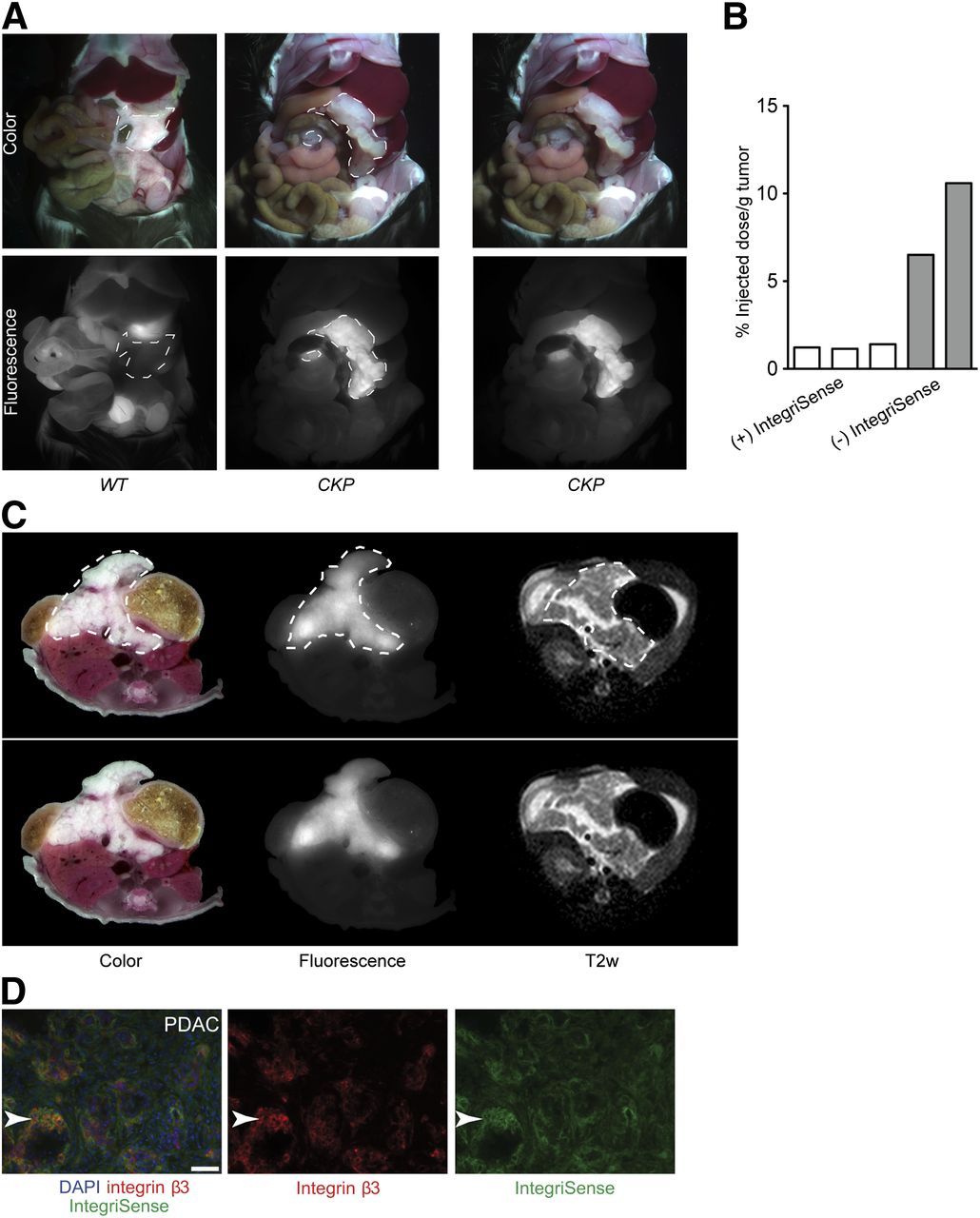
In vivo intraoperative and ex vivo cryoslicing optical imaging of IntegriSense in murine PDAC. (A) Representative planar epi-illumination whole-body color autopsy and original fluorescence images of wild-type and CKP animals. Tumor or pancreas area is lined with dotted white line (left), and tumor images are repeated without delineation (right). Strong equally distributed IntegriSense 680 fluorescence signals are detectable at tumor surface in CKP animals and no signals from wild-type pancreata. In wild-type animals, bladder visibly fluoresces due to probe clearance through the urinary system. (B) Injection of IntegriSense 24 h before injection of 68Ga-NODAGA-RGD decreases uptake of 68Ga-NODAGA-RGD into murine PDAC. Every bar represents 1 animal. (C) Representative whole-body axial cryoslicer images: color, fluorescence, and corresponding T2w images for CKP animal. Tumor is delineated with white dotted line. In CKP animals, axial T2w and fluorescence images correlated well, but delineation of tumor borders was better on fluorescence images. Images without delineation for readers' comfort (lower). (D) Microscopic analysis of IntegriSense distribution in PDAC. In CKP animals, IntegriSense accumulated in tumors. IntegriSense fluorescence (green) correlated well with β3 staining (red), demonstrating specific IntegriSense accumulation in β3+ areas. Scale bar: 50 μm. WT= wild-type.
To confirm that IntegriSense 680 indeed binds to αvβ3 integrin in PDAC, we performed a competitive binding experiment with 68Ga-NODAGA-RGD (Fig. 4B). The preinjection of IntegriSense dramatically decreased the amount of 68Ga-NODAGA-RGD that accumulated in PDAC.
For validation of intraoperative and FMT-XCT imaging results, we used a recently developed ex vivo multispectral cryoslicing system (24). In animals with PDAC, fluorescence images of cryosections demonstrated strong and specific fluorescence signals in the tumor (Fig. 4C). Fluorescence accumulation at the tumor rims was not observed, and axial fluorescent images of the tumor correlated well with the axial T2w MR images, suggesting good probe penetration and binding in the tumor. Furthermore, cryoslicing confirmed the intraoperative observations of enhanced demarcation of tumor borders (Fig. 4C; Supplemental Fig. 1). The tumor was distinguishable from the surrounding organs. Some fluorescence was observable in various parts of the gut. However, tumor fluorescence was consistently stronger than that of the gut.
Microscopic analysis of cryoslides confirmed specific accumulation of IntegriSense in PDAC (Fig. 4D). Fluorescence signals for IntegriSense were observable around the β3+ cells in PDAC, suggesting specific probe deposition in β3+ areas of the tumor.
DISCUSSION
Recent breakthroughs in oncologic research have highlighted that traditional approaches to cancer as a whole organ must be remodeled and adapted to the fact that cancers are extremely complex and heterogeneous, necessitating deeper molecular characterization for personalized therapeutic approaches (26). Molecular imaging approaches that allow imaging and delineation of tumors and identification of clinically relevant subtypes are thus promising new imaging strategies. Our study suggests αvβ3 integrin to be one of the molecular targets suitable for such approaches as substantiated with the several imaging results presented in this work.
The clinical relevance of αvβ3 integrin is mirrored in the fact that patients whose cancer cells express αvβ3 have increased metastasis to lymph nodes and worse survival than those without αvβ3 expression (14,15). This was further confirmed in animal experiments, in which overexpression of αvβ3 led to enhanced primary tumor growth of orthotopic transplanted pancreatic cancer cells and enhanced metastatic potential, whereas αvβ3 knock-down diminished these actions (14).
For analysis of αvβ3 integrin expression in human PDAC, we performed immunohistochemistry for the integrin β3 subunit. β3 integrin dimerizes only with 2 different α integrins, αv and αIIb. αIIb integrin is specifically expressed on blood platelets (27), and it is thus generally accepted that β3 expression pattern in other tissues represents the expression of the αvβ3 dimer. Similar to previously published data on β3 expression found in 58% of human PDACs (15), we also found an increased expression of αvβ3 in a subpopulation of human PDAC (31%). In previously published studies and our study, the staining was found in cancer cells. The differences in the percentage of β3+ PDAC can probably be attributed to limited total number of samples used in both studies (50 and 77). Heterogeneity in expression level and in localization of β3 among PDAC samples was observed as described previously (14,15). With the previously mentioned role of αvβ3 in metastatic potential of pancreatic cancer cells, this result suggests that the αvβ3 receptor might have an important biologic function and is potentially suitable for personalized targeted therapeutic approaches based on αvβ3 status.
As in humans, increased expression of αvβ3 was observed in murine PDAC. CKP animals used in this study recapitulate the human disease well, developing moderately differentiated, invasive PDAC with dense stroma (28). Most of the β3 expression in murine PDACs could be attributed to malignant (tumor) cells or reactive stroma. The different expression pattern of β3 and CD31, a marker of blood vessels, suggested that only a minor part of β3+ cells actually belongs to the vascular compartment. Thus, although molecular imaging of αvβ3 is usually used for detection of tumor angiogenesis (13), targeting αvβ3 in murine PDAC would rather detect the tumor itself.
As a clinically relevant and applicable approach, we used PET imaging for αvβ3 detection in murine PDAC. Of many available αvβ3-targeting RGD tracers, a novel 68Ga-labeled NODAGA-conjugated RGD peptide was chosen because of its easy accessibility, high stability, and previously reported good imaging properties (17,29). Furthermore, it has already been demonstrated that 68Ga-labeled NODAGA-RGD accumulates to a small degree into the wild-type pancreas (29), making this agent suitable for specific detection of tumorous pancreatic tissue. PET imaging with 68Ga-labeled NODAGA-RGD allowed good identification of murine PDAC. Imaging was further confirmed by accumulation of 68Ga-labeled NODAGA-RGD in PDAC in the biodistribution study. Furthermore, efficient block of 68Ga-NODAGA-RGD binding to αvβ3 by cilengitide confirmed the specificity of 68Ga-NODAGA-RGD for αvβ3 integrin in PDAC. Accumulation ranged from 2% to 10% of injected dose in different experiments, and we assume that these small differences do not reflect heterogeneity in αvβ3 expression levels but can rather be attributed to varying size of the tumors and technical aspects.
In light of the recent success of fluorescence-based technologies for detection of αvβ3-positive (αvβ3+) xenotransplanted cancers (30,31), we investigated whether better detection of PDAC is achievable with optical imaging, namely a novel noninvasive hybrid imaging method combining CT (XCT) with FMT (21) using IntegriSense 680 as a probe. Optical imaging with αvβ3-targeted probes was also successful in identification of PDAC. Although FMT is not yet widely adopted for clinical use, the future application of nonradiating, inexpensive, fluorescent probes is easy to envisage. The FMT-XCT offers superposition of anatomic and molecular information, the latter obtained from fluorescence agents, and has recently been reported as successful for the detection of murine lung tumors (21). By using XCT information in the FMT inversion, this hybrid approach also offers the most accurate FMT performance to date (21). In our study, FMT performance was superior to PET, with specific signals detected from PDAC, suggesting that this method is a promising method of choice for future noninvasive identification of patients with αvβ3+ cancers and thus identification of PDAC with higher metastatic potential.
The clear definition of surgical margins and complete operative removal of residual tissue are keys to PDAC treatment. Residual cancer tissues missed during surgery present a potential for cancer relapse. In the case of patients with resectable PDAC, translation of PET or FMT images into the operating room is a major challenge for the performing surgeon who needs on-site imaging assistance for surgery. Recently, van Dam et al. performed the first fluorescence-guided surgical approach in ovarian cancer patients and provided the first proof of principle for implementation of this technique in surgical practice (7). For this reason, we optimized an optical imaging approach suitable for potential intraoperative purposes with a fluorescent molecular probe targeting the αvβ3 integrin. Several αvβ3-targeting fluorescently labeled RGD molecules with different chemical and molecular properties have been developed and used for optical imaging in various xenograft cancer mouse models (32–34). IntegriSense 680 used in this study is commercially available and with good described specificity for αvβ3 integrin (35). Our competitive studies with 68Ga-NODAGA-RGD also suggested specific binding of IntegriSense for integrin αvβ3.
IntegriSense has been previously used for fluorescence-guided surgery in xenograft cancer models (30,31). To our knowledge, it has not been evaluated in endogenous cancer mouse models. Intraoperative optical imaging of the abdomen showed specific IntegriSense signals at the tumor surface. Some unspecific fluorescence was observed on the surface of other organs, mostly gut, probably due to unspecific probe accumulation and food autofluorescence. Furthermore, intraoperative and FMT images were confirmed by ex vivo fluorescence imaging under cryosectioning that enables the detailed evaluation of probe biodistribution inside the tissue and comparison to other surrounding organs. Multispectral cryoslicing confirmed good probe penetration and specific intratumoral distribution and no unspecific probe accumulation at the tumor rims that could account for false-positive signals from tumor surface in intraoperative imaging. Thus, IntegriSense-guided intraoperative imaging may be of use for endoscopic evaluation and to navigate removal of PDAC aiming to minimize the amount of residual cancer tissue.
CONCLUSION
We showed that αvβ3 integrin is expressed in human and murine PDAC and can be detected by molecular imaging technologies in PDAC of GEMM. This strategy can further be exploited for identification of patients with αvβ3+ PDAC, fluorescence-guided surgical removal of PDAC, and application of αvβ3-targeted therapies.
DISCLOSURE
The costs of publication of this article were defrayed in part by the payment of page charges. Therefore, and solely to indicate this fact, this article is hereby marked “advertisement” in accordance with 18 USC section 1734. This work was supported by the collaborative research center SFB 824, German Research Foundation (DFG) “Imaging for Selection, Monitoring and Individualization of Cancer Therapies.” No other potential conflict of interest relevant to this article was reported.
Acknowledgments
We thank Rosel Oos, Christina Lesti, Sybelle Reder, Claudia Meyerhofer, and Matilde Neuhofer for excellent technical assistance.
Footnotes
Published online Feb. 17, 2014.
- © 2014 by the Society of Nuclear Medicine and Molecular Imaging, Inc.
REFERENCES
- Received for publication July 18, 2013.
- Accepted for publication October 24, 2013.


