


Article Figures & Data
Figures
- FIGURE 1.
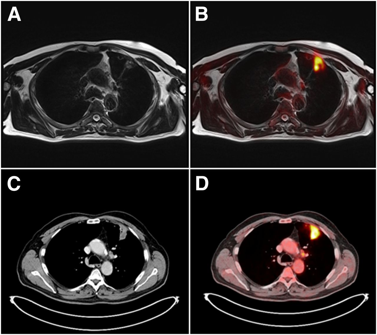
18F-FDG–avid, histologically proven NSCLC in left upper lobe of 59-y-old female patient on T2-weighted blade MR image with measured maximum diameter of 3.7 cm (A) and on fused 18F-FDG PET/MR image (B). Identical tumor mass of same patient in left upper lobe on CT image with measured maximum diameter of 3.8 cm (C) and on 18F-FDG PET/CT image (D). Primary tumor was correctly staged as T2a tumor in 18F-FDG PET/MR imaging and 18F-FDG PET/CT.
- FIGURE 2.
Not pathologically enlarged suprahilar lymph node on left side in 71-y-old male patient with histologically proven NSCLC in left upper lung with short-axis diameter of 0.7 cm on T2-weighted blade MR image (A) and on fused 18F-FDG PET/MR image (B). Identical suprahilar lymph node of same patient on CT image with short-axis diameter of 0.7 cm (C) and on 18F-FDG PET/CT image (D). Histopathologically, lymph node was rated as benign (inflammatory changes).
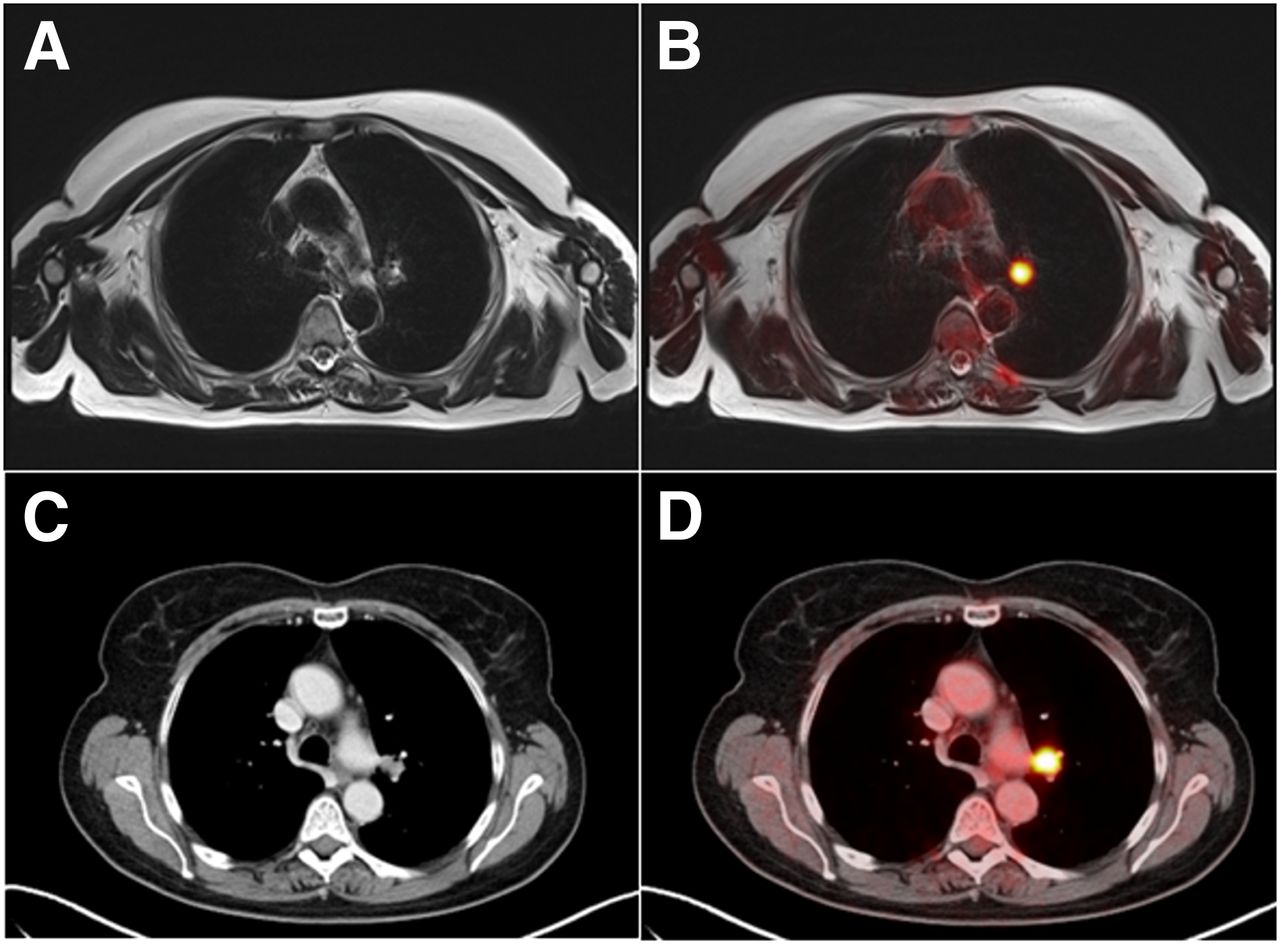
- FIGURE 3.
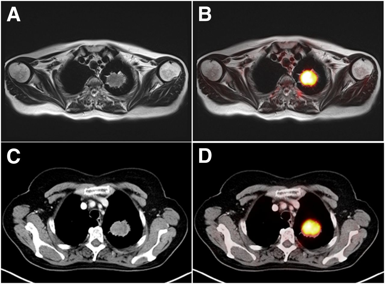
Hilar lymph node on left side in 59-y-old female patient with histologically proven NSCLC in left upper lung on T2-weighted blade MR image with short-axis diameter of 1.3 cm (A). This lymph node shows pathologic 18F-FDG uptake on fused 18F-FDG PET/MR image (B). Identical hilar lymph node of same patient on CT image with short-axis diameter of 1.3 cm (C), clearly visible on 18F-FDG PET/CT image (D). Histopathologically, lymph node was rated as benign (inflammatory changes).
- FIGURE 4.
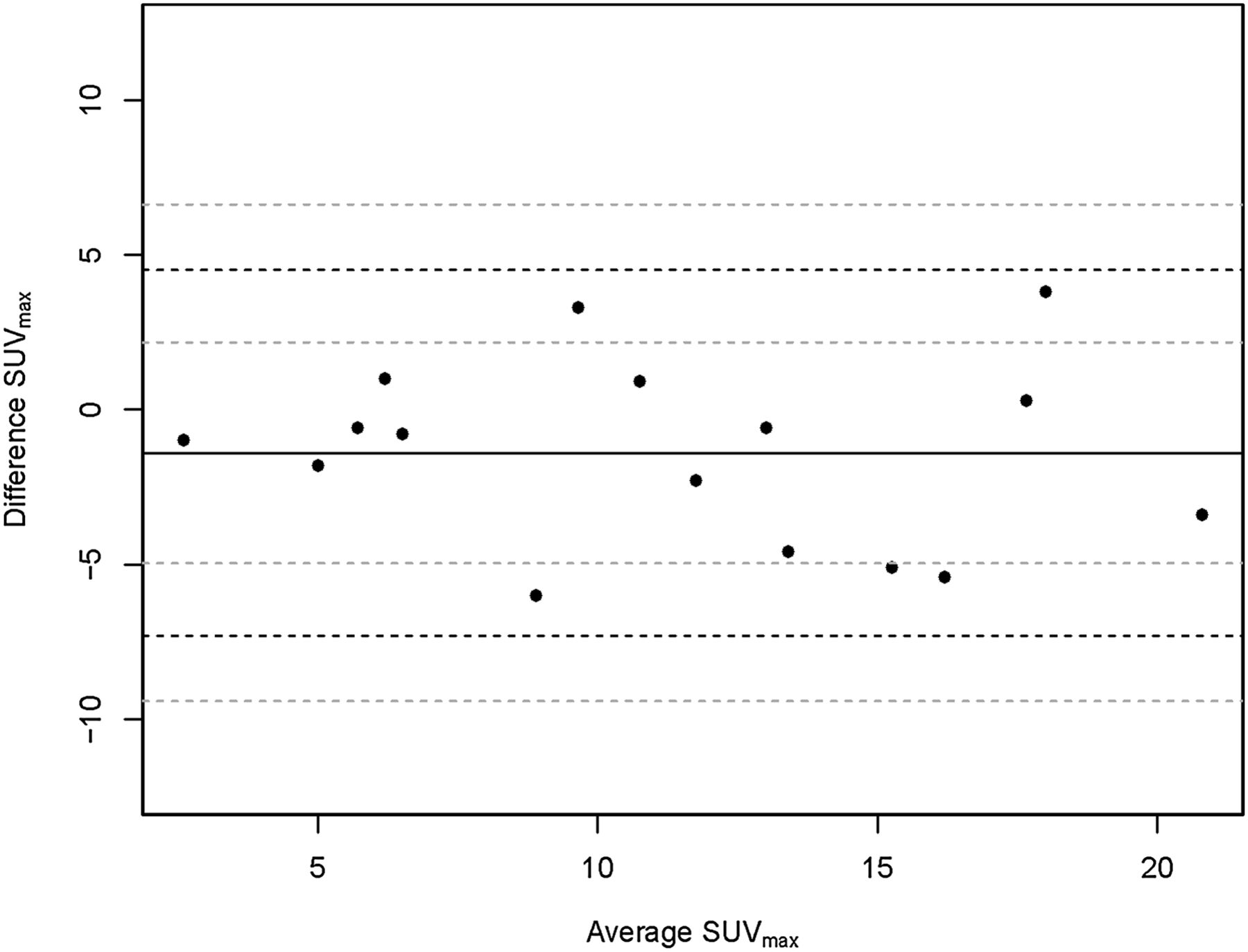
Bland–Altman analysis showing lower and upper limits of agreement between 18F-FDG PET/CT and 18F-FDG PET/MR imaging of −7.42 to 4.40 for SUVmax.
- FIGURE 5.
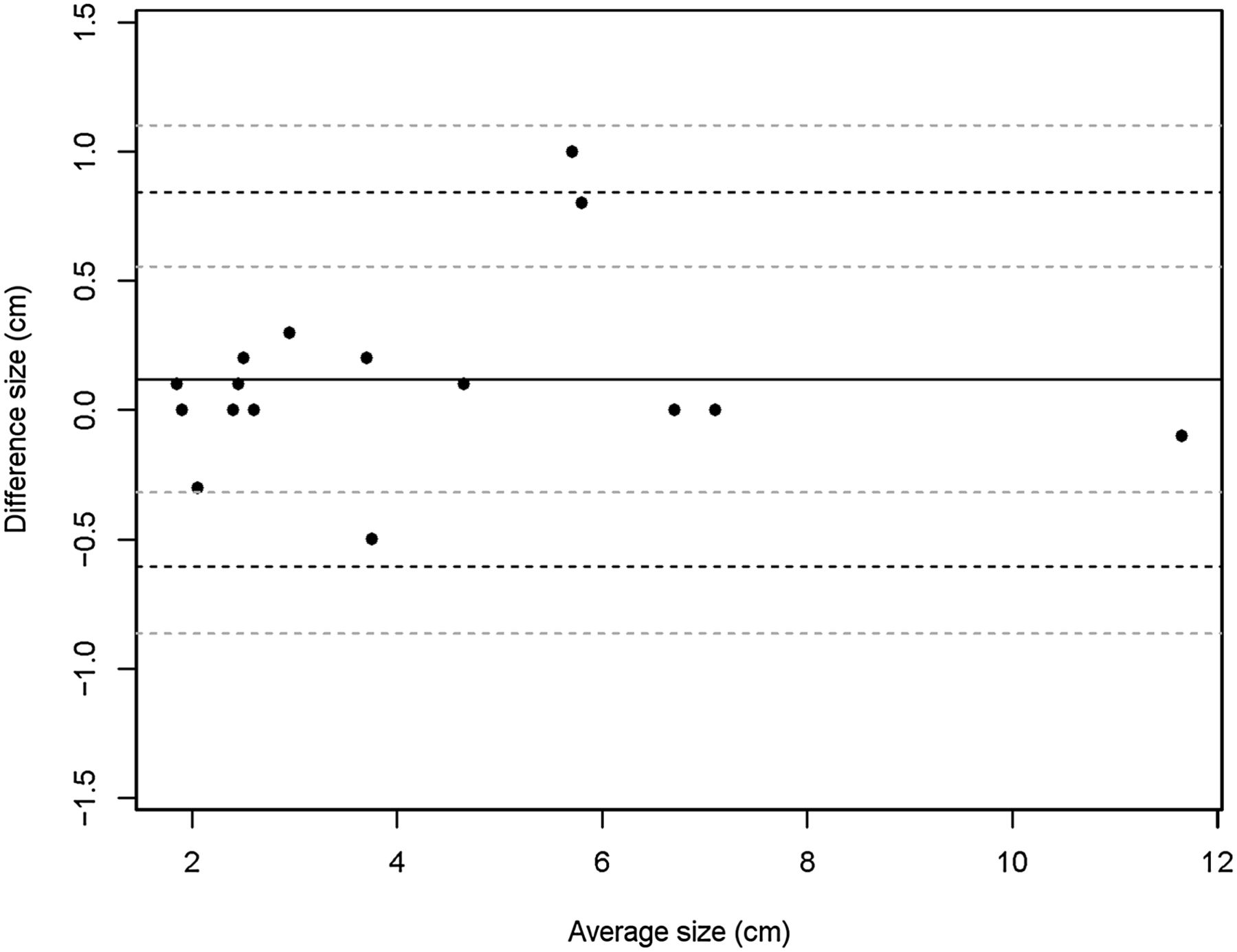
Bland–Altman analysis showing lower and upper limits of agreement between 18F-FDG PET/CT and 18F-FDG PET/MR imaging of −0.59 to 0.83 for tumor size.


Jump to section
Related Articles
Cited By...
- PET/MRI Versus PET/CT for Whole-Body Staging: Results from a Single-Center Observational Study on 1,003 Sequential Examinations
- Decoding Intratumoral Heterogeneity of Breast Cancer by Multiparametric In Vivo Imaging: A Translational Study
- Comparative Performance of 18F-FDG PET/MRI and 18F-FDG PET/CT in Detection and Characterization of Pulmonary Lesions in 121 Oncologic Patients
- 18F-FDG PET/CT and PET/MRI Perform Equally Well in Cancer: Evidence from Studies on More Than 2,300 Patients
- TNM Staging of Non-Small Cell Lung Cancer: Comparison of PET/MR and PET/CT