
Main menu

  • Home
  • Content
    • Current
    • Ahead of print
    • Past Issues
    • JNM Supplement
    • SNMMI Annual Meeting Abstracts
    • Continuing Education
    • JNM Podcasts
  • Subscriptions
    • Subscribers
    • Institutional and Non-member
    • Rates
    • Journal Claims
    • Corporate & Special Sales
  • Authors
    • Submit to JNM
    • Information for Authors
    • Assignment of Copyright
    • AQARA requirements
  • Info
    • Reviewers
    • Permissions
    • Advertisers
  • About
    • About Us
    • Editorial Board
    • Contact Information
  • More
    • Alerts
    • Feedback
    • Help
    • SNMMI Journals
  • SNMMI
    • JNM
    • JNMT
    • SNMMI Journals
    • SNMMI

User menu

  • Subscribe
  • My alerts
  • Log in
  • My Cart

Search

  • Advanced search
Journal of Nuclear Medicine
  • SNMMI
    • JNM
    • JNMT
    • SNMMI Journals
    • SNMMI
  • Subscribe
  • My alerts
  • Log in
  • My Cart
Journal of Nuclear Medicine

Advanced Search

  • Home
  • Content
    • Current
    • Ahead of print
    • Past Issues
    • JNM Supplement
    • SNMMI Annual Meeting Abstracts
    • Continuing Education
    • JNM Podcasts
  • Subscriptions
    • Subscribers
    • Institutional and Non-member
    • Rates
    • Journal Claims
    • Corporate & Special Sales
  • Authors
    • Submit to JNM
    • Information for Authors
    • Assignment of Copyright
    • AQARA requirements
  • Info
    • Reviewers
    • Permissions
    • Advertisers
  • About
    • About Us
    • Editorial Board
    • Contact Information
  • More
    • Alerts
    • Feedback
    • Help
    • SNMMI Journals
  • View or Listen to JNM Podcast
  • Visit JNM on Facebook
  • Join JNM on LinkedIn
  • Follow JNM on Twitter
  • Subscribe to our RSS feeds
Research ArticleBasic Science Investigations

Adenosine 2A Receptor Occupancy by Tozadenant and Preladenant in Rhesus Monkeys

Olivier Barret, Jonas Hannestad, David Alagille, Christine Vala, Adriana Tavares, Caroline Papin, Thomas Morley, Krista Fowles, Hsiaoju Lee, John Seibyl, Dominique Tytgat, Marc Laruelle and Gilles Tamagnan
Journal of Nuclear Medicine October 2014, 55 (10) 1712-1718; DOI: https://doi.org/10.2967/jnumed.114.142067
Olivier Barret
1Molecular NeuroImaging, LLC, New Haven, Connecticut
  • Find this author on Google Scholar
  • Find this author on PubMed
  • Search for this author on this site
Jonas Hannestad
2UCB Pharma, Braine-l’Alleud, Belgium; and
  • Find this author on Google Scholar
  • Find this author on PubMed
  • Search for this author on this site
David Alagille
1Molecular NeuroImaging, LLC, New Haven, Connecticut
  • Find this author on Google Scholar
  • Find this author on PubMed
  • Search for this author on this site
Christine Vala
1Molecular NeuroImaging, LLC, New Haven, Connecticut
  • Find this author on Google Scholar
  • Find this author on PubMed
  • Search for this author on this site
Adriana Tavares
1Molecular NeuroImaging, LLC, New Haven, Connecticut
  • Find this author on Google Scholar
  • Find this author on PubMed
  • Search for this author on this site
Caroline Papin
1Molecular NeuroImaging, LLC, New Haven, Connecticut
  • Find this author on Google Scholar
  • Find this author on PubMed
  • Search for this author on this site
Thomas Morley
1Molecular NeuroImaging, LLC, New Haven, Connecticut
  • Find this author on Google Scholar
  • Find this author on PubMed
  • Search for this author on this site
Krista Fowles
3Yale PET Center, New Haven, Connecticut
  • Find this author on Google Scholar
  • Find this author on PubMed
  • Search for this author on this site
Hsiaoju Lee
1Molecular NeuroImaging, LLC, New Haven, Connecticut
  • Find this author on Google Scholar
  • Find this author on PubMed
  • Search for this author on this site
John Seibyl
1Molecular NeuroImaging, LLC, New Haven, Connecticut
  • Find this author on Google Scholar
  • Find this author on PubMed
  • Search for this author on this site
Dominique Tytgat
2UCB Pharma, Braine-l’Alleud, Belgium; and
  • Find this author on Google Scholar
  • Find this author on PubMed
  • Search for this author on this site
Marc Laruelle
2UCB Pharma, Braine-l’Alleud, Belgium; and
  • Find this author on Google Scholar
  • Find this author on PubMed
  • Search for this author on this site
Gilles Tamagnan
1Molecular NeuroImaging, LLC, New Haven, Connecticut
  • Find this author on Google Scholar
  • Find this author on PubMed
  • Search for this author on this site
  • Article
  • Figures & Data
  • Supplemental
  • Info & Metrics
  • PDF
Loading

Article Figures & Data

Figures

  • Tables
  • Additional Files
  • FIGURE 1.
    • Download figure
    • Open in new tab
    • Download powerpoint
    FIGURE 1.

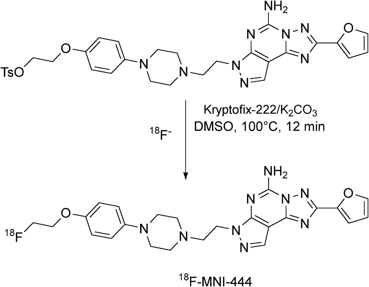
    Radiosynthesis of 18F-MNI-444 from its tosylate precursor. DMSO = dimethyl sulfoxide.

  • FIGURE 2.
    • Download figure
    • Open in new tab
    • Download powerpoint
    FIGURE 2.

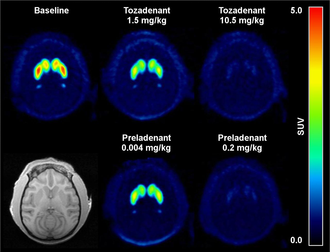
    Average 18F-MNI-444 PET images over 180 min for rhesus macaque in transverse plane at baseline and after dosing with tozadenant at 1.5 and 10.5 mg/kg (occupancy of 47% and 95%, respectively) or with preladenant at 0.004 and 0.2 mg/kg (occupancy of 32% and 90%, respectively). Monkey individual MR image is also shown. SUV = standardized uptake value.

  • FIGURE 3.
    • Download figure
    • Open in new tab
    • Download powerpoint
    FIGURE 3.

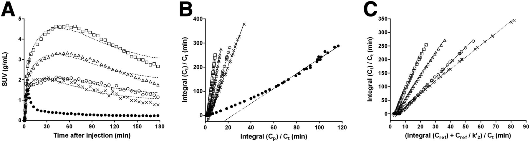
    (A) Representative time–activity curves at baseline for rhesus macaque in some brain regions after bolus injection of 18F-MNI-444. Dashed lines represent SRTM fit. (B) LGA with plasma input function. Dashed lines represent linear regression with t* set to 60 min. (C) NI-LGA with reference region input function. Dashed lines represent linear regression with t* set to 60 min and k′2 set to 0.35 min−1. △ = caudate; □ = putamen; ○ = globus pallidus; × = nucleus accumbens; ● = cerebellum.

  • FIGURE 4.
    • Download figure
    • Open in new tab
    • Download powerpoint
    FIGURE 4.

    Correlation of SRTM and NI-LGA BPND with LGA BPND for acquisitions of 120 min (A) or 180 min (B). ○ = SRTM; △ = NI-LGA; solid line = line of identity; dashed line = SRTM linear regression fit; dotted line = NI-LGA linear regression fit.

  • FIGURE 5.
    • Download figure
    • Open in new tab
    • Download powerpoint
    FIGURE 5.

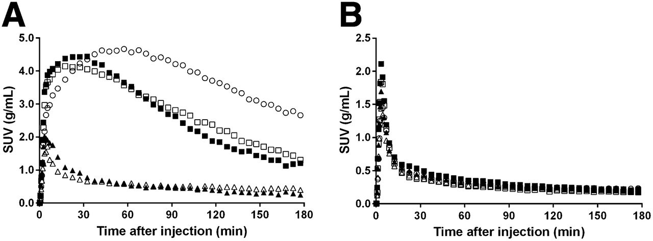
    Time–activity curves for rhesus macaque in putamen (A) and cerebellum (B) after bolus injection of 18F-MNI-444 at baseline and after dosing with tozadenant or preladenant. ○ = baseline scan; ■ = tozadenant, 1.5 mg/kg; □ = preladenant, 0.004 mg/kg; ▲ = tozadenant, 10.5 mg/kg; △ = preladenant, 0.2 mg/kg.
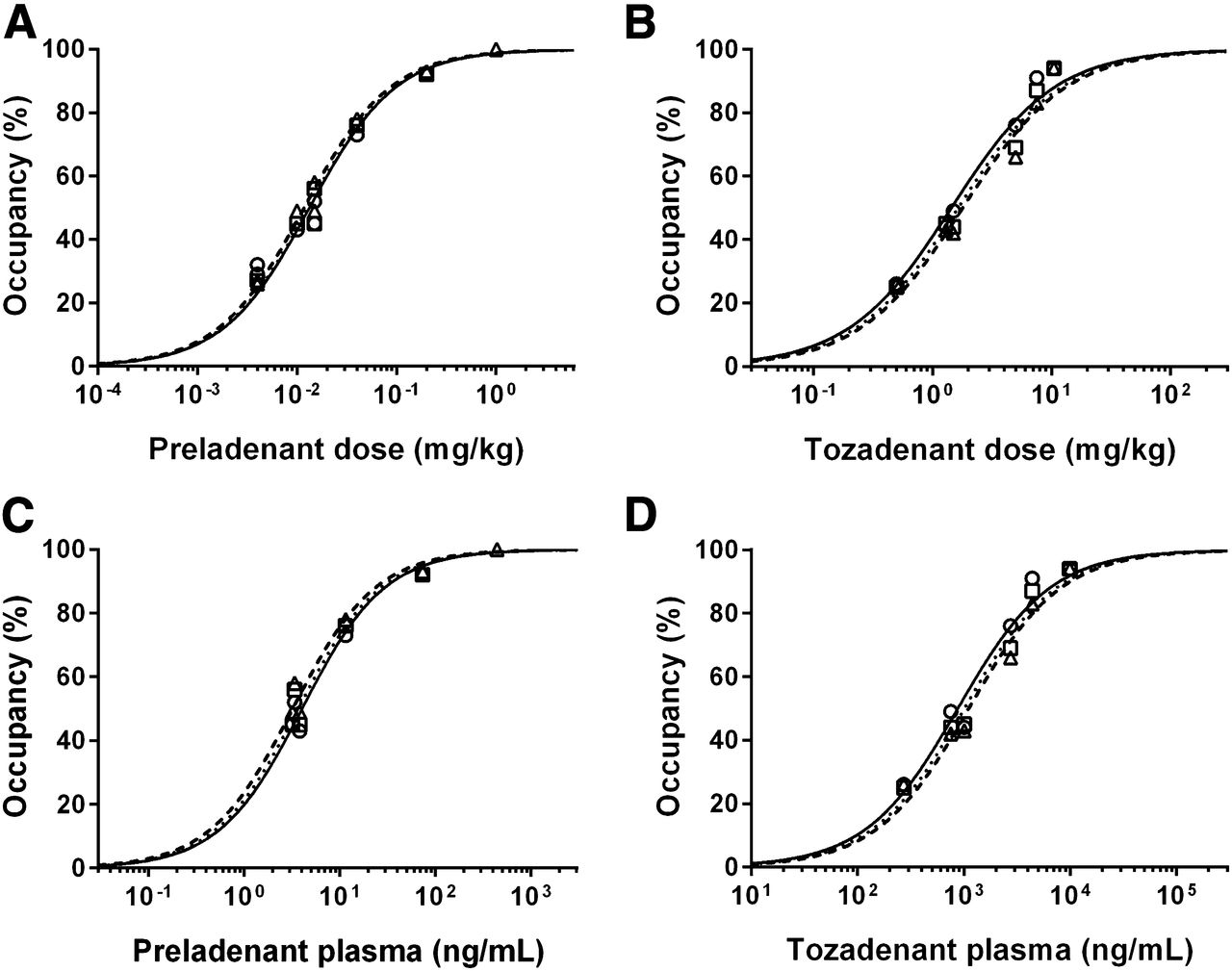
  • FIGURE 6.
    • Download figure
    • Open in new tab
    • Download powerpoint
    FIGURE 6.

    Striatal A2A RO against preladenant (A) or tozadenant (B) administered doses, and against preladenant (C) or tozadenant (D) plasma levels at beginning of scan. Symbols represent occupancy estimates using LGA (○), SRTM (△), or NI-LGA (□) for 120 min of data. Lines represent model fits using LGA (solid line), SRTM (dashed line), or NI-LGA (dotted lines) occupancy estimates.
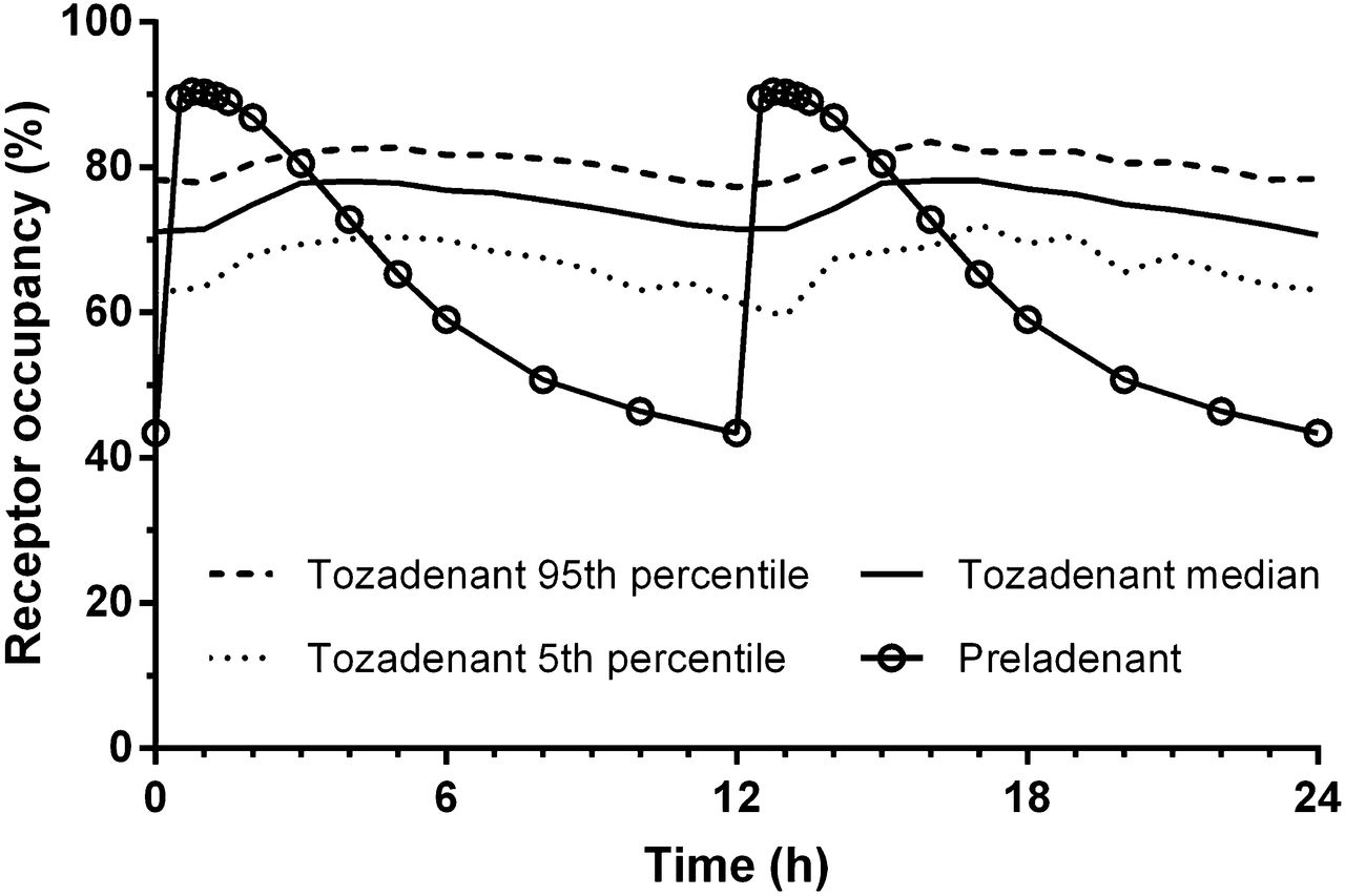
  • FIGURE 7.
    • Download figure
    • Open in new tab
    • Download powerpoint
    FIGURE 7.

    Pharmacokinetic modeling of tozadenant and preladenant plasma concentrations in humans was used to predict A2A RO after repeated dosing (180 mg BID of tozadenant and 10 mg BID of preladenant).

Tables

  • Figures
  • Additional Files
    • View popup
    TABLE 1

    18F-MNI-444 VT and BPND at Baseline for Acquisitions of 180 Minutes

    RegionVT LGABPND LGABPND SRTMBPND NI-LGA
    Caudate21.9 ± 3.05.5 ± 1.56.8 ± 1.06.3 ± 1.0
    Putamen30.3 ± 5.88.0 ± 2.79.6 ± 1.99.1 ± 2.0
    Globus pallidus16.1 ± 3.93.6 ± 0.74.5 ± 0.94.2 ± 0.9
    Nucleus accumbens12.4 ± 2.12.6 ± 0.43.5 ± 0.43.2 ± 0.3
    Frontal cortex4.7 ± 1.30.3 ± 0.10.2 ± 0.10.2 ± 0.1
    Temporal cortex3.5 ± 0.60.0 ± 0.00.0 ± 0.00.0 ± 0.0
    Occipital cortex3.6 ± 0.30.1 ± 0.10.1 ± 0.10.1 ± 0.1
    Cerebellum3.5 ± 0.7NANANA
    • NA = not applicable.

    • Data are mean ± SD (n = 6).

    • View popup
    TABLE 2

    Preladenant and Tozadenant A2A RO

    Occupancy (%) for 180 min of dataOccupancy (%) for 120 min of data
    DrugDose (mg/kg)LGASRTMNI-LGALGASRTMNI-LGA
    Preladenant1.0103100101102100101
    0.2909291929392
    0.04697673737876
    0.015394542454945
    0.015535655525856
    0.010354440434945
    0.004322627322727
    0.004232525292627
    Tozadenant10.5959494949494
    7.5938488918387
    5.0766871766669
    1.5474244494244
    1.3434446444345
    0.5282627262625

Additional Files

  • Figures
  • Tables
  • Supplemental Data

    Files in this Data Supplement:

    • Supplemental Data
PreviousNext
Back to top

In this issue

Journal of Nuclear Medicine: 55 (10)
Journal of Nuclear Medicine
Vol. 55, Issue 10
October 1, 2014
  • Table of Contents
  • Table of Contents (PDF)
  • About the Cover
  • Index by author
Print
Download PDF
Article Alerts
Sign In to Email Alerts with your Email Address
Email Article

Thank you for your interest in spreading the word on Journal of Nuclear Medicine.

NOTE: We only request your email address so that the person you are recommending the page to knows that you wanted them to see it, and that it is not junk mail. We do not capture any email address.

Enter multiple addresses on separate lines or separate them with commas.
Adenosine 2A Receptor Occupancy by Tozadenant and Preladenant in Rhesus Monkeys
(Your Name) has sent you a message from Journal of Nuclear Medicine
(Your Name) thought you would like to see the Journal of Nuclear Medicine web site.
Citation Tools
Adenosine 2A Receptor Occupancy by Tozadenant and Preladenant in Rhesus Monkeys
Olivier Barret, Jonas Hannestad, David Alagille, Christine Vala, Adriana Tavares, Caroline Papin, Thomas Morley, Krista Fowles, Hsiaoju Lee, John Seibyl, Dominique Tytgat, Marc Laruelle, Gilles Tamagnan
Journal of Nuclear Medicine Oct 2014, 55 (10) 1712-1718; DOI: 10.2967/jnumed.114.142067

Citation Manager Formats

  • BibTeX
  • Bookends
  • EasyBib
  • EndNote (tagged)
  • EndNote 8 (xml)
  • Medlars
  • Mendeley
  • Papers
  • RefWorks Tagged
  • Ref Manager
  • RIS
  • Zotero
Share
Adenosine 2A Receptor Occupancy by Tozadenant and Preladenant in Rhesus Monkeys
Olivier Barret, Jonas Hannestad, David Alagille, Christine Vala, Adriana Tavares, Caroline Papin, Thomas Morley, Krista Fowles, Hsiaoju Lee, John Seibyl, Dominique Tytgat, Marc Laruelle, Gilles Tamagnan
Journal of Nuclear Medicine Oct 2014, 55 (10) 1712-1718; DOI: 10.2967/jnumed.114.142067
Twitter logo Facebook logo LinkedIn logo Mendeley logo
  • Tweet Widget
  • Facebook Like
  • Google Plus One
Bookmark this article

Jump to section

  • Article
    • Abstract
    • MATERIALS AND METHODS
    • RESULTS
    • DISCUSSION
    • CONCLUSION
    • DISCLOSURE
    • Footnotes
    • REFERENCES
  • Figures & Data
  • Supplemental
  • Info & Metrics
  • PDF

Related Articles

  • This Month in JNM
  • PubMed
  • Google Scholar

Cited By...

  • In Vivo Evaluation of 11C-Preladenant for PET Imaging of Adenosine A2A Receptors in the Conscious Monkey
  • Preclinical Evaluation and Quantification of 18F-Fluoroethyl and 18F-Fluoropropyl Analogs of SCH442416 as Radioligands for PET Imaging of the Adenosine A2A Receptor in Rat Brain
  • Characterization in Humans of 18F-MNI-444, a PET Radiotracer for Brain Adenosine 2A Receptors
  • Google Scholar

More in this TOC Section

  • 11C-Methionine PET of Myocardial Inflammation in a Rat Model of Experimental Autoimmune Myocarditis
  • Counting Rate Characteristics and Image Distortion in Preclinical PET Imaging During Radiopharmaceutical Therapy
  • Design and Fabrication of Kidney Phantoms for Internal Radiation Dosimetry Using 3D Printing Technology
Show more Basic Science Investigations

Similar Articles

Keywords

  • A2A receptors PET imaging
  • Parkinson’s disease
  • receptor occupancy
  • tozadenant
  • preladenant
SNMMI

© 2025 SNMMI

Powered by HighWire