
Main menu

  • Home
  • Content
    • Current
    • Ahead of print
    • Past Issues
    • JNM Supplement
    • SNMMI Annual Meeting Abstracts
    • Continuing Education
    • JNM Podcasts
  • Subscriptions
    • Subscribers
    • Institutional and Non-member
    • Rates
    • Journal Claims
    • Corporate & Special Sales
  • Authors
    • Submit to JNM
    • Information for Authors
    • Assignment of Copyright
    • AQARA requirements
  • Info
    • Reviewers
    • Permissions
    • Advertisers
  • About
    • About Us
    • Editorial Board
    • Contact Information
  • More
    • Alerts
    • Feedback
    • Help
    • SNMMI Journals
  • SNMMI
    • JNM
    • JNMT
    • SNMMI Journals
    • SNMMI

User menu

  • Subscribe
  • My alerts
  • Log in
  • My Cart

Search

  • Advanced search
Journal of Nuclear Medicine
  • SNMMI
    • JNM
    • JNMT
    • SNMMI Journals
    • SNMMI
  • Subscribe
  • My alerts
  • Log in
  • My Cart
Journal of Nuclear Medicine

Advanced Search

  • Home
  • Content
    • Current
    • Ahead of print
    • Past Issues
    • JNM Supplement
    • SNMMI Annual Meeting Abstracts
    • Continuing Education
    • JNM Podcasts
  • Subscriptions
    • Subscribers
    • Institutional and Non-member
    • Rates
    • Journal Claims
    • Corporate & Special Sales
  • Authors
    • Submit to JNM
    • Information for Authors
    • Assignment of Copyright
    • AQARA requirements
  • Info
    • Reviewers
    • Permissions
    • Advertisers
  • About
    • About Us
    • Editorial Board
    • Contact Information
  • More
    • Alerts
    • Feedback
    • Help
    • SNMMI Journals
  • View or Listen to JNM Podcast
  • Visit JNM on Facebook
  • Join JNM on LinkedIn
  • Follow JNM on Twitter
  • Subscribe to our RSS feeds
Research ArticleClinical Investigations

Comparison of the Amino Acid Tracers 18F-FET and 18F-DOPA in High-Grade Glioma Patients

Constantin Lapa, Thomas Linsenmann, Camelia Maria Monoranu, Samuel Samnick, Andreas K. Buck, Christina Bluemel, Johannes Czernin, Almuth F. Kessler, Gyoergy A. Homola, Ralf-Ingo Ernestus, Mario Löhr and Ken Herrmann
Journal of Nuclear Medicine October 2014, 55 (10) 1611-1616; DOI: https://doi.org/10.2967/jnumed.114.140608
Constantin Lapa
1Department of Nuclear Medicine, University Hospital Würzburg, Würzburg, Germany
  • Find this author on Google Scholar
  • Find this author on PubMed
  • Search for this author on this site
Thomas Linsenmann
2Department of Neurosurgery, University Hospital Würzburg, Würzburg, Germany
  • Find this author on Google Scholar
  • Find this author on PubMed
  • Search for this author on this site
Camelia Maria Monoranu
3Department of Neuropathology, Institute of Pathology, University of Würzburg, Würzburg, Germany
  • Find this author on Google Scholar
  • Find this author on PubMed
  • Search for this author on this site
Samuel Samnick
1Department of Nuclear Medicine, University Hospital Würzburg, Würzburg, Germany
  • Find this author on Google Scholar
  • Find this author on PubMed
  • Search for this author on this site
Andreas K. Buck
1Department of Nuclear Medicine, University Hospital Würzburg, Würzburg, Germany
  • Find this author on Google Scholar
  • Find this author on PubMed
  • Search for this author on this site
Christina Bluemel
1Department of Nuclear Medicine, University Hospital Würzburg, Würzburg, Germany
  • Find this author on Google Scholar
  • Find this author on PubMed
  • Search for this author on this site
Johannes Czernin
4Ahmanson Translational Imaging Division, Department of Molecular and Medical Pharmacology, David Geffen School of Medicine at UCLA, Los Angeles, California; and
  • Find this author on Google Scholar
  • Find this author on PubMed
  • Search for this author on this site
Almuth F. Kessler
2Department of Neurosurgery, University Hospital Würzburg, Würzburg, Germany
  • Find this author on Google Scholar
  • Find this author on PubMed
  • Search for this author on this site
Gyoergy A. Homola
5Department of Neuroradiology, University Hospital Würzburg, Würzburg, Germany
  • Find this author on Google Scholar
  • Find this author on PubMed
  • Search for this author on this site
Ralf-Ingo Ernestus
2Department of Neurosurgery, University Hospital Würzburg, Würzburg, Germany
  • Find this author on Google Scholar
  • Find this author on PubMed
  • Search for this author on this site
Mario Löhr
2Department of Neurosurgery, University Hospital Würzburg, Würzburg, Germany
  • Find this author on Google Scholar
  • Find this author on PubMed
  • Search for this author on this site
Ken Herrmann
1Department of Nuclear Medicine, University Hospital Würzburg, Würzburg, Germany
4Ahmanson Translational Imaging Division, Department of Molecular and Medical Pharmacology, David Geffen School of Medicine at UCLA, Los Angeles, California; and
  • Find this author on Google Scholar
  • Find this author on PubMed
  • Search for this author on this site
  • Article
  • Figures & Data
  • Info & Metrics
  • PDF
Loading

Article Figures & Data

Figures

  • Tables
  • FIGURE 1.
    • Download figure
    • Open in new tab
    • Download powerpoint
    FIGURE 1.

    Schematic outline of study. Within 35 d before tumor surgery or biopsy, MR imaging (mean, 15 d prior) and 18F-FET PET/CT (mean, 8 d) and 18F-DOPA PET/CT (mean, 6 d) were performed. Histopathology served as gold standard. Mean intervals to surgery are noted in time scale.
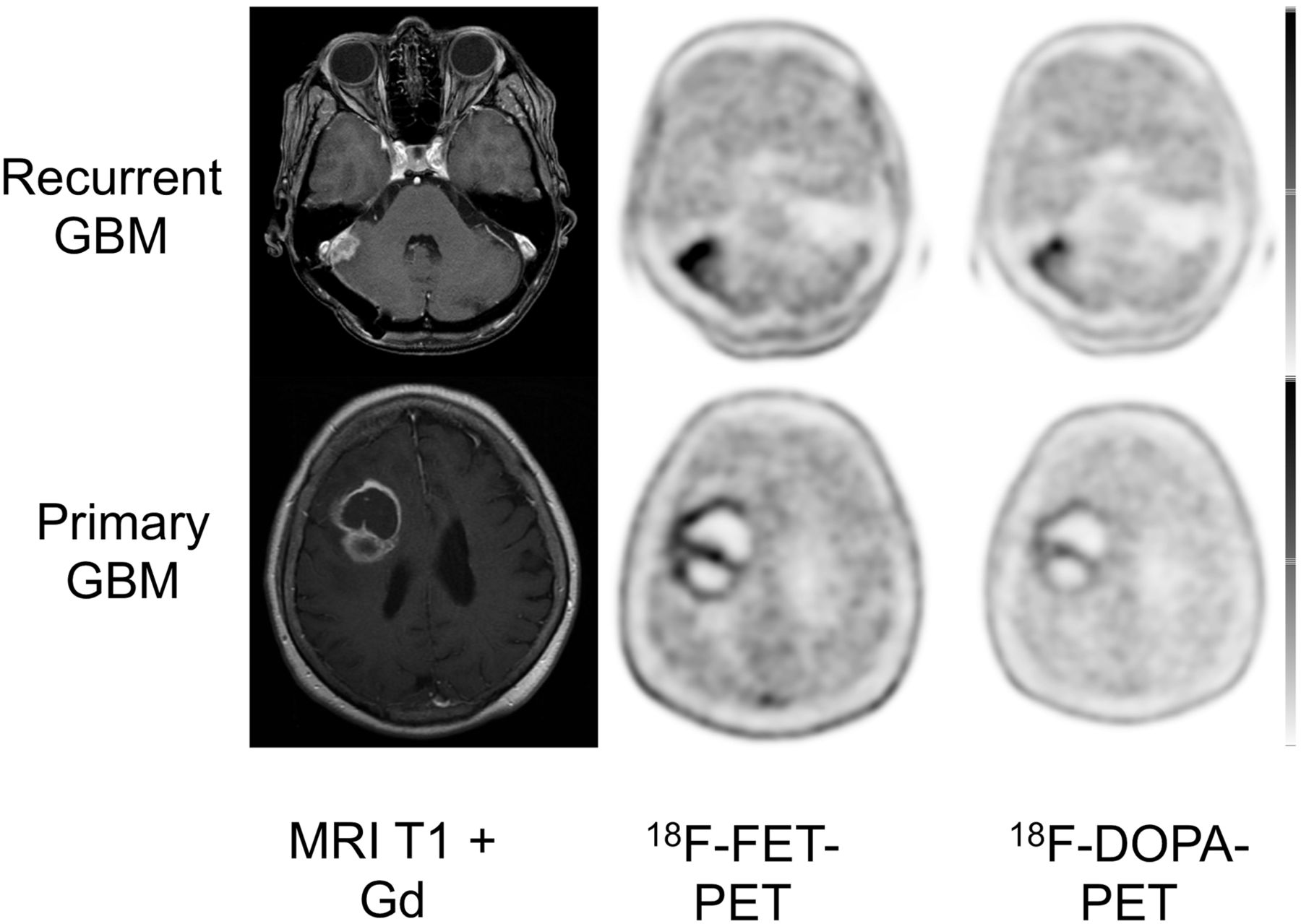
  • FIGURE 2.
    • Download figure
    • Open in new tab
    • Download powerpoint
    FIGURE 2.

    Display of transaxial contrast-enhanced T1-weighted MR imaging, 18F-FET, and 18F-DOPA PET/CT scans of patient with recurrent GBM and primary GBM. In both patients, 18F-FET uptake (SUVmax and SUVmean, 6.1 and 5.6, respectively) was higher than that of 18F-DOPA (SUVmax and SUVmean, 4.4 and 2.5, respectively).
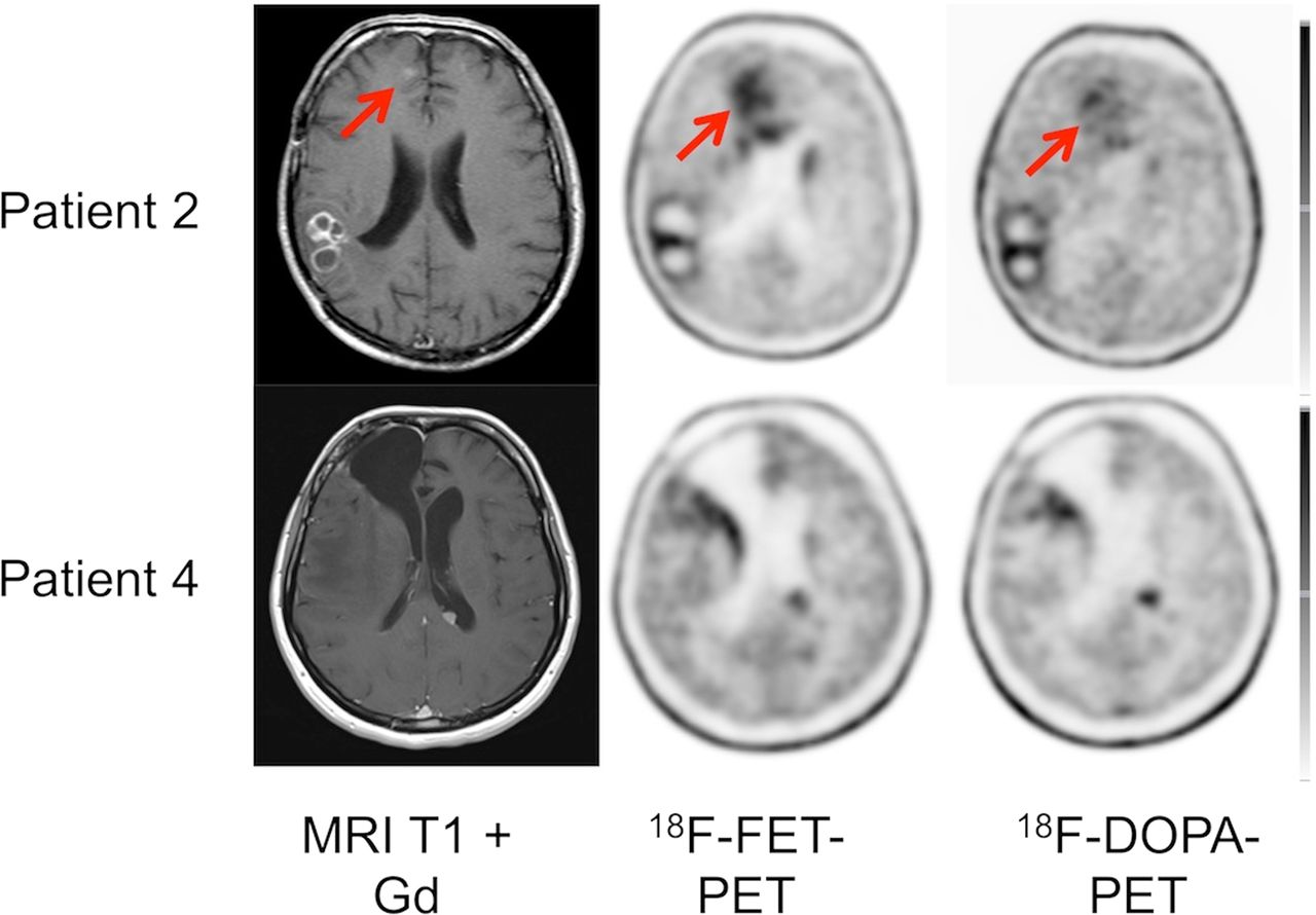
  • FIGURE 3.
    • Download figure
    • Open in new tab
    • Download powerpoint
    FIGURE 3.

    Display of transaxial contrast-enhanced T1-weighted MR imaging, 18F-FET, and 18F-DOPA PET/CT scans of 2 patients with multifocal GBM. In both patients, 18F-FET and 18F-DOPA depicted all lesions.

  • FIGURE 4.
    • Download figure
    • Open in new tab
    • Download powerpoint
    FIGURE 4.

    Display of transaxial 18F-FET and 18F-DOPA PET/CT scans of patient with primary glioma affecting basal ganglia. Striatal uptake does not significantly compromise tumor delineation.

Tables

  • Figures
    • View popup
    TABLE 1

    Patients’ Characteristics

    Patient no.Age (y)SexPrimary/recurrent HGGHistologyLesion sitePrevious therapy
    147MRPrimary GBMLeft temporalSurgery, radiochemotherapy
    245FRSecondary GBMMultifocalSurgery, radiochemotherapy
    349MRPrimary GBMLeft frontalSurgery, radiochemotherapy
    450MRPrimary GBMMultifocalSurgery, radiochemotherapy
    573MRPrimary GBMLeft frontalSurgery, radiochemotherapy
    642MRPrimary GBMRight parietooccipitalSurgery, radiochemotherapy
    743MRPrimary GBMLeft parietalSurgery, radiochemotherapy
    841FRAnaplastic astrocytoma IIILeft temporalSurgery, radiochemotherapy
    944MRAnaplastic astrocytoma IIIRight temporalSurgery, radiochemotherapy
    1070FRPrimary GBMLeft temporalSurgery, radiochemotherapy
    1151FRPrimary GBMLeft parietalSurgery, radiochemotherapy
    1260MRPrimary GBMLeft temporalSurgery, radiochemotherapy
    1367MRPrimary GBMLeft frontalSurgery, radiochemotherapy
    1455MRPrimary GBMLeft temporalSurgery, radiochemotherapy
    1554FRPrimary GBMRight cerebellarSurgery, radiochemotherapy
    1633MROligoastrocytoma IIILeft frontalSurgery, radiotherapy
    1757MRPrimary GBMRight parietalSurgery, radiochemotherapy
    1858MRPrimary GBMRight temporooccipitalSurgery, radiochemotherapy
    1961MRPrimary GBMRight parietalSurgery, radiochemotherapy
    14/255MRPrimary GBMLeft parietalSurgery, radiochemotherapy
    17/255MRPrimary GBMRight parietalSurgery, radiochemotherapy
    2025MRSecondary GBMRight temporalSurgery, radiotherapy
    2175MRPrimary GBMRight temporalSurgery, radiochemotherapy
    10/271FRPrimary GBMLeft temporalSurgery, radiochemotherapy
    2233MRPrimary GBMRight frontalSurgery, radiochemotherapy
    2377FPPrimary GBMRight frontalNone
    2480FPPrimary GBMLeft parietooccipitalNone
    2551MPPrimary GBMRight frontalNone
    2665FPPrimary GBMLeft temporalNone
    2740FPPilocytic astrocytoma iRight basal gangliaNone
    • 10/2; 14/2; 17/2 = 3 patients were imaged twice.

    • View popup
    TABLE 2

    Semiquantitative Analysis of 18F-DOPA and 18F-FET

    Parameter18F-FET SUVmean18F-DOPA SUVmean18F-FET SUVmax18F-DOPA SUVmax18F-FET TBR SUVmean18F-DOPA TBR SUVmean18F-FET TBR SUVmax18F-DOPA TBR SUVmax
    All
     Mean4.03.54.94.33.83.43.33.0
     SD2.01.62.32.01.71.21.61.1
     Range2.0–11.92.0–9.22.2–13.32.3–10.32.0–10.82.1–8.41.8–9.52.0–6.9
     P<0.001<0.0010.0040.086
    Newly diagnosed HGG
     Mean4.43.55.64.74.33.63.63.3
     SD1.41.72.02.71.41.21.31.3
     Range3.2–6.82.5–6.54.0–9.02.9–9.42.9–6.22.6–5.42.7–6.02.2–5.5
     P0.0430.080.0430.225
    Recurrent HGG
     Mean3.93.54.74.23.73.43.23.0
     SD2–11.72.41.81.81.21.61.0
     Range2.0–11.92.0–9.22.2–13.32.3–10.32.0–10.82.1–8.41.8–9.52.0–6.9
     P0.0080.0030.0340.201
    • Respective SUVmax and SUVmean and tumor-to-background values for 18F-DOPA and 18F-FET for entire cohort and both subgroups (newly diagnosed and recurrent HGG).

PreviousNext
Back to top

In this issue

Journal of Nuclear Medicine: 55 (10)
Journal of Nuclear Medicine
Vol. 55, Issue 10
October 1, 2014
  • Table of Contents
  • Table of Contents (PDF)
  • About the Cover
  • Index by author
Print
Download PDF
Article Alerts
Sign In to Email Alerts with your Email Address
Email Article

Thank you for your interest in spreading the word on Journal of Nuclear Medicine.

NOTE: We only request your email address so that the person you are recommending the page to knows that you wanted them to see it, and that it is not junk mail. We do not capture any email address.

Enter multiple addresses on separate lines or separate them with commas.
Comparison of the Amino Acid Tracers 18F-FET and 18F-DOPA in High-Grade Glioma Patients
(Your Name) has sent you a message from Journal of Nuclear Medicine
(Your Name) thought you would like to see the Journal of Nuclear Medicine web site.
Citation Tools
Comparison of the Amino Acid Tracers 18F-FET and 18F-DOPA in High-Grade Glioma Patients
Constantin Lapa, Thomas Linsenmann, Camelia Maria Monoranu, Samuel Samnick, Andreas K. Buck, Christina Bluemel, Johannes Czernin, Almuth F. Kessler, Gyoergy A. Homola, Ralf-Ingo Ernestus, Mario Löhr, Ken Herrmann
Journal of Nuclear Medicine Oct 2014, 55 (10) 1611-1616; DOI: 10.2967/jnumed.114.140608

Citation Manager Formats

  • BibTeX
  • Bookends
  • EasyBib
  • EndNote (tagged)
  • EndNote 8 (xml)
  • Medlars
  • Mendeley
  • Papers
  • RefWorks Tagged
  • Ref Manager
  • RIS
  • Zotero
Share
Comparison of the Amino Acid Tracers 18F-FET and 18F-DOPA in High-Grade Glioma Patients
Constantin Lapa, Thomas Linsenmann, Camelia Maria Monoranu, Samuel Samnick, Andreas K. Buck, Christina Bluemel, Johannes Czernin, Almuth F. Kessler, Gyoergy A. Homola, Ralf-Ingo Ernestus, Mario Löhr, Ken Herrmann
Journal of Nuclear Medicine Oct 2014, 55 (10) 1611-1616; DOI: 10.2967/jnumed.114.140608
Twitter logo Facebook logo LinkedIn logo Mendeley logo
  • Tweet Widget
  • Facebook Like
  • Google Plus One
Bookmark this article

Jump to section

  • Article
    • Abstract
    • MATERIALS AND METHODS
    • RESULTS
    • DISCUSSION
    • CONCLUSION
    • DISCLOSURE
    • Footnotes
    • REFERENCES
  • Figures & Data
  • Info & Metrics
  • PDF

Related Articles

  • This Month in JNM
  • PubMed
  • Google Scholar

Cited By...

  • Diagnostic Accuracy of PET Tracers for the Differentiation of Tumor Progression from Treatment-Related Changes in High-Grade Glioma: A Systematic Review and Metaanalysis
  • Evaluation of 11C-Methionine PET and Anatomic MRI Associations in Diffuse Intrinsic Pontine Glioma
  • Google Scholar

More in this TOC Section

  • Feasibility of Ultra-Low-Activity 18F-FDG PET/CT Imaging Using a Long–Axial-Field-of-View PET/CT System
  • Cardiac Presynaptic Sympathetic Nervous Function Evaluated by Cardiac PET in Patients with Chronotropic Incompetence Without Heart Failure
  • Validation and Evaluation of a Vendor-Provided Head Motion Correction Algorithm on the uMI Panorama PET/CT System
Show more Clinical Investigations

Similar Articles

Keywords

  • Glioblastoma
  • Positron Emission Tomography
  • PET
  • amino acids
  • FET
  • F-DOPA
SNMMI

© 2025 SNMMI

Powered by HighWire